Jump to content
In this section we have spot diagnoses posted on a daily basis since June 2010, now over 4000! You can review the archived cases and read the suggested diagnoses by users and the final comment by the contributors.
Case are uploaded each week day by 10 am UK time with the correct diagnosis will generally be posted at 8 pm UK time. Why not view the most recent spot diagnosis and proffer a diagnosis?

Case Number : Case 712 - 8 Mar Posted By: Guest

Please read the clinical history and view the images by clicking on them before you proffer your diagnosis.
Submitted Date :
   (0 reviews)

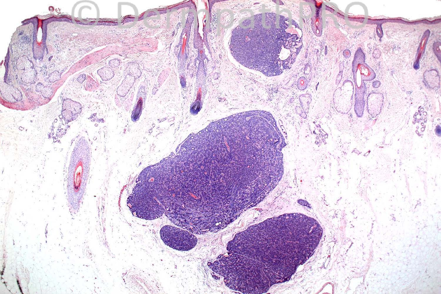
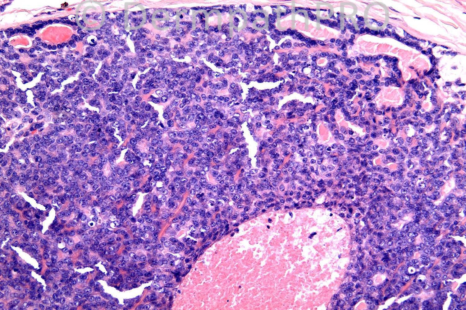
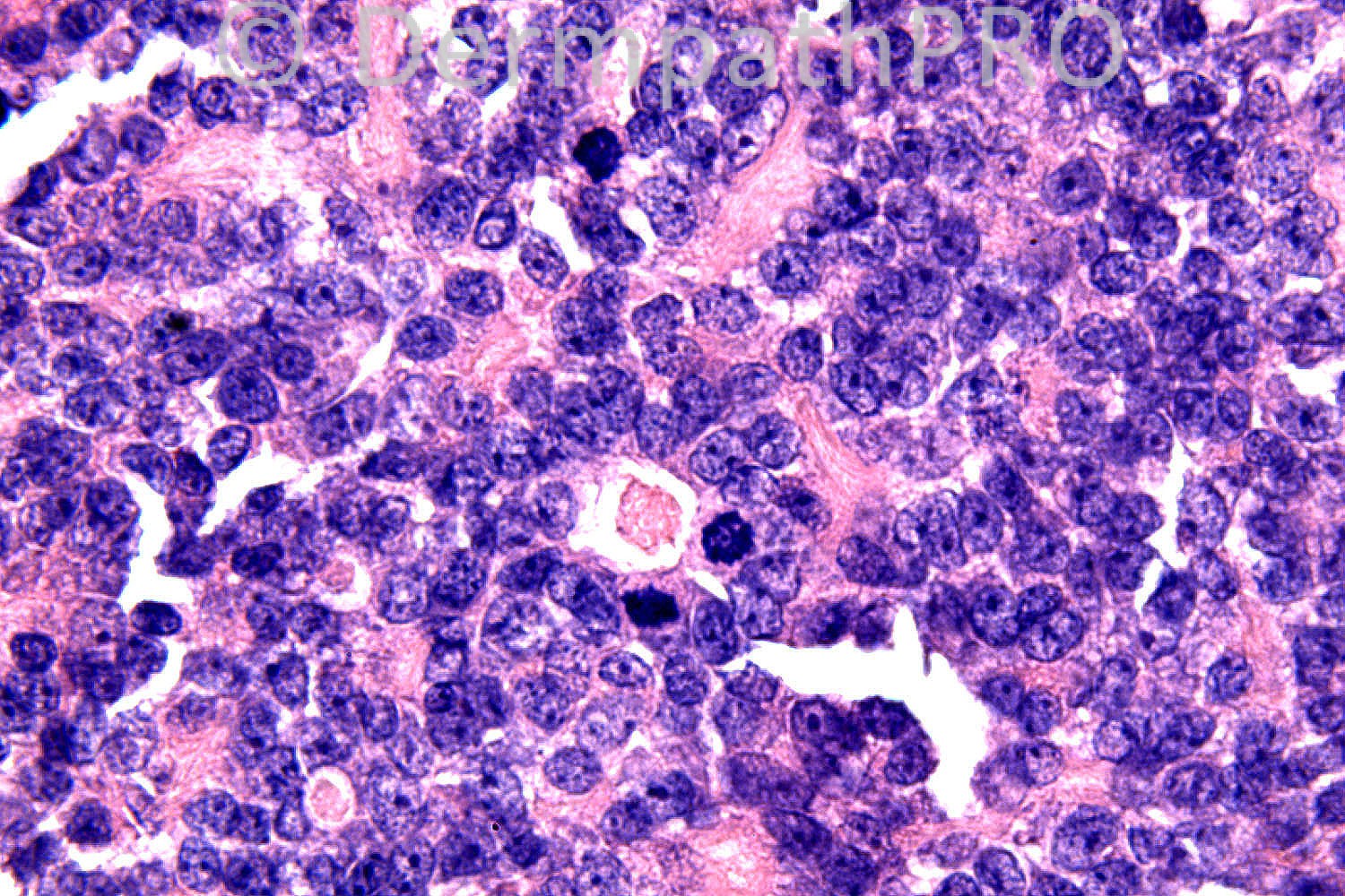
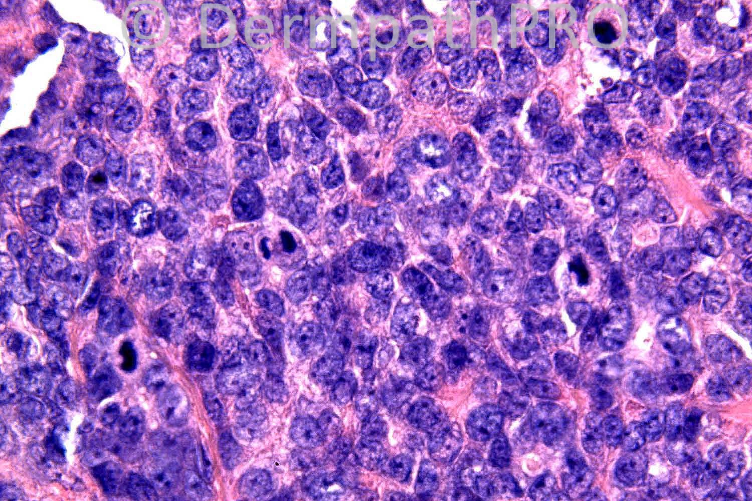
54 years-old male. Scalp. Tumour at vertex. ?Appendageal.

Case posted by Dr. Richard Carr


  Report Record

User Feedback


Most probable a metastatic adenoCa

Share this comment


Link to comment
share_externally

Guest Guillermo Solis

Posted

Agree with metastatic adenocarcinoma. Spiradenocarcinoma as differential diagnosis.

Share this comment


Link to comment
share_externally

Azza Esmat

Posted

Spiradenocarcinoma

Share this comment


Link to comment
share_externally

Robledo F. Rocha

Posted

Large pale cells and small basaloid ones forming solid cords and ductal structures embedded in hyalinized stroma. Frequent mitotic figures, foci of necrosis and atypical nuclei confirm this as a malignant tumor.
I favor malignant eccrine spiradenoma. Given this is an exceedingly rare tumor, metastatic adenocarcinoma must be ruled out.

Share this comment


Link to comment
share_externally

Dr. Mona Abdel-Halim

Posted

Spiradenocarcinoma

Share this comment


Link to comment
share_externally

Agree entirely with Dr Rocha.

Share this comment


Link to comment
share_externally

Dr. Richard Carr

Posted

This was a 5mm diameter solitary lesion on the scalp. You are seeing the lesion
in it's entirety and the lesion lacks cytological atypia. Perhpas a re-think?
I am not saying this is easy as very high power images of slightly thick
sections can play tricks!!

Share this comment


Link to comment
share_externally

nick turnbull

Posted

Malignant trichoblastoma?

Share this comment


Link to comment
share_externally

Sasi Attili

Posted

Trust you to come up with a googly on a Friday Richard!! I thought about Trichoblastoma, but did not see any PMC's. How about a BCC?

Share this comment


Link to comment
share_externally

nick turnbull

Posted

small blue cell? moulding? Merkel?

Share this comment


Link to comment
share_externally

Guest Maria AntoniaPastor

Posted

Spiradenocarcinoma

Share this comment


Link to comment
share_externally

Guest Jim Davie MD

Posted

Agree with Dr. Rocha's description. For me, the dual layer acinar architecture with eosinophilic secretions, prominent vasculature, hint of infiltrative architecture at the edges of the nodules, mild-moderate nuclear pleomorphism/atypia and high mitotic rate would have me side with the opinions of malignant eccrine adenocarcinoma (eccrine spiradenocarcinoma). Difficult to call on the image: necrosis vs. central vessel. Not sure I could call this benign on these pics.

Share this comment


Link to comment
share_externally

Dr. Mona Abdel-Halim

Posted

The scanning image is typical of spiradenoma, but the many mitoses and the appearence of the cells in the 4th and 5th images made me consider malignant transformation. Seems, i was tricked by the high magnification of slightly thick sections!!!!! So may be this is just spiradenoma!!!!!!

Share this comment


Link to comment
share_externally

Guest Bansal_

Posted

? spiradenoma

Share this comment


Link to comment
share_externally

Robledo F. Rocha

Posted

So, I was tricked by thick high-power sections, which made me interpret nuclei as atypical.
Anyway, frequent mitotic figures really caught my eyes. According to a paper (Cooper PH. Mitotic figures in sweat gland adenomas. J Cutan Pathol 1987;14:10-14.), less than 1 / 10 HPF would be expected in such this benign tumor.
So, I will agree with Dr. Attili and alternatively suggest basal cell carcinoma with appendageal differentiation. On scanning magnification, the uppermost tumor island shows retraction artifact between it and the surrounding stroma.

Share this comment


Link to comment
share_externally

Spiradenocarcinoma

Share this comment


Link to comment
share_externally

Guest Marcia

Posted

Agree with spiradenoma

Share this comment


Link to comment
share_externally

Eman El-Nabarawy

Posted

Spiradenoma.

Share this comment


Link to comment
share_externally

Hanan Vaknine MD

Posted

Spiradenoma with an atypical adenomatous component [b]consistent with an in situ lesion [/b]

Share this comment


Link to comment
share_externally

Guest Dr Engin Sezer

Posted

Conspicious vasculature consistent with spiradenoma

Share this comment


Link to comment
share_externally

Dr. Richard Carr

Posted

Really great contributions. Thank you all for very good contributions. For me I still like to see features of an invasive lesion before calling outright malignant. I entirely agree the silhouette here is just typical spiradenoma. There were 18 mitotic figures per mm squared! Now there does not appear to be any exacting criteria for mitotic activity and malignancy in this setting but I have seen otherwise typical spiradenoma with mitoses (this case is exceptional in being so mitotic). Lymphocytes were reduced. Despite the rather sheet-like growth CK7 and p63 were quite interesting and revealed a nice biphasic population of peripheral basaloid cells (p63) and slightly plumper lumina cells (CK7) throughout, neither population outgrowing the other. p53 was wild type and Ki67 around 30%. The referring pathologist like many contributors here favoured eccrine spiradenocarcinoma. I did not think there were criteria for outright malignancy here (for me mitotic activity alone in an adnexal neoplasm is insufficient) and favoured a mitotically active spiradenoma but recommended follow-up given slight uncertainty over the behaviour. Also cellular necrosis (or degeneration) is not unique to malignant lesions being occasioanlly seen in the centre of large lobules in benign tumours. I shared the case (more images thay you had) with Dr Dmitry Kazakov and Dr Thomas Brenn and neither considered this lesion to be clearly malignant. Yet again I would suggest admitting uncertainty in such difficult cases and certainly sharing the cases with colleagues and other experts.
Regards to all.

Share this comment


Link to comment
share_externally

Dr. Mona Abdel-Halim

Posted

Thank you Dr Carrr, your cases as usual are always highly informative and educational...

Share this comment


Link to comment
share_externally

Dr. Richard Carr

Posted

Thank you both

Share this comment


Link to comment
share_externally


×
×
  • Create New...