Jump to content
In this section we have spot diagnoses posted on a daily basis since June 2010, now over 4000! You can review the archived cases and read the suggested diagnoses by users and the final comment by the contributors.
Case are uploaded each week day by 10 am UK time with the correct diagnosis will generally be posted at 8 pm UK time. Why not view the most recent spot diagnosis and proffer a diagnosis?

Case Number : Case 713 - 11 Mar Posted By: Guest

Please read the clinical history and view the images by clicking on them before you proffer your diagnosis.
Submitted Date :
   (0 reviews)

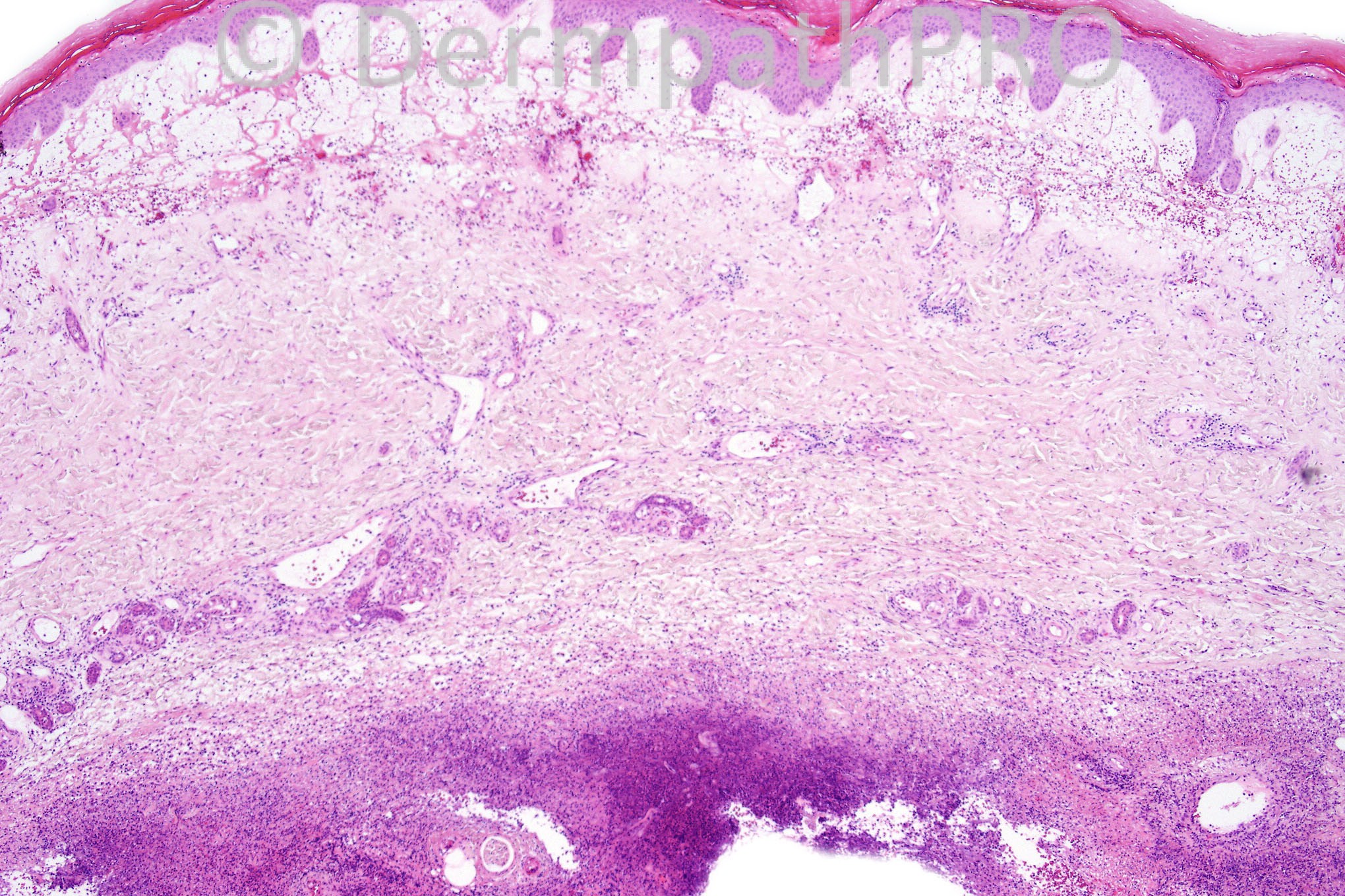
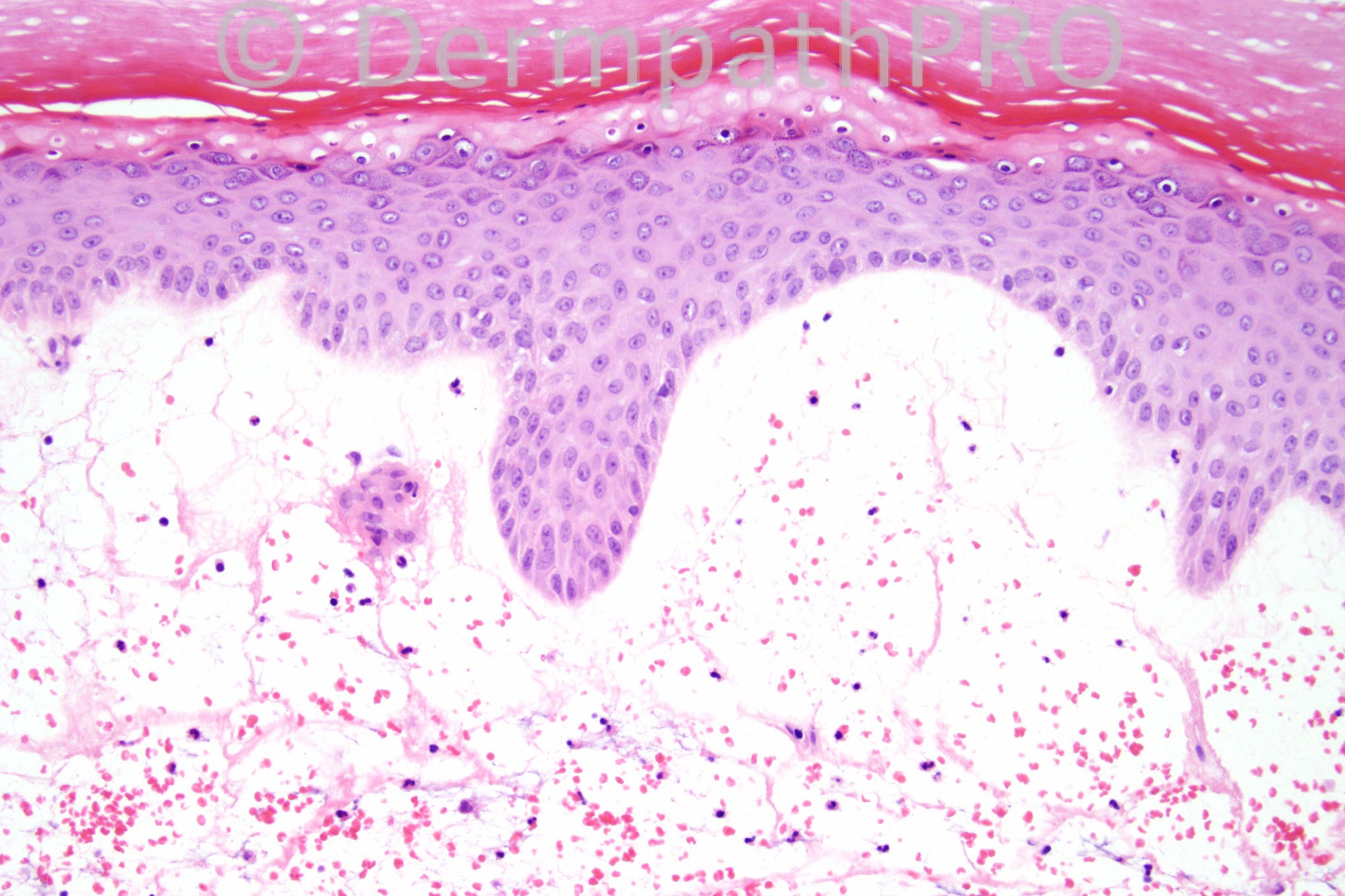
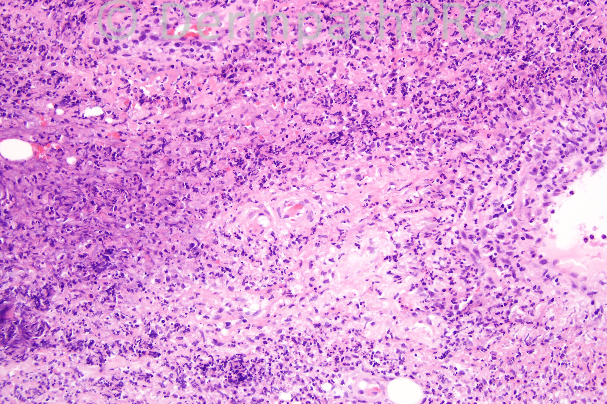
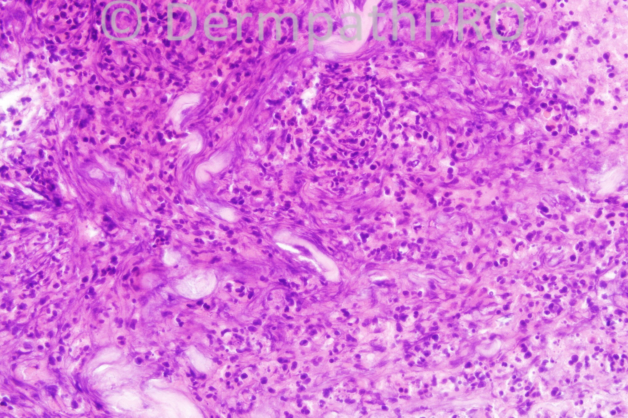
42 years-old male with erythema and edema of the hand.


  Report Record

User Feedback


Eman El-Nabarawy

Posted

Cellulitis/ infective panniculitis?

Share this comment


Link to comment
share_externally

Robledo F. Rocha

Posted

A Lynch-Barrett’s “blue granuloma” in deep reticular dermis composed of dense neutrophilic infiltrate with nuclear dusting and degenerated collagen bundles. A large vessel with inflammatory cells in the wall surrounded by subtle hemorrhage can be found on Picture #1 (bottom right corner). Minimal palisading of histiocytes is present in Picture #3. There’s also a striking edema of papillary dermis with some extravasated erythrocytes.
Wegener’s granulomatosis is my hypothesis.

Share this comment


Link to comment
share_externally

Iskander H. Chaudhry

Posted

Hi All, today's case is [font=arial, sans-serif][size=3]dissecting cellulitis/fasciitis.[/size][/font]

Share this comment


Link to comment
share_externally


×
×
  • Create New...