Jump to content
In this section we have spot diagnoses posted on a daily basis since June 2010, now over 4000! You can review the archived cases and read the suggested diagnoses by users and the final comment by the contributors.
Case are uploaded each week day by 10 am UK time with the correct diagnosis will generally be posted at 8 pm UK time. Why not view the most recent spot diagnosis and proffer a diagnosis?

Case Number : Case 714 - 12 Mar Posted By: Guest

Please read the clinical history and view the images by clicking on them before you proffer your diagnosis.
Submitted Date :
   (0 reviews)

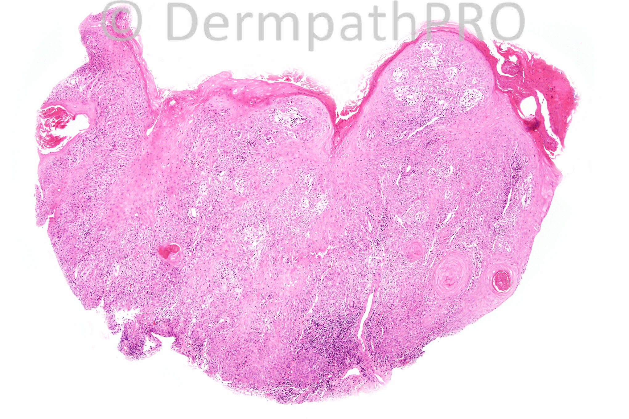
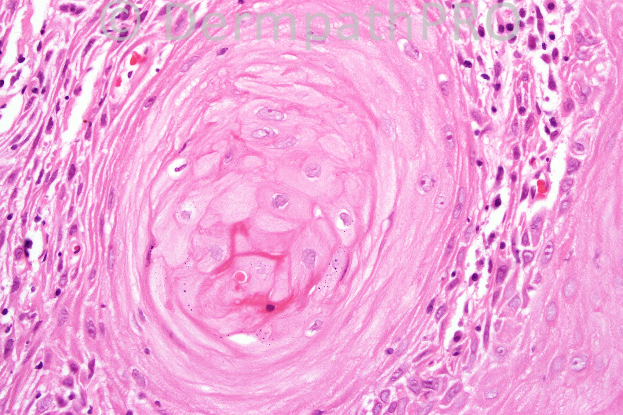
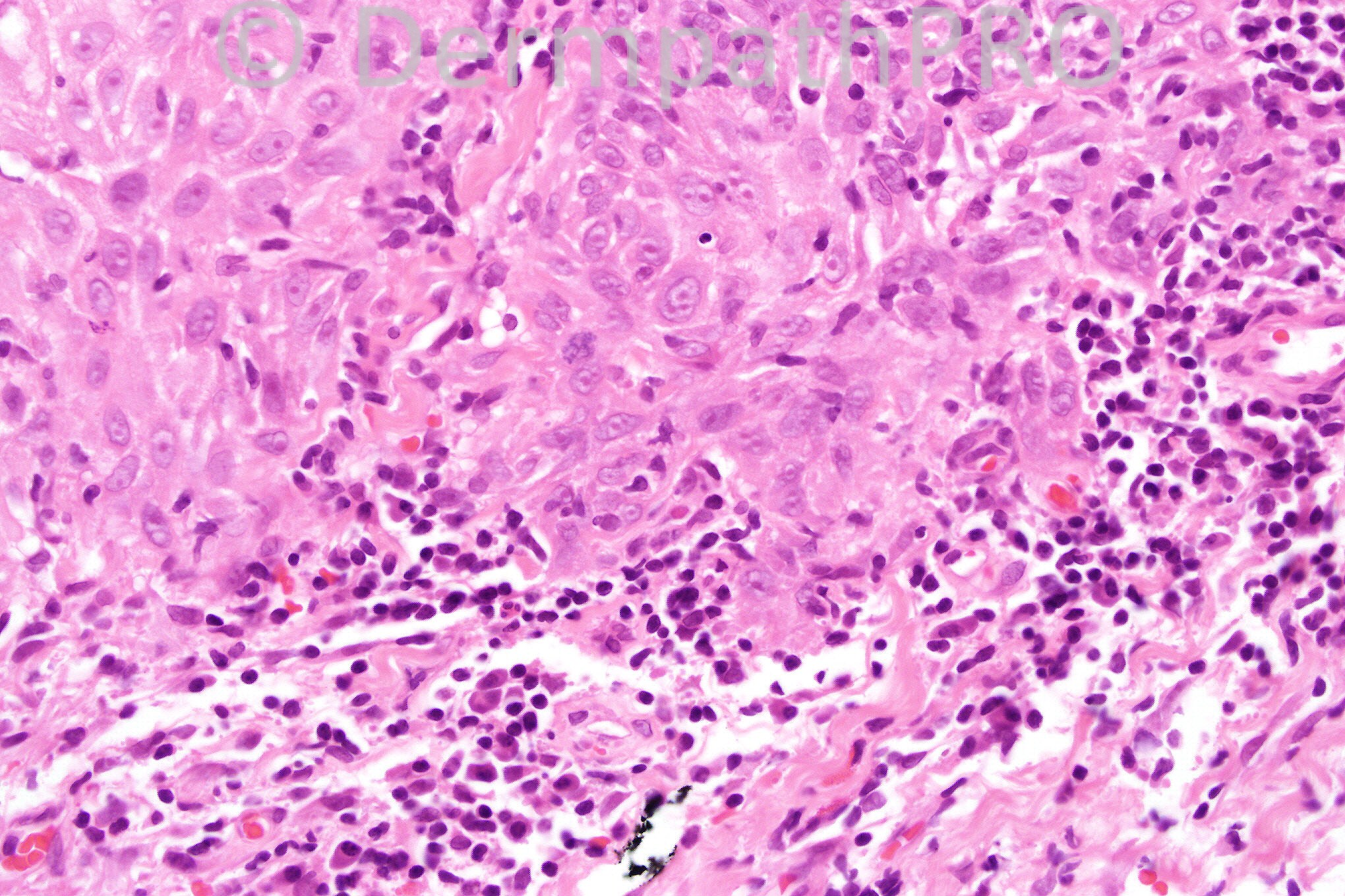
62 years-old female with a rapidly growing 3x12 mm nodule with a central cavity on the ear.


  Report Record

User Feedback


Dr. Mona Abdel-Halim

Posted

Well differentiated keratoacanthomatous like SCC.

Share this comment


Link to comment
share_externally

Eman El-Nabarawy

Posted

Keratoacanthoma.

Share this comment


Link to comment
share_externally

Robledo F. Rocha

Posted

[font=arial, helvetica, sans-serif]Pictures show a lesion that can be a keratoacanthoma or a squamous-cell carcinoma. Differentiate between both is a difficult task that hardly could be achieved on microscopic examination alone. In this particular case, history is more effective than images.[/font]
[font=arial, helvetica, sans-serif]I favor keratoacanthoma after a history of a rapidly growing nodule with a central cavity. However, some microscopic features of keratoacanthoma can be found: exo-endophytic silhouette, absence of ulceration, neoplastic keratinocytes with abundant pale-staining cytoplasm.[/font]

Share this comment


Link to comment
share_externally

Guest Dr Engin Sezer

Posted

İnvasive SCC (well differentiated), I couldn't observe symmetry, central plugging as well as neutrophilic microabscesses for KA.

Share this comment


Link to comment
share_externally

Guest Dr. David Morán

Posted

Keratoacanthoma

Share this comment


Link to comment
share_externally

Dr. Phillip McKee

Posted

It is nice to be back after having been travelling a lot for the past weeks.
This case is always a difficult diagnostic dilemma. I have never been comfortable with the diagnosis of KA since (A) I regard KA as a variant of SCC and (B) some perfect so-called KA's that I have personally expereinced have metastasized and killed the patient. I would regard this lesion as a KA variant of SCC and have it completely excised. I am well aware that many authorities and experts completely disagree with me but at the end of the day you have to decide how you yourself view KA and act accordingly.

Share this comment


Link to comment
share_externally

Dr. Mona Abdel-Halim

Posted

Thank u Dr Philip,we have missed you :-)

Share this comment


Link to comment
share_externally

Guest Dr. Carlos De la Torre

Posted

I agree with the diagnsis of well differenciated SCC.
To add:
No lips, no central plugging
No cup-like endo-exophitic shape

Share this comment


Link to comment
share_externally

Guest Dr. Francisco Vílchez

Posted

SCC

Share this comment


Link to comment
share_externally

Guest Telmo Valença

Posted

[color=#1C2837][font=arial, verdana, tahoma, sans-serif][size=4]Well differentiated SCC[/size][/font][/color]

Share this comment


Link to comment
share_externally

Guest Dr.Pedro Muriel

Posted

I think It's a well differentiated SCC

Share this comment


Link to comment
share_externally


×
×
  • Create New...