Jump to content
In this section we have spot diagnoses posted on a daily basis since June 2010, now over 4000! You can review the archived cases and read the suggested diagnoses by users and the final comment by the contributors.
Case are uploaded each week day by 10 am UK time with the correct diagnosis will generally be posted at 8 pm UK time. Why not view the most recent spot diagnosis and proffer a diagnosis?

Case Number : Case 717 - 15 Mar Posted By: Guest

Please read the clinical history and view the images by clicking on them before you proffer your diagnosis.
Submitted Date :
   (0 reviews)

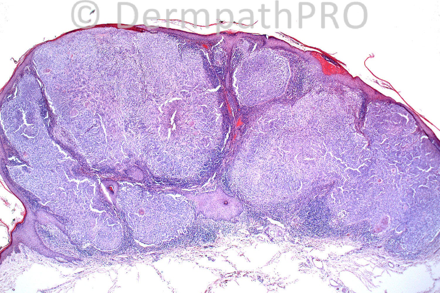
64 years-old female. Lateral thigh. Increased size over one year. Pigmented. Amelanotic melanoma? Clear cell acanthoma? Pyogenic granuloma?

Case posted by Dr. Richard Carr


  Report Record

User Feedback


Dr. Mona Abdel-Halim

Posted

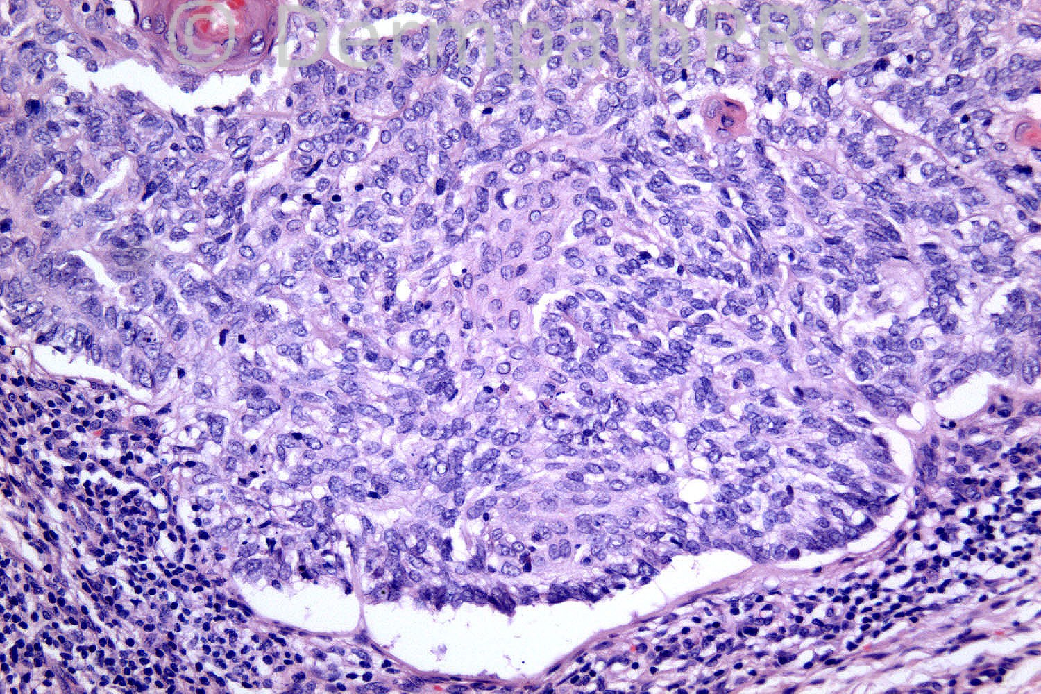
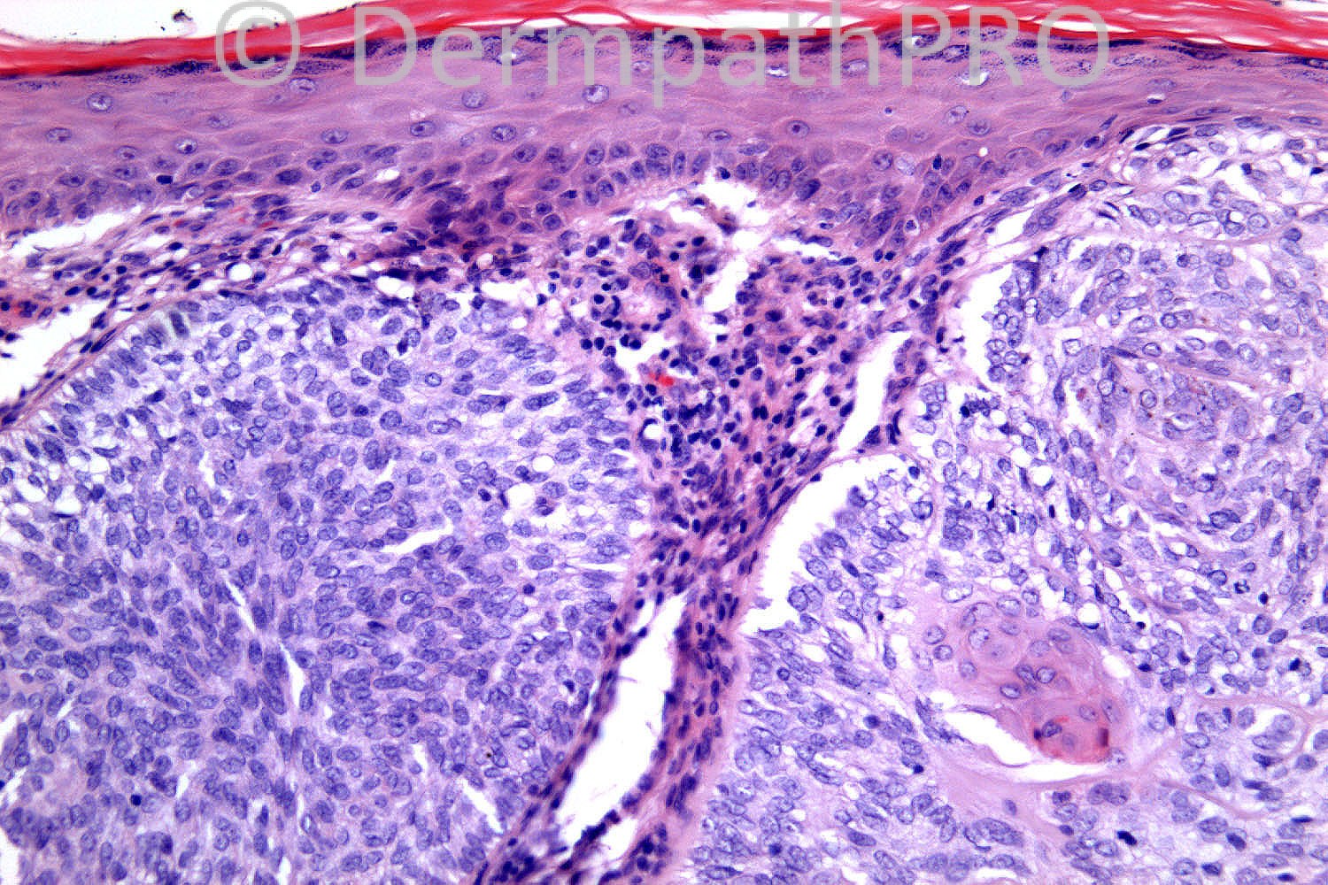
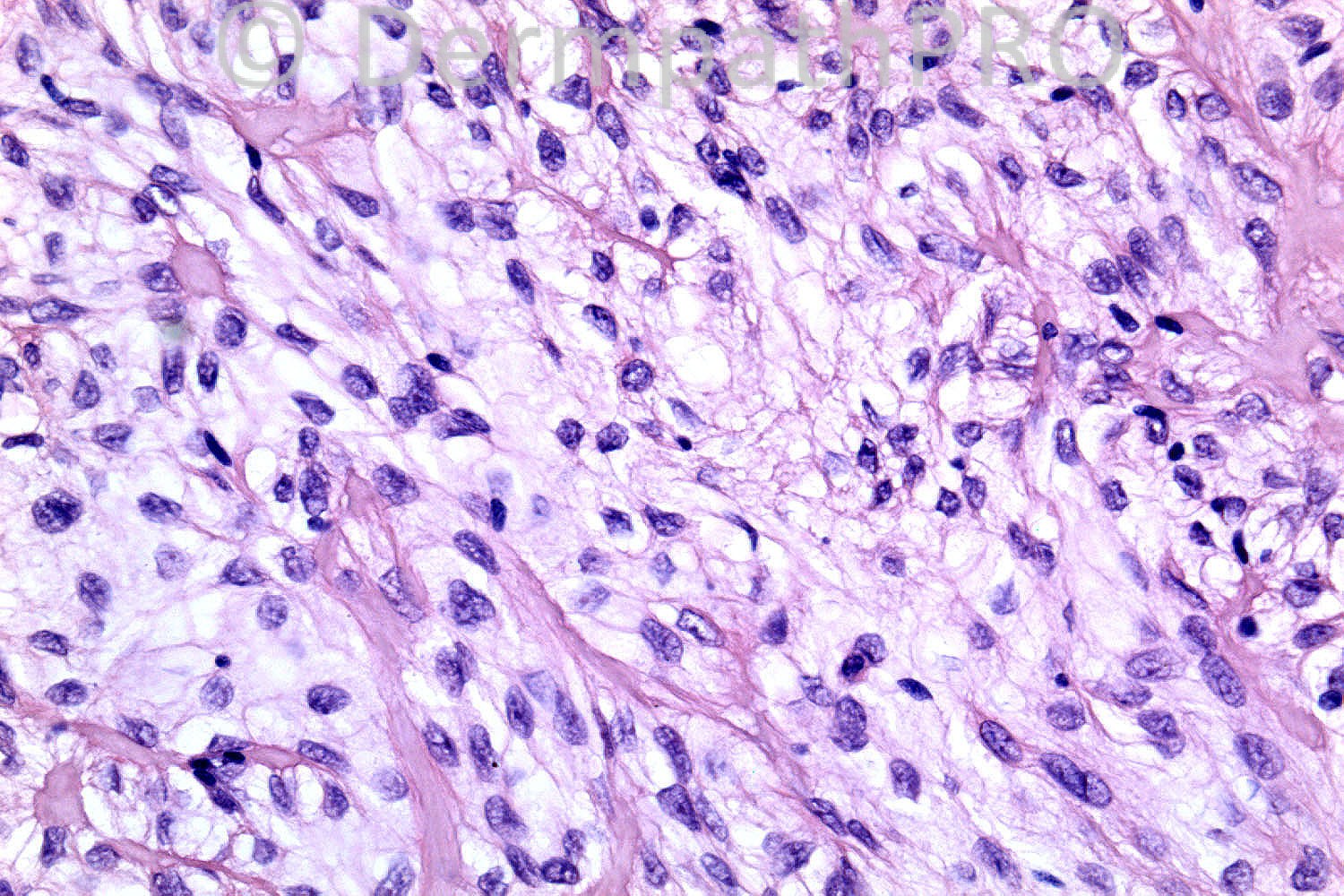
Peripheral palisading in Fig 3, peritumoral clefting in Fig 2 and Fig 3, clear cell change in Fig 4, BCC with clear cell change.

Share this comment


Link to comment
share_externally

BCC with clear cell change, common things being common.

Share this comment


Link to comment
share_externally

Eman El-Nabarawy

Posted

First impression BCC with clear cell change.

Share this comment


Link to comment
share_externally

Robledo F. Rocha

Posted

[size=4][font=Palatino Linotype, serif]Expansive nests of basaloid cells showing epidermal attachment, peripheral palisading and cleft-like retraction. There are neoplastic cells with prominent clear cytoplasm.[/font]
[font=Palatino Linotype, serif]I agree with my colleagues: basal cell carcinoma, clear cell variant.[/font][/size]

Share this comment


Link to comment
share_externally

Azza Esmat

Posted

Agree on BCC with clear cells

Share this comment


Link to comment
share_externally

Dr. Richard Carr

Posted

You are all right common things being common this should be BCC. This case however is not in my opinion a BCC. Please note the "retraction" in BCC should be between the intact basal lamina of the peripheral cells and the stroma and should (almost always) have a hint of blue tinged stromal mucin in the retraction space. In this case the "retraction" is artefactual through the basal cytoplasm of the cells and a very distinct basement membrane is clearly seen beyond the artefact. Please try again!!

Share this comment


Link to comment
share_externally

Robledo F. Rocha

Posted

Trichilemmoma, basaloid variant? Squamous eddies and prominent basement membrane are clues.

Share this comment


Link to comment
share_externally

Guest Bansal_

Posted

? trichilemmoma.

Share this comment


Link to comment
share_externally

Guest Maria george

Posted

Clear cell Hidradenoma.

Share this comment


Link to comment
share_externally

Guest Catarina Shaletich

Posted

Neoplasia epitelial sem aparente conexão com a epiderme, na primeira e na segunda fotos dá a impressão (sem segurança) de presença de pequenos ductos, inflamação associada relativamente proeminente.
Acho que pode ser uma neoplasia anexial sudorípara (se forem ductos mesmo), não tenho certeza de sua benignidade e sem mais fotos não considero possível diagnóstico seguro.
Gostaria de ver mais fotos em grande aumento de outras áreas e da transição da lesão com a epiderme.

Share this comment


Link to comment
share_externally

Dr. Mona Abdel-Halim

Posted

Then it is trichilemmoma ..

Share this comment


Link to comment
share_externally

Dr. Richard Carr

Posted

Well done Robledo this is a basaloid variant of tricholemmoma. We have a moderate database of tricholemmomas (61 cases) we have performed immuno on. This includes a series of 10 cases with prominent basaloid change that can easily be confused with BCC. This is the only case of 61 that did not arise on the head! and it took me a while to realise this was not just a BCC. Put this tumour on the central face and I am sure we would all have considered tricholemmoma as it has all the typcial features i.e. rather rounded lobulated pushing borders, subtle palisading with reversed polarity (Fig 3 with nuclei away from the basement membrane), prominent basement membrane and pale / clear cell changes. Not the lesion lacks pleomorphism. It turns out immunostains can be quite helpful we found all tricholemmomas expressing moderate to strong CD34 in >10% of tumour area in all tricholemmomas and only focal BerEP4 (moderate to strong) in up to 20% of the basaloid epithelium in one case and 10% of the tumour epithelium in a second case. BerEP4 is moderately to strongly expressed in >20% of the epithelium in 97.6% of our series of nodular BCC that includes rare variants including small biopsies and cases with clear cell changes. None of the nodular BCC in our database expressed CD34. A PAS stain can help accentuate the basement membranes in tricholemmoma (might be prominent in Fig 4. I am sure the basaloid variant is under-recognised and many cases will be signed out as BCC. Textbooks do not highlight this variant and we hope to publish our series in the near future. Regards to all, hope you are enjoying your weekends.

Share this comment


Link to comment
share_externally

Guest Dr. Francisco Vílchez

Posted

BCC

Share this comment


Link to comment
share_externally

Dr. Richard Carr

Posted

Thank you for reminding me of this reference. If thier study was repeated it
would certainly need to include BerEP4 in the analysis. Difficult on the
images in the paper but their case in Fig 1 I would accept as a tricholemmoma
if BerEP4 negative and CD34 positive. We have a few cases we could not decide
in our database but the case I illustrated was not one of those. I did not
include the images of the desmoplastic tricholemmomatous areas as I wanted you
all to call it a BCC to prove a point!

I made some additional comments in my slide collection files as follows:
Nice for basaloid tricholemmoma study. CD34 ++15%, Neg BerEP4 support
tricholemmoma. Dendritic melanocytic cells throughout. PAS showing masses
of glycogen (diastase labile) and prominent BM on DPAS. Focal area of
desmoplastic trichlemmoma.
Retraction is inside the basemement membrane probably in the basal cytoplasm.
No interstitial mucin.

Your comments and the reference made me search my slide collection and immuno
databast for BCC with prominent basement membrane and 3 cases I have seen
(out of literally thousands of BCC over the years although I may not have
collected them all as I was aware of this occasional finding in BCC).
We also noted prominent BM in one or two
lymphadenoma and trichoblastomas (apart from tricholemmomas and cylindromas
of course).

Share this comment


Link to comment
share_externally


×
×
  • Create New...