Jump to content
In this section we have spot diagnoses posted on a daily basis since June 2010, now over 4000! You can review the archived cases and read the suggested diagnoses by users and the final comment by the contributors.
Case are uploaded each week day by 10 am UK time with the correct diagnosis will generally be posted at 8 pm UK time. Why not view the most recent spot diagnosis and proffer a diagnosis?

Case Number : Case 718 - 18 Mar Posted By: Guest

Please read the clinical history and view the images by clicking on them before you proffer your diagnosis.
Submitted Date :
   (0 reviews)

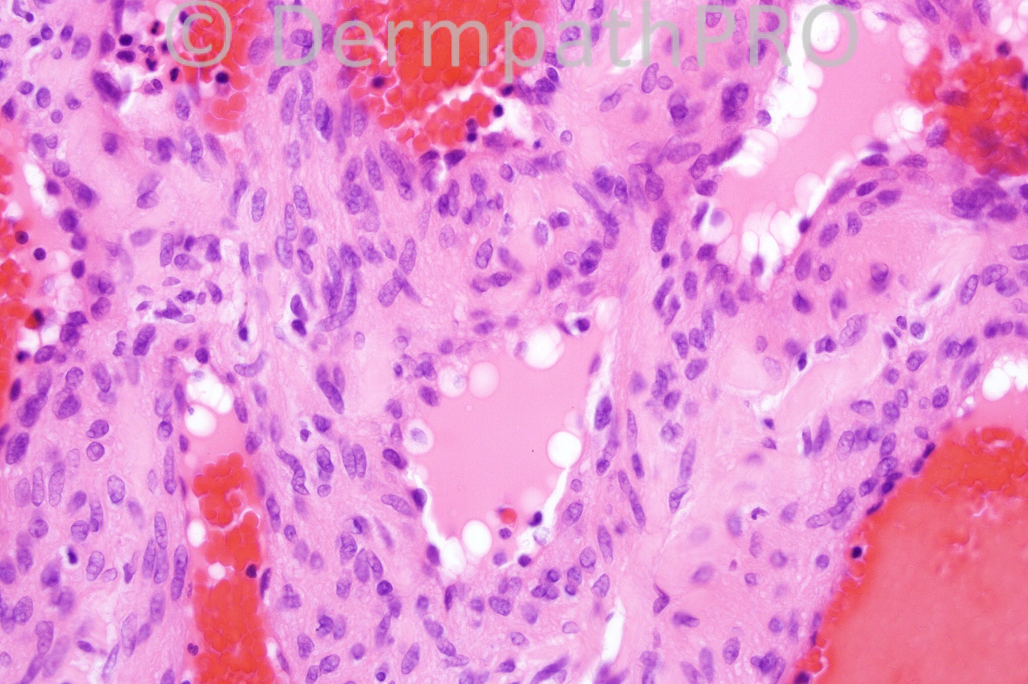
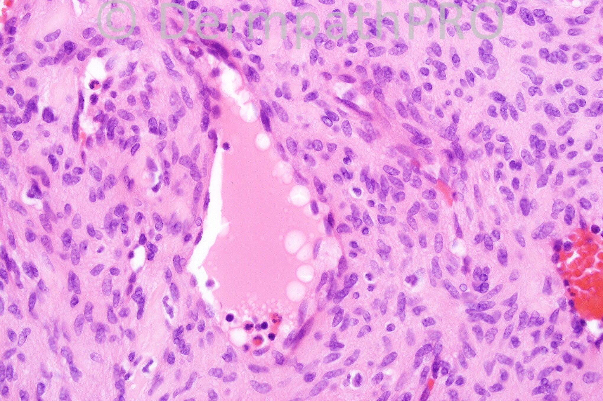
30 years-old male, DF-like lesion right lower leg.


  Report Record

User Feedback


nick turnbull

Posted

Aneurysmal fibrous histiocytoma?

Share this comment


Link to comment
share_externally

Guest Dr Engin Sezer

Posted

Spindle cell hemangioma

Share this comment


Link to comment
share_externally

Sasi Attili

Posted

Quite an interesting relatively circumscribed dermal tumour composed of spindle and epithelioid cells, associated with dilated vascular spaces with red cells. On low power I considered Spindle cell hemangioma, Aneurysmal FH and nodular Kaposi's. However I am not sure that the high power views have made me any wiser and I find this tough. The relatively well circumscribed nature and the lack of obvious muscular channels or AVM like areas in the periphery of the lesio,n is against a spindle cell hemangioma. I do not see any Eosinophilic globules (KS).

Therefore I favour this being an Aneurysmal FH. However, the vascular channels seem to be lined in places by a true endothelial lining, which is confusing me. But maybe I am just looking too closely!!

Share this comment


Link to comment
share_externally

Guest Bansal_

Posted

Looks benign but unsure of diagnosis. Considered spindle cell haemangioma, angioleiomyoma and glomangiomyoma but morphology not typical. Look forward to answer.

Share this comment


Link to comment
share_externally

Arif Usmani

Posted

Agree with Dr. Bansal, could be angioleiomyoma and possibly glomangiomyoma

Share this comment


Link to comment
share_externally

Dr. Phillip McKee

Posted

The spindled cells are positive for smooth muscle markers. I diagnosed the lesion as a superficial angioleiomyoma.

Share this comment


Link to comment
share_externally

Dr. Mona Abdel-Halim

Posted

Very late, but I also thought of angioleiomyoma.

Share this comment


Link to comment
share_externally

Nada Macaron

Posted

How about myopericytoma? Curious to know if desmin and caldesmon were positive.

Share this comment


Link to comment
share_externally


×
×
  • Create New...