Jump to content
In this section we have spot diagnoses posted on a daily basis since June 2010, now over 4000! You can review the archived cases and read the suggested diagnoses by users and the final comment by the contributors.
Case are uploaded each week day by 10 am UK time with the correct diagnosis will generally be posted at 8 pm UK time. Why not view the most recent spot diagnosis and proffer a diagnosis?

Case Number : Case 725 - 27 Mar Posted By: Guest

Please read the clinical history and view the images by clicking on them before you proffer your diagnosis.
Submitted Date :
   (0 reviews)

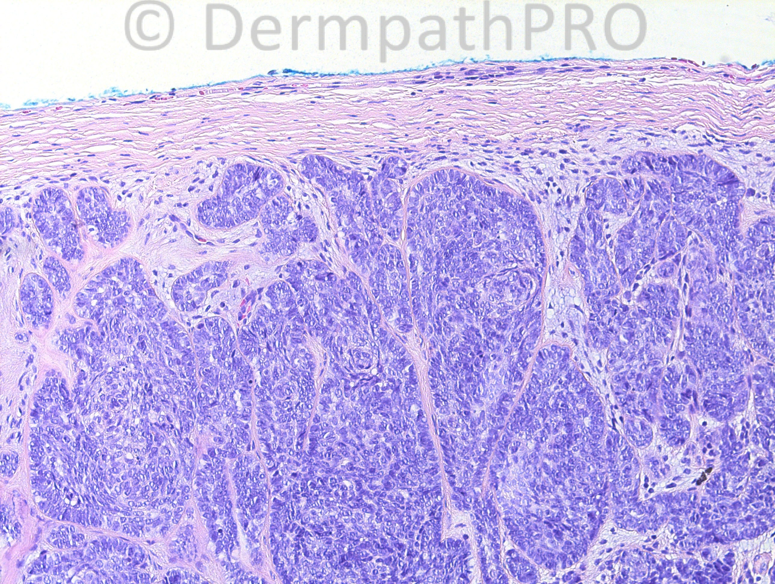
33 years-old female with right neck mass for 5 years. Clinical impression: rule out cyst.

Case posted by Dr Hafeez Diwan.


  Report Record

User Feedback


Robledo F. Rocha

Posted

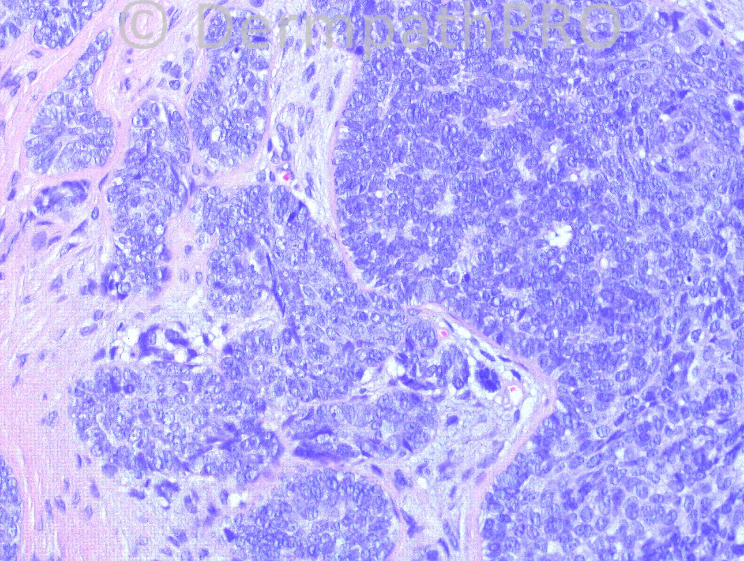
Irregular nests of basaloid cells surrounded by cellular fibrous stroma. Hair differentiation take form in bud-like follicular germ structures.
I think it´s trichoblastoma, since there´s no mucinous alteration of the intervening stroma nor retractions between tumor nests and peritumoral stroma, as might be expected if it were basal cell carcinoma.

Share this comment


Link to comment
share_externally

Sasi Attili

Posted

Tricky one. Benign!! Favour Trichoblastoma but I am not able to appreciate well formed papillary mesenchymal cells surrounding the lobules. The germ like structure in the bottom right has a fibrous acellular core! Therefore Trichoblastoma is a difficult diagnosis to make in this circumstance. There is a suggestion of sebaceous differentation in ONE cell in the image on the bottom left! Therefore I would consider a sebaceoma (though sebaceous differentiation can occur within trichoblastoma's!). Adipophilin and Ber-EP4/ EMA would be helpful.

Share this comment


Link to comment
share_externally

Dr. Mona Abdel-Halim

Posted

Trichoblastoma.

Share this comment


Link to comment
share_externally

Dr. Richard Carr

Posted

Not my case but I think a clue is the way the lesion has shelled out beyond
the stroma in Fig 1. There are subtle stromal condensations.
Favour trichoblastoam. Occasional large nodular trichoblastomas and
trichogerminomas have relatively sparse stromal component. I would do CD10
(usually stromal predominent in TE/TB and epithelial predominant in BCC) and
CK20 (about 40% of TE/TB have numerous MC which is exceedingly rare in BCC.
However the diagnosis is usually acheived by careful examination of the H&E.

Share this comment


Link to comment
share_externally

Eman El-Nabarawy

Posted

Trichoblastoma.

Share this comment


Link to comment
share_externally

Guest Jim Davie MD

Posted

Giant trichoblastoma (giant solitary trichoepithelioma), if this is as big as the first image suggest, and given clues of years' long history, young adult, absence of obvious mitotic activity.

Share this comment


Link to comment
share_externally

Dr. Hafeez Diwan

Posted

I diagnosed this as trichoblastoma.

Share this comment


Link to comment
share_externally

Guest Amr Gohar

Posted

[color=#231F20][size=3]Distinction between [color=red]trichoblastoma[/color] and basal cell carcinoma can be difficult, particularly in small biopsies. [b]Mitotic figures[/b] can be frequent in trichoblastomas.[/size][/color][color=black][size=3] Hence the need to see the [b]low-power view[/b](in trichoblastomas the tumour is not usually connected to the epidermis and [/size][/color][color=#231F20][size=3]a deep infiltrative pattern is noticed in basal cell carcinoma)[/size][/color][color=black][size=3], to search for [b]Merkel cells[/b] by [/size][/color][color=#231F20][size=3]immunohistochemical markers ([/size][/color][color=black][size=3]Merkel cells are quite common in trichoblastomas, while basal cell carcinomas generally lack these cells) and to test for[/size][/color][color=#231F20][size=3]the immunohistochemical expression of [b]androgen receptor[/b](usually negative in trichoblastomas and positive in basal cell carcinomas).[/size][/color]

Share this comment


Link to comment
share_externally


×
×
  • Create New...