Jump to content
In this section we have spot diagnoses posted on a daily basis since June 2010, now over 4000! You can review the archived cases and read the suggested diagnoses by users and the final comment by the contributors.
Case are uploaded each week day by 10 am UK time with the correct diagnosis will generally be posted at 8 pm UK time. Why not view the most recent spot diagnosis and proffer a diagnosis?

Case Number : Case 750 - 1st May Posted By: Guest

Please read the clinical history and view the images by clicking on them before you proffer your diagnosis.
Submitted Date :
   (0 reviews)

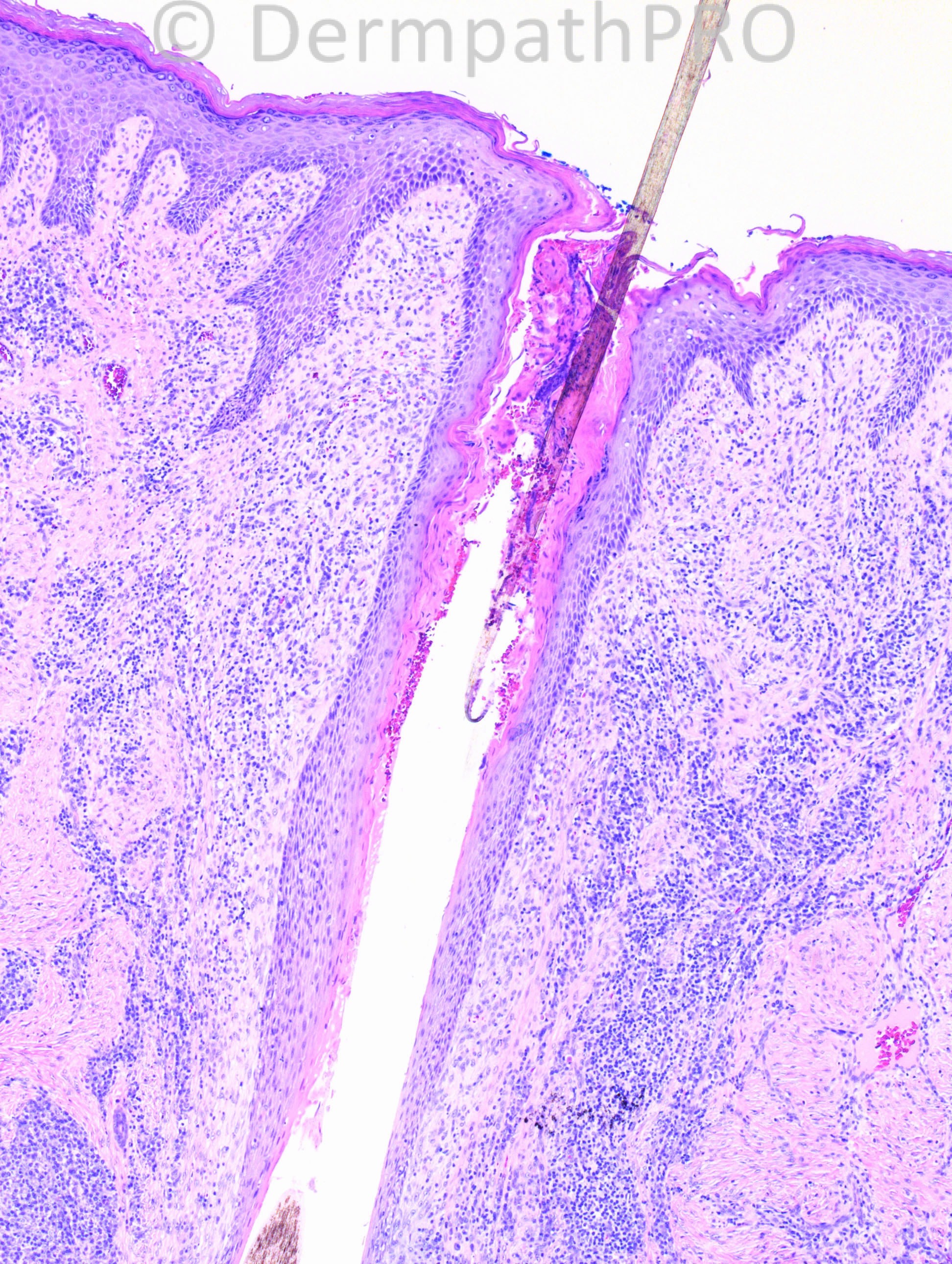
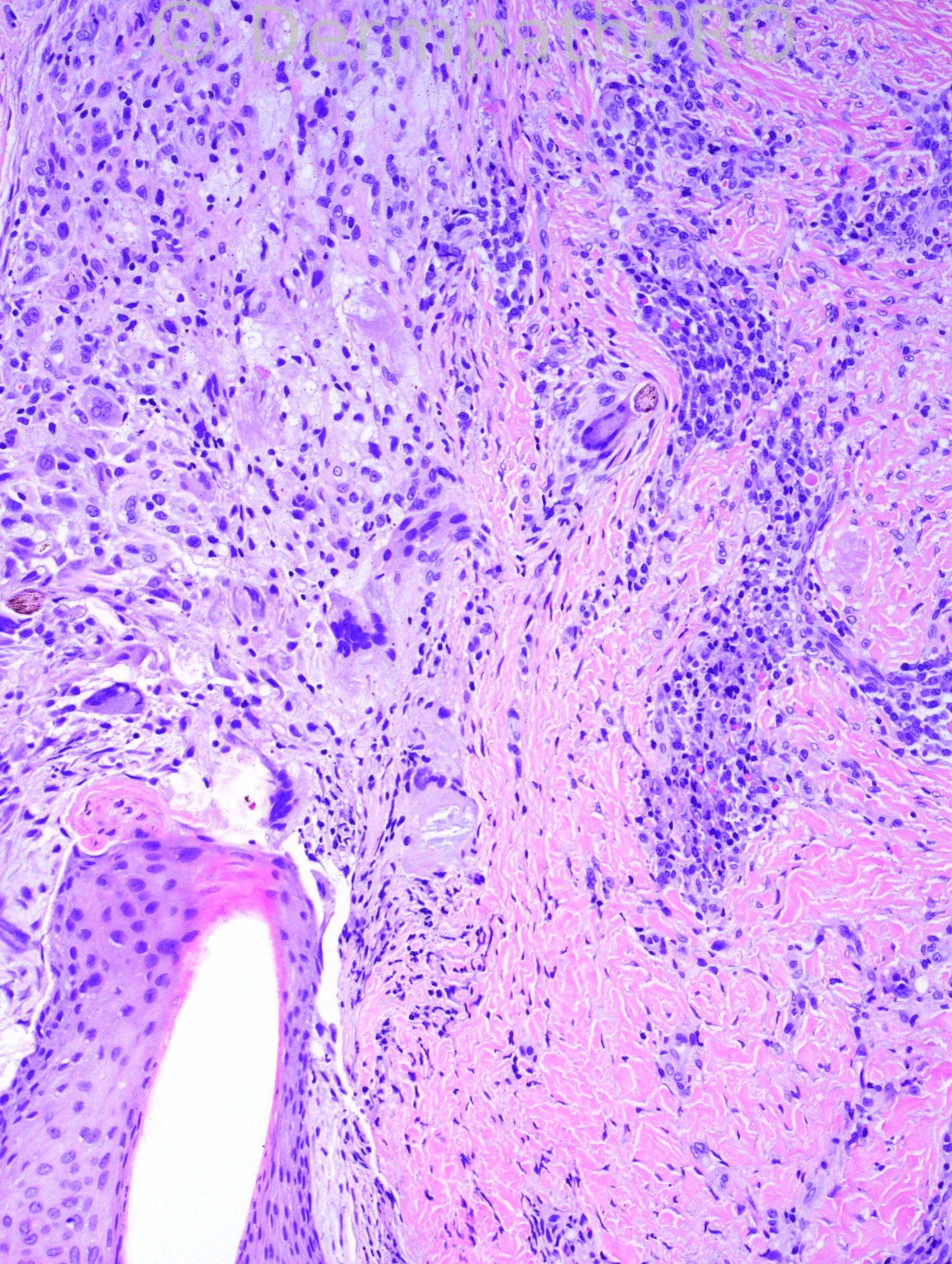
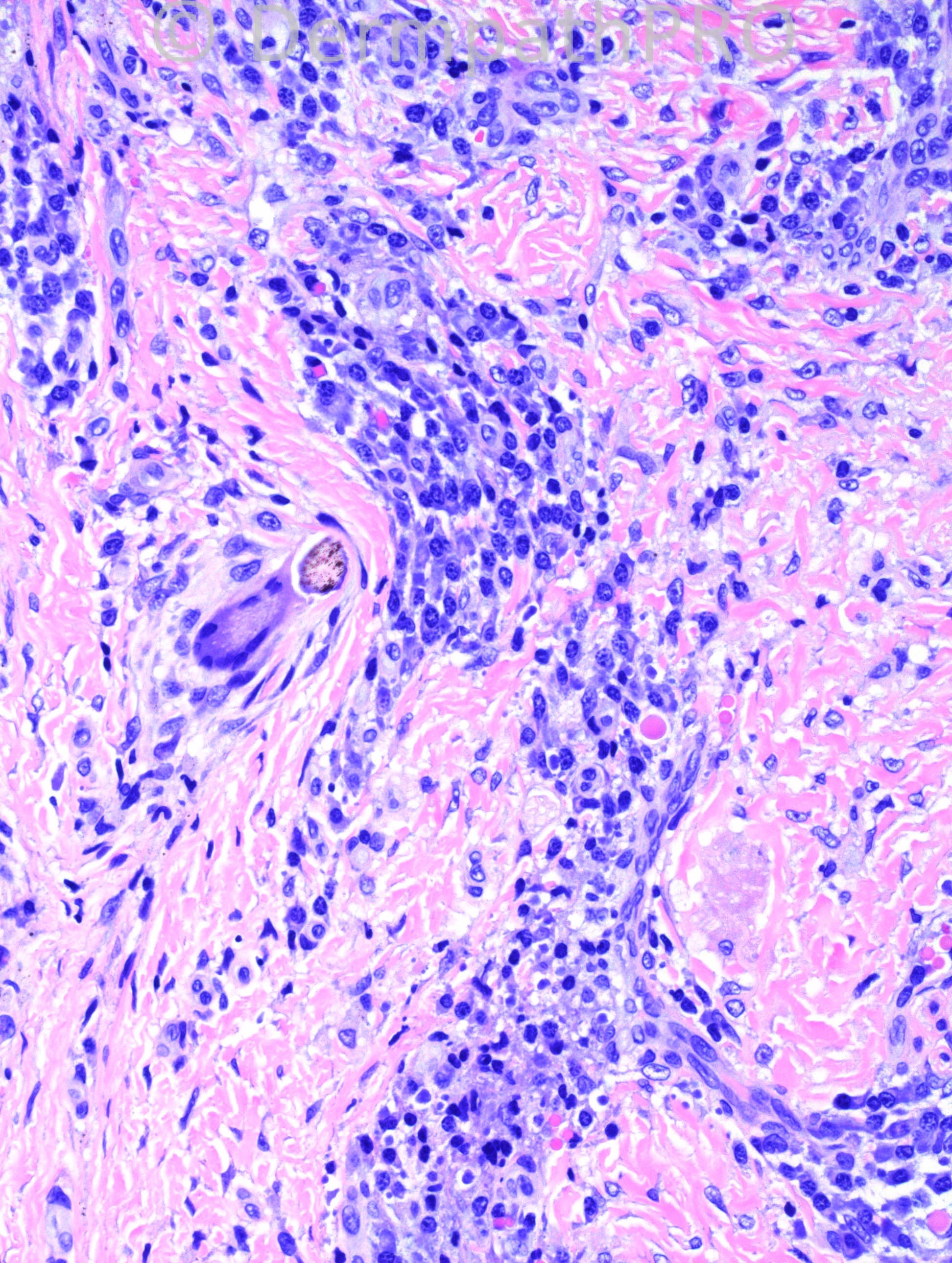
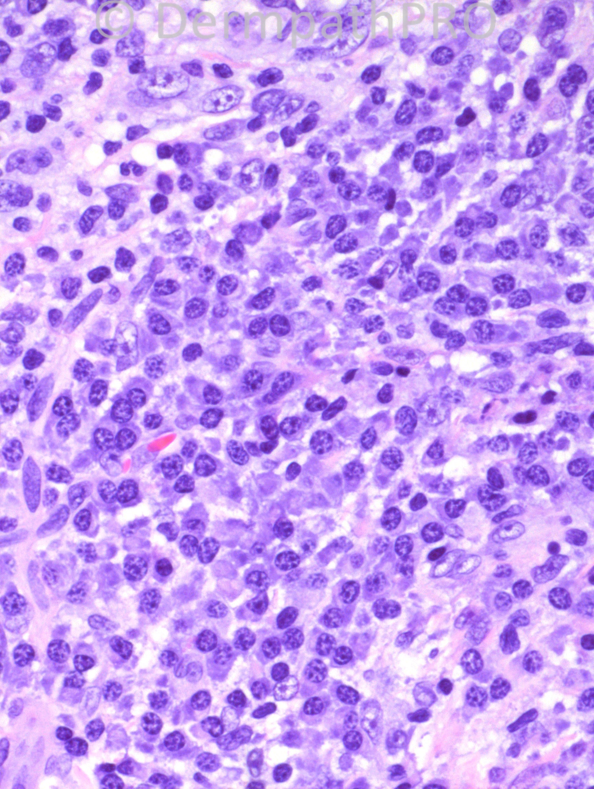
40 years-old Hispanic male with 5mm firm lesion on posterior left scalp. Clinical impression: nevus sebaceus.

Case posted by Dr. Hafeez Diwan.


  Report Record

User Feedback


Dr. Mona Abdel-Halim

Posted

Chronic folliculitis with ruptured follicle and secondary granulomatous reaction to released hair shaft structures. The lesion is associated with dense plasma cell collections demonstrating beautifully Russel bodies. Plasma cells are commonly encountered in association with chronic folliculitis, do not think the whole pattern of the plasma cell proliferation fits with associated plasma cell granuloma, though it is a possibility. Will do PAS to exclude fungus as a cause of this folliculitis (although age is less likely), and will do kappa and lambda staining to be sure of the polyclonal nature of the plasma cell proliferation and to exclude plasmacytoma.

Share this comment


Link to comment
share_externally

Guest Dr Engin Sezer

Posted

Ruptured follicular cyst

Share this comment


Link to comment
share_externally

Guest Romualdo C. L. Filho

Posted

[u]Chronic folliculitis. I am not concerned about neoplastic conditions but would order special stains to exclude fungal and bacterial infections.[/u]

Share this comment


Link to comment
share_externally

Robledo F. Rocha

Posted

I go with acneiform syphilis due to granulomatous plasma cell-rich perifolliculitis. Alternatively, late phase of acne keloidalis nuchae due to suggestion of scarring.

Share this comment


Link to comment
share_externally

Guest Bansal_

Posted

Granulomatous folliculitis and associated inflammation rich in plasma cells suggestive of infectious aetiology.

Share this comment


Link to comment
share_externally

Guest Jim Davie MD

Posted

I favor a chronic lesion of [b]acne keloidalis nuchae[/b], in the context of a papular occipital lesion.

The central follicle shows: tapering exclamation-point features in the more superficial, dysmorphic hair shaft, polytrichia, concentric lamellar fibroplasia and atrophy extending deep into the infundibulum. There are numerous naked hair shaft foreign body granulomas (some with partially degraded hair shafts). There are numerous plasma cells and fibrosis, with absence of significant acute inflammatory component.

Share this comment


Link to comment
share_externally

Dr. Hafeez Diwan

Posted

I called this acne keloidalis nuchae.

Share this comment


Link to comment
share_externally


×
×
  • Create New...