Jump to content
In this section we have spot diagnoses posted on a daily basis since June 2010, now over 4000! You can review the archived cases and read the suggested diagnoses by users and the final comment by the contributors.
Case are uploaded each week day by 10 am UK time with the correct diagnosis will generally be posted at 8 pm UK time. Why not view the most recent spot diagnosis and proffer a diagnosis?

Case Number : Case 752 - 3rd May Posted By: Guest

Please read the clinical history and view the images by clicking on them before you proffer your diagnosis.
Submitted Date :
   (0 reviews)

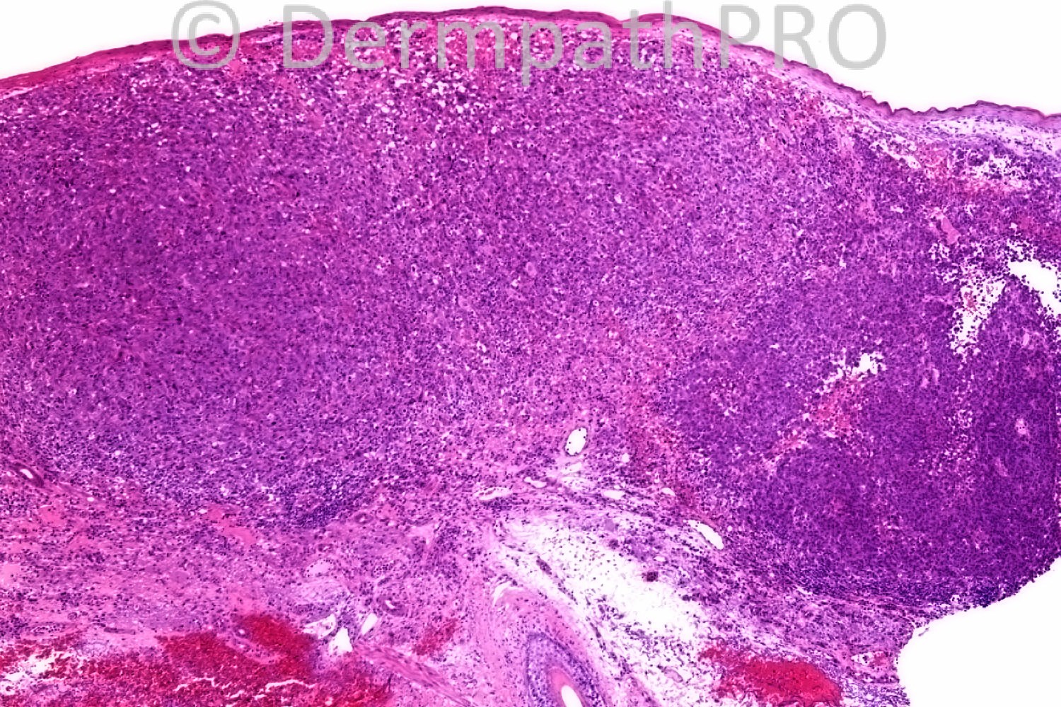
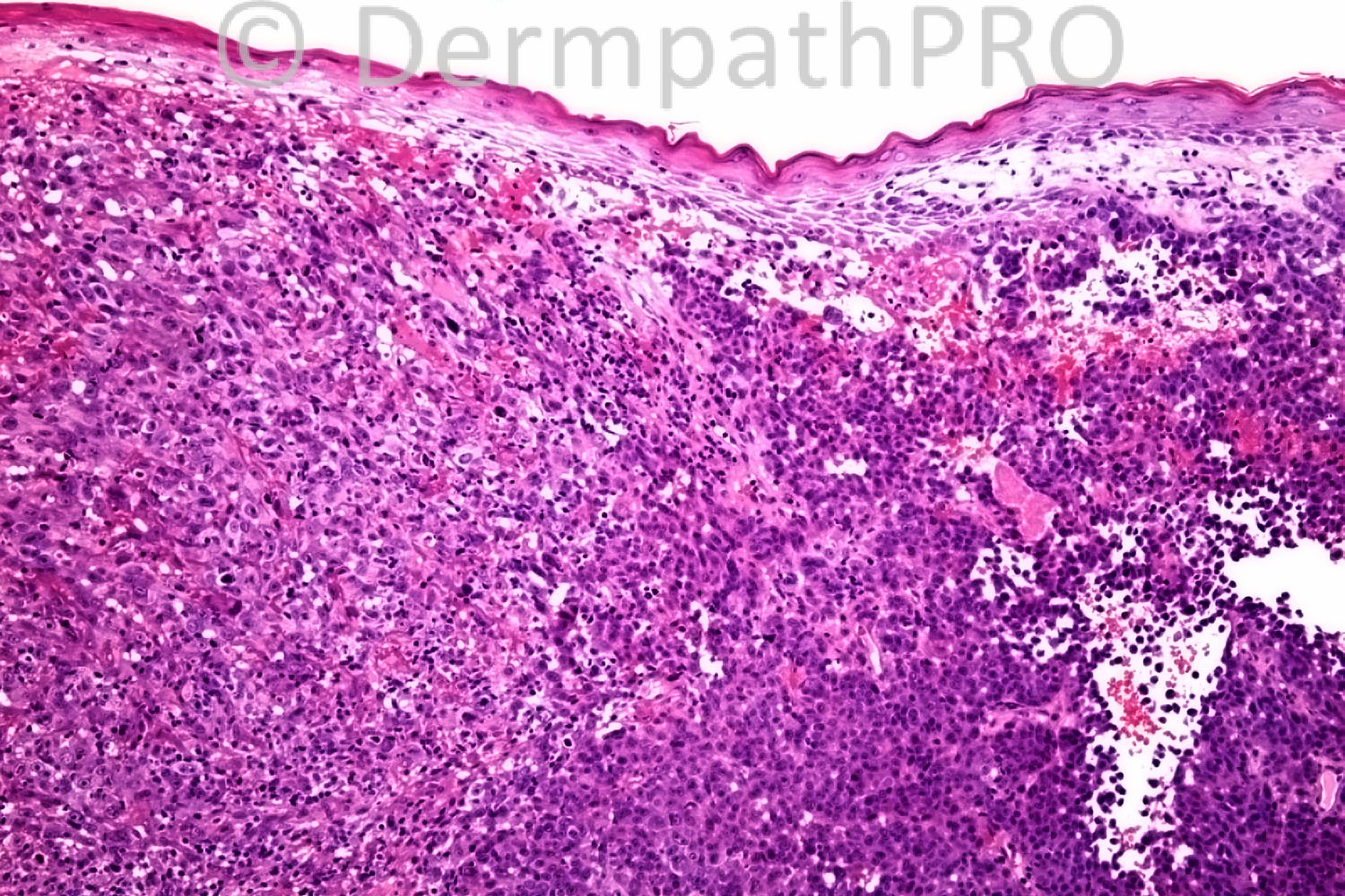
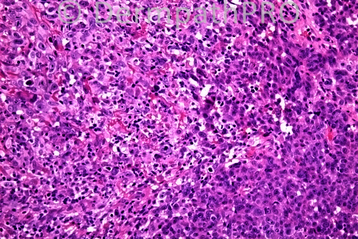
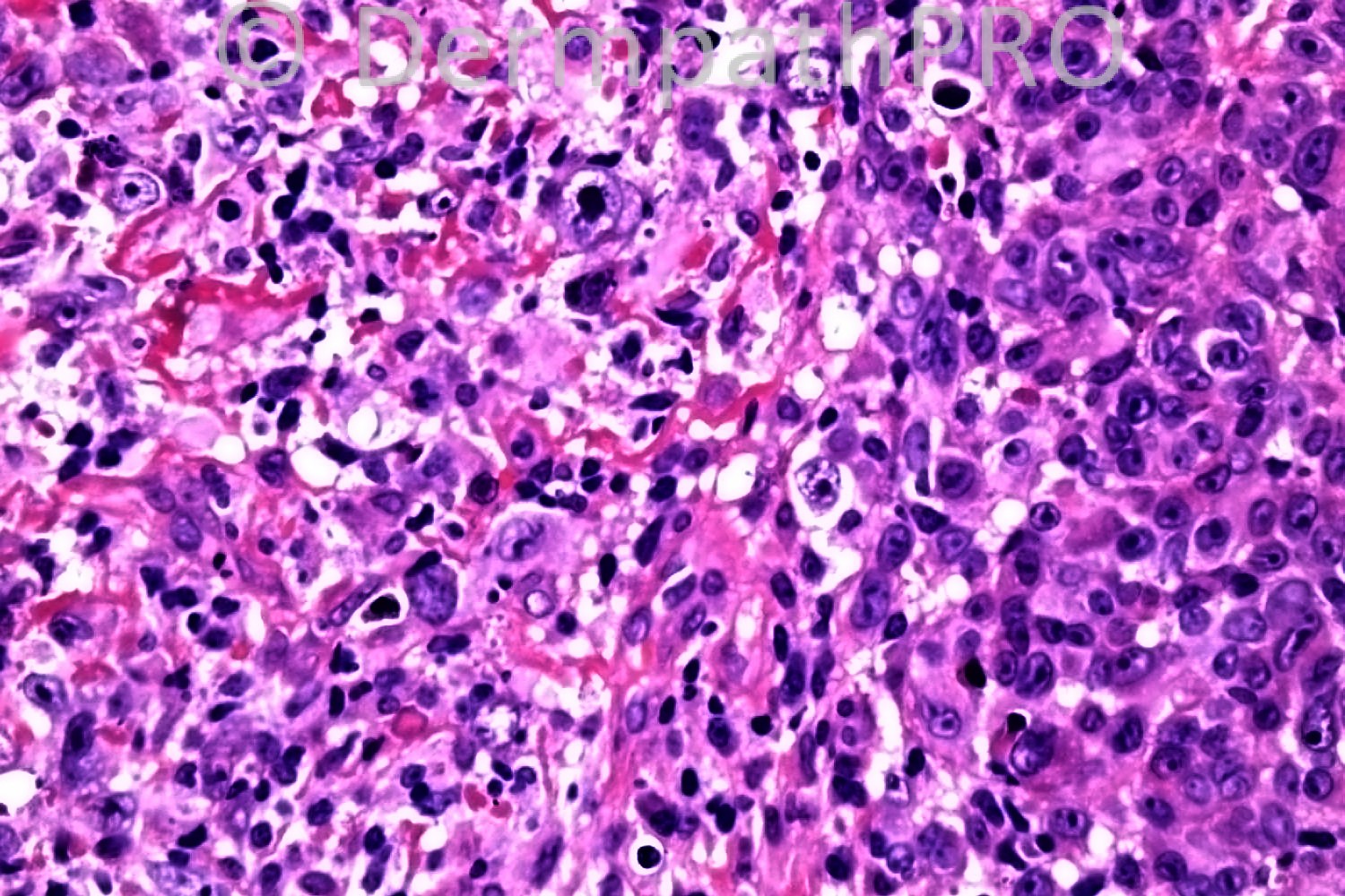
76 years-old female. Left arm, bleeding nodule ?BCC ?PG

Case posted by Dr. Richard Carr.


  Report Record

User Feedback


Guest Dr Engin Sezer

Posted

DDX: epithelioid angiosarcoma, AFX, acantholytic poor differentiated SCC

Share this comment


Link to comment
share_externally

Guest Romualdo C. L. Filho

Posted

There is a grenz zone between nonatypical epidermis and the neoplasia, against a diagnosis of SCC. Also, the great cellularity and absence of marked pleomorphism do not favor a diagnosis of atypical fibroxantoma. Epithelioid angiosarcoma is my first choice too.

Share this comment


Link to comment
share_externally

Guest Bansal_

Posted

Agree, likely to be an angiosarcoma. Would do immunos: C31, CD34, AE1/AE3, S100 protein.

Share this comment


Link to comment
share_externally

Anaplastic large cell lymphoma

Share this comment


Link to comment
share_externally

Dr. Mona Abdel-Halim

Posted

My first impression is epithelioid angiosarcoma.

Share this comment


Link to comment
share_externally

Guest Rodrigo Restrepo

Posted

[color=#1C2837][font=arial, verdana, tahoma, sans-serif][size=4]Anaplastic large cell lymphoma[/size][/font][/color]

Share this comment


Link to comment
share_externally

Robledo F. Rocha

Posted

There are limited examples of soft tissue tumors with clinical presentation of a hemorrhagic mass. Taking into account the case history and the H&E pictures, I favor epithelioid angiosarcoma, although this is rare as a primary cutaneous tumor and I didn't find any focus of irregular vascular channels inside the solid sheets.
Immunohistochemistry will rule out or confirm the hypothesis put forward here.

Share this comment


Link to comment
share_externally

Dr. Richard Carr

Posted

This was two for the price of one.
The peripheral darker epithelioid shoulders expressed melanA and S100 strongly and were CD10 negative.
The central larger pleomorphic epithelioid cells were completely negative for melanA and S100 and strongly diffusely positive for CD10.
So my diagnosis: Collision of nodular malignant melanoma and AFX. You might just think the melanoma de-differentiated centrally (and acquired CD10) I guess it is academic!
Vascular, muscle, cytokeratin and p63 markers were all negative. I think ruling out anaplastic large cell lymphoma is a good suggestion I am still awaiting the CD30.

Share this comment


Link to comment
share_externally

Guest Jim Davie MD

Posted

[font=arial,helvetica,sans-serif][size=4]Sorry...too late to join the fun discussion today!

Looking at the photos, I didn't get the impression this would be lymphoma given:
- its well-circumscribed nesting architecture superficially, in right hand smaller cell component.
- Its origin in the superficial dermis, as suggested by the downwards displacement of nodular solar elastosis to a position below the expansile tumor mass.

(Look forward to see if the CD30 shoots this down!).


I don't envy your dilemma of choosing between two very unlikely events (collision of two rare tumors vs. chance of melanoma losing S100 and MelanA simultaneously)...but a dilemma amenable to possible resolution via additional stains. Maybe a SOX-10 or MITF (melanoma), and/or procollagen (AFX) stain would help reveal the true deriation of the CD10 positive anaplastic cells. But academic, as you said, if it makes no clinical treatment difference.

If there is low mitotic activity (from photos, at least) in the anaplastic CD10 population, I would slightly favor the dedifferentiated melanoma option.[/size][/font]

Share this comment


Link to comment
share_externally

Dr. Richard Carr

Posted

CD30 is negative

Share this comment


Link to comment
share_externally


×
×
  • Create New...