Jump to content
In this section we have spot diagnoses posted on a daily basis since June 2010, now over 4000! You can review the archived cases and read the suggested diagnoses by users and the final comment by the contributors.
Case are uploaded each week day by 10 am UK time with the correct diagnosis will generally be posted at 8 pm UK time. Why not view the most recent spot diagnosis and proffer a diagnosis?

Case Number : Case 754 - 8th May Posted By: Guest

Please read the clinical history and view the images by clicking on them before you proffer your diagnosis.
Submitted Date :
   (0 reviews)

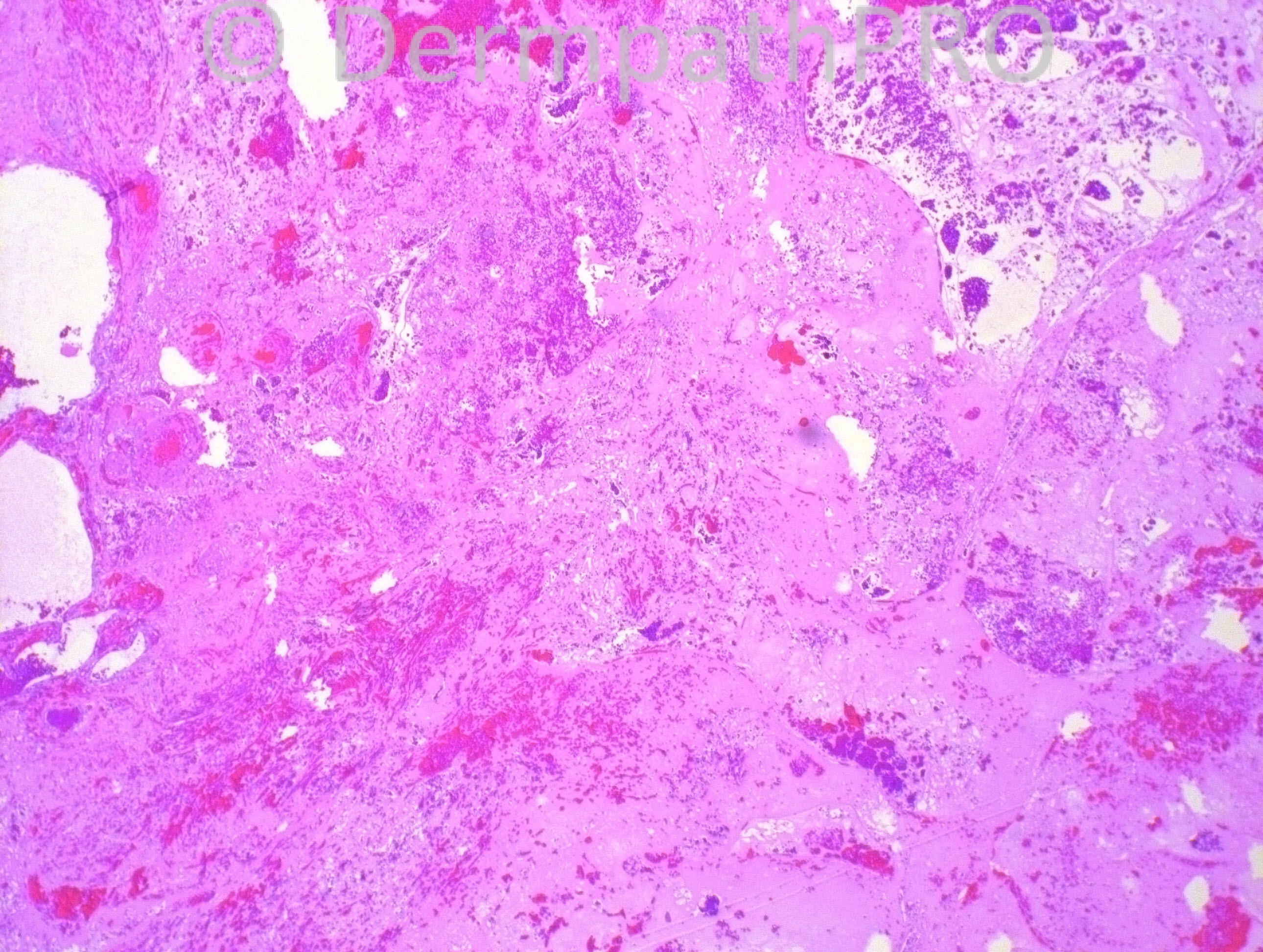
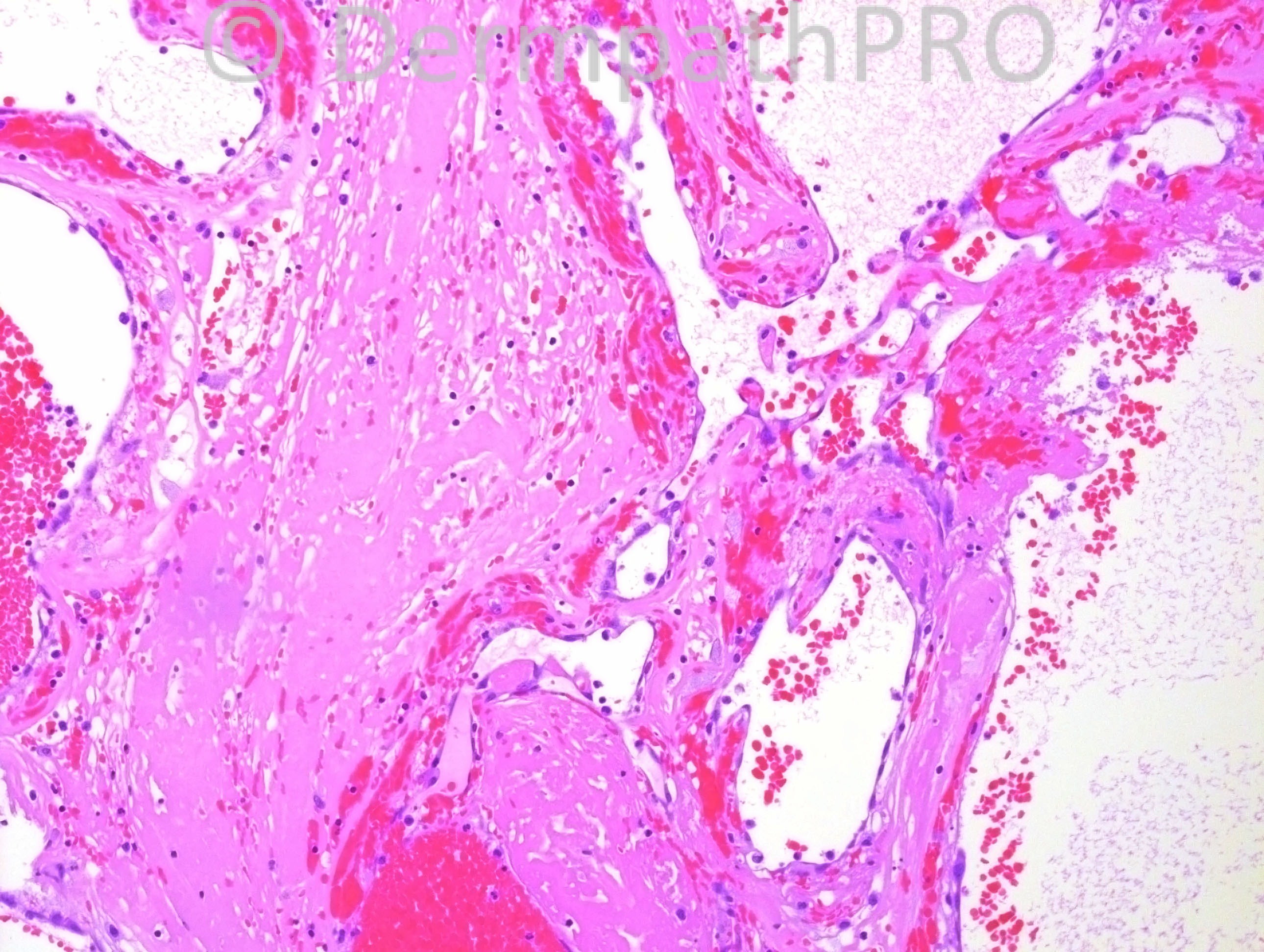
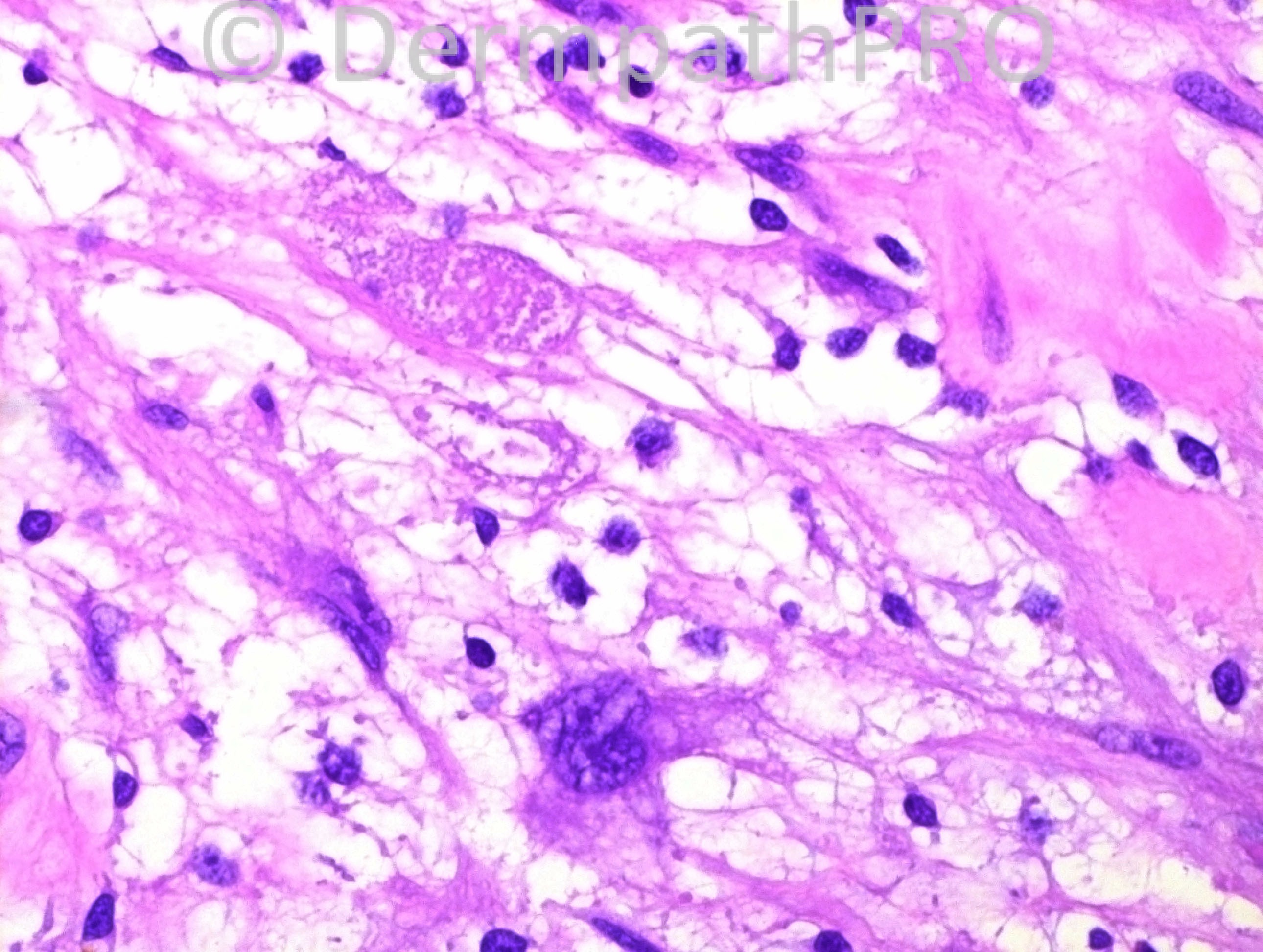
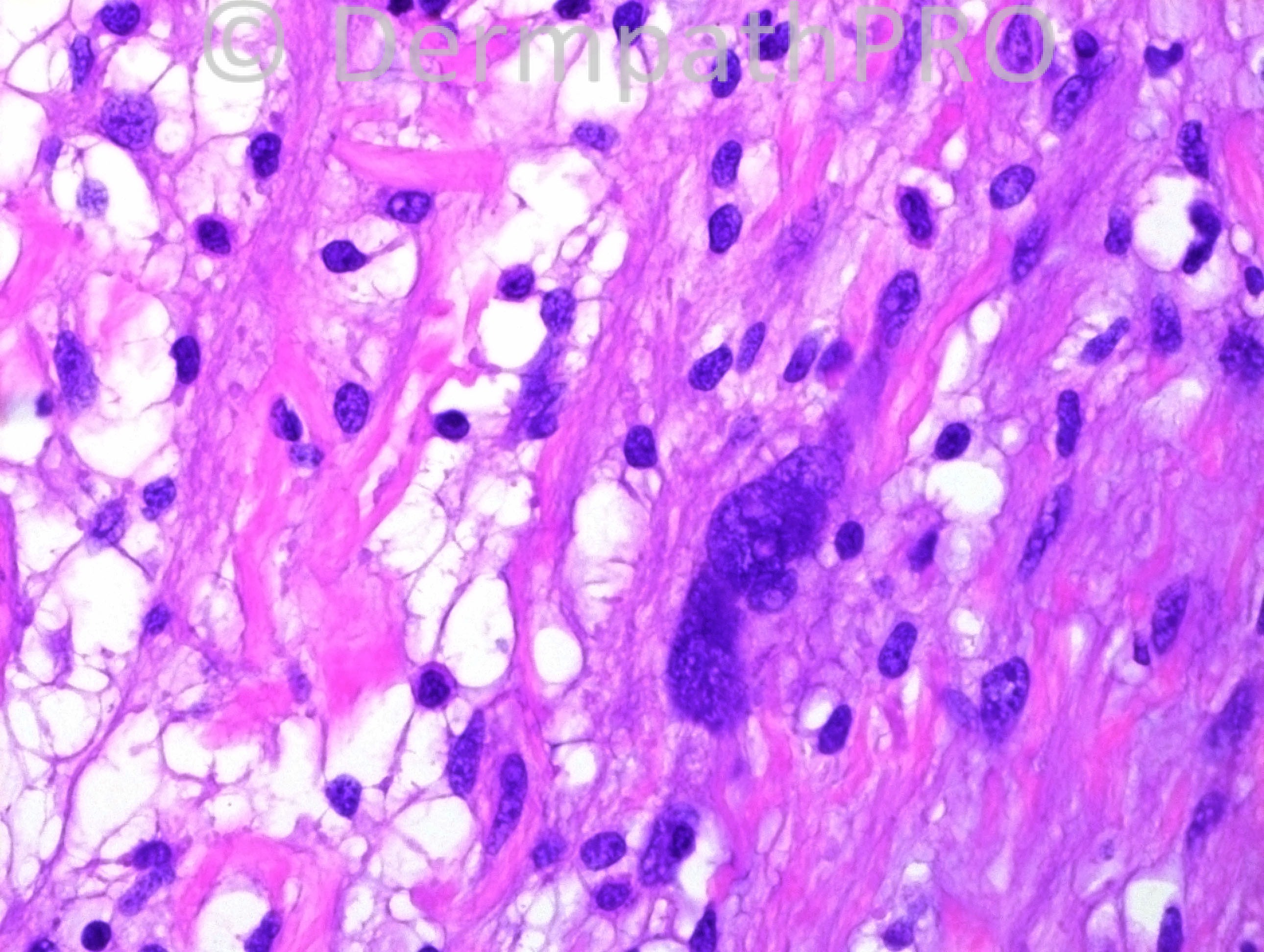
76 years-old Hispanic female with right thigh 5.8 x 5.5 cm mass, present for 15 years.

Case posted by Dr. Hafeez Diwan.
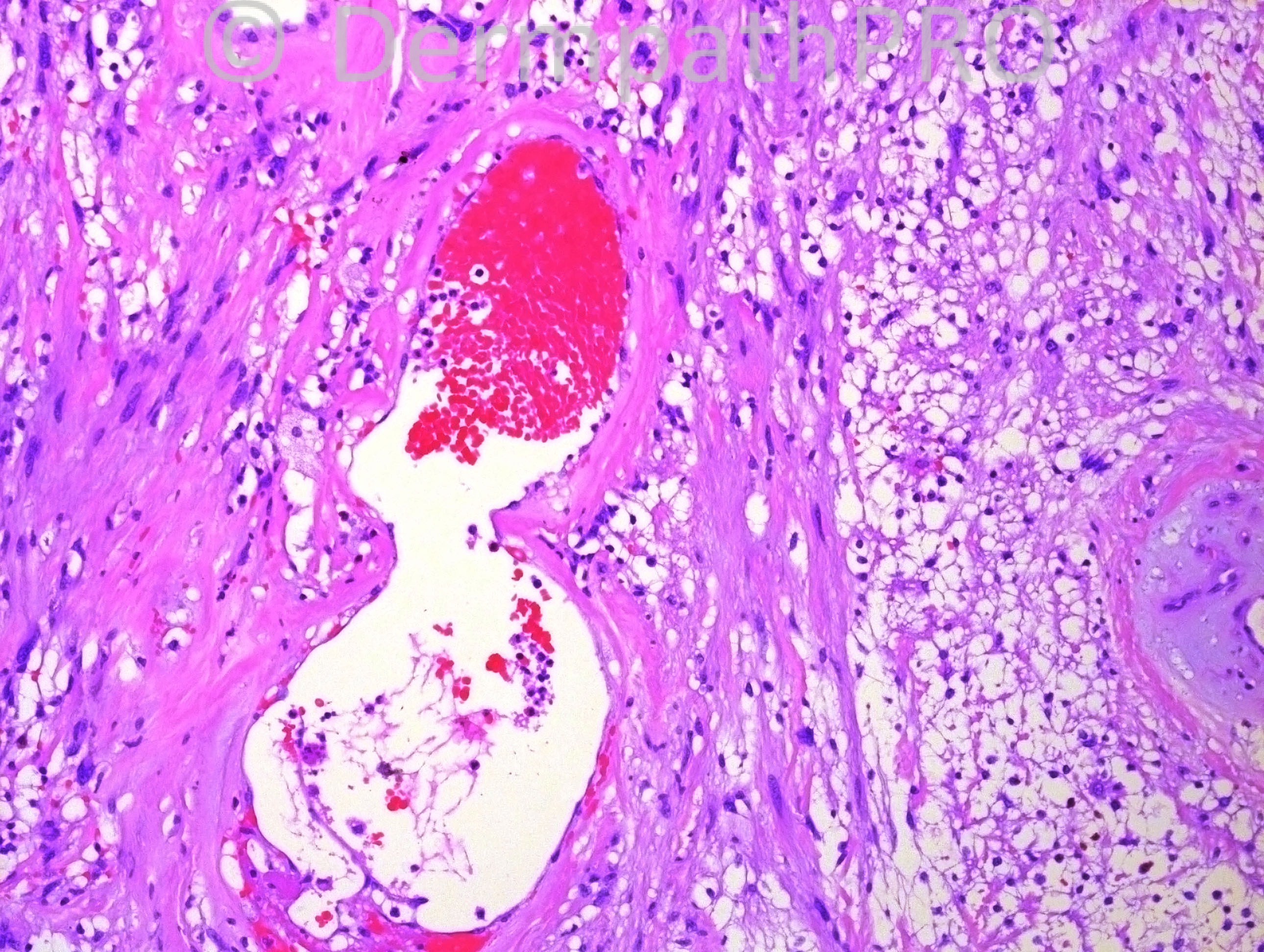

  Report Record

User Feedback


Dr. Mona Abdel-Halim

Posted

I am thinking of pleomorphic hyalinizing angiectatic tumor of soft tissue.

Share this comment


Link to comment
share_externally

IgorSC

Posted

Great case! I also agree with Mona´s diagnosis, Pleomorphic hyalinizing angiectatic tumor with their viral-like cells similar to those seen in Myxoinflammatory fibroblastic sarcoma (some authors say there´s a relationship between them). I had 2 cases like this.

Share this comment


Link to comment
share_externally

Guest Romualdo C. L. Filho

Posted

PHAT.

Share this comment


Link to comment
share_externally

Robledo F. Rocha

Posted

I encountered frequent neoplastic cells with too pronounced nuclear pleomorphism and I didn’t find any convincing intranuclear pseudoinclusion. Clear stromal areas may be reminiscent of myxoid matrix.
So, until immunohistochemical studies prove otherwise, I go with myxofibrosarcoma (or other related tumor) with PHAT-like vascular changes. Favor this hypothesis the history of a slowly growing tumor. Probably mitotic figures and thin-walled curvilinear vessels can be found elsewhere.

Share this comment


Link to comment
share_externally

Guest Bansal_

Posted

Agree, pleomorphic hyalinising angiectatic tumour.

Share this comment


Link to comment
share_externally

Guest Rodrigo Restrepo

Posted

Pleomorphic hyalinizing angiectatic tumor

Share this comment


Link to comment
share_externally

Guest Jim Davie MD

Posted

There are large floret-type vs. multinucleated atypical cells in the two center photos, and large telangiectatic vessels with a little hemosiderin. Fat necrosis with foamy histiocytes, fibrosis, and possibly dystrophic basophilic calcification (top left photo) seems to be a prevalent--if somewhat non-specific--finding.
Given the long-term 15 yr history, and low cellularity, I would favor trauma-related fat necrosis, or trauma-related hemosiderotic fibrohistiocytic lipomatous lesion (has floret cells, but usually distal extremity), in the differential diagnosis with PHAT. A stain for CD34 and CD68 may be of utility for differentiation.

Share this comment


Link to comment
share_externally

Ancient schwannoma.

Share this comment


Link to comment
share_externally

Dr. Hafeez Diwan

Posted

I called this pleomorphic hyalinizing angiectatic tumor.

Share this comment


Link to comment
share_externally


×
×
  • Create New...