Jump to content
In this section we have spot diagnoses posted on a daily basis since June 2010, now over 4000! You can review the archived cases and read the suggested diagnoses by users and the final comment by the contributors.
Case are uploaded each week day by 10 am UK time with the correct diagnosis will generally be posted at 8 pm UK time. Why not view the most recent spot diagnosis and proffer a diagnosis?

Case Number : Case 755 - 9th May Posted By: Guest

Please read the clinical history and view the images by clicking on them before you proffer your diagnosis.
Submitted Date :
   (0 reviews)

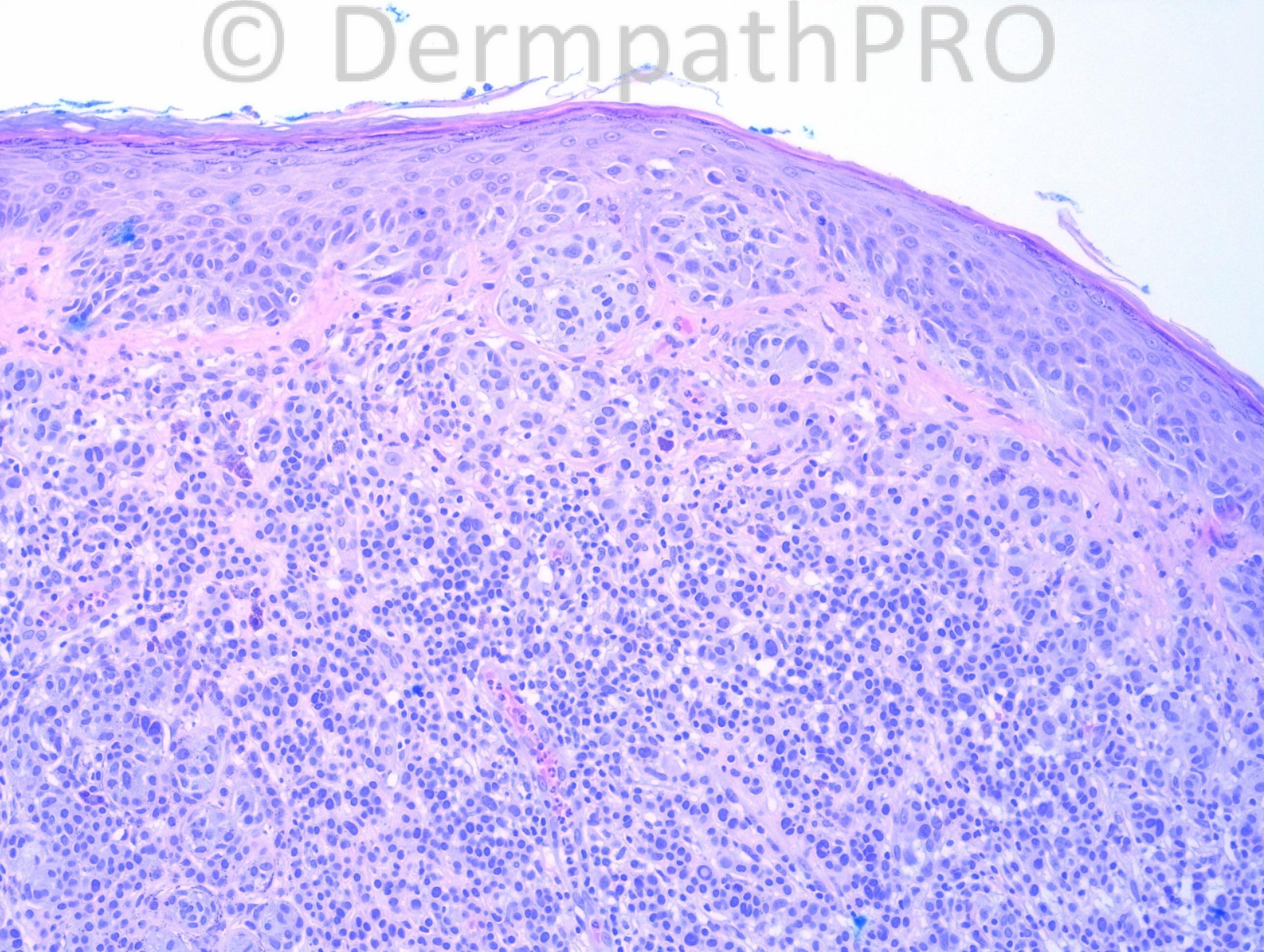
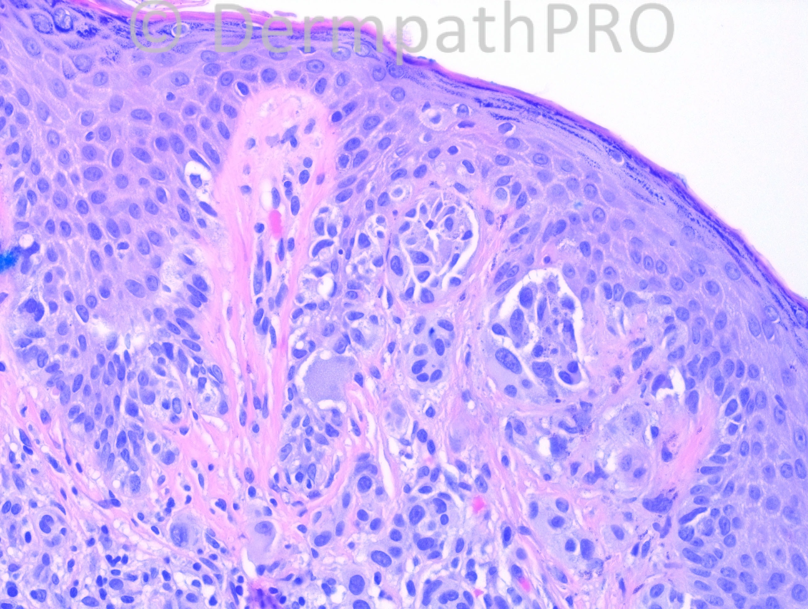
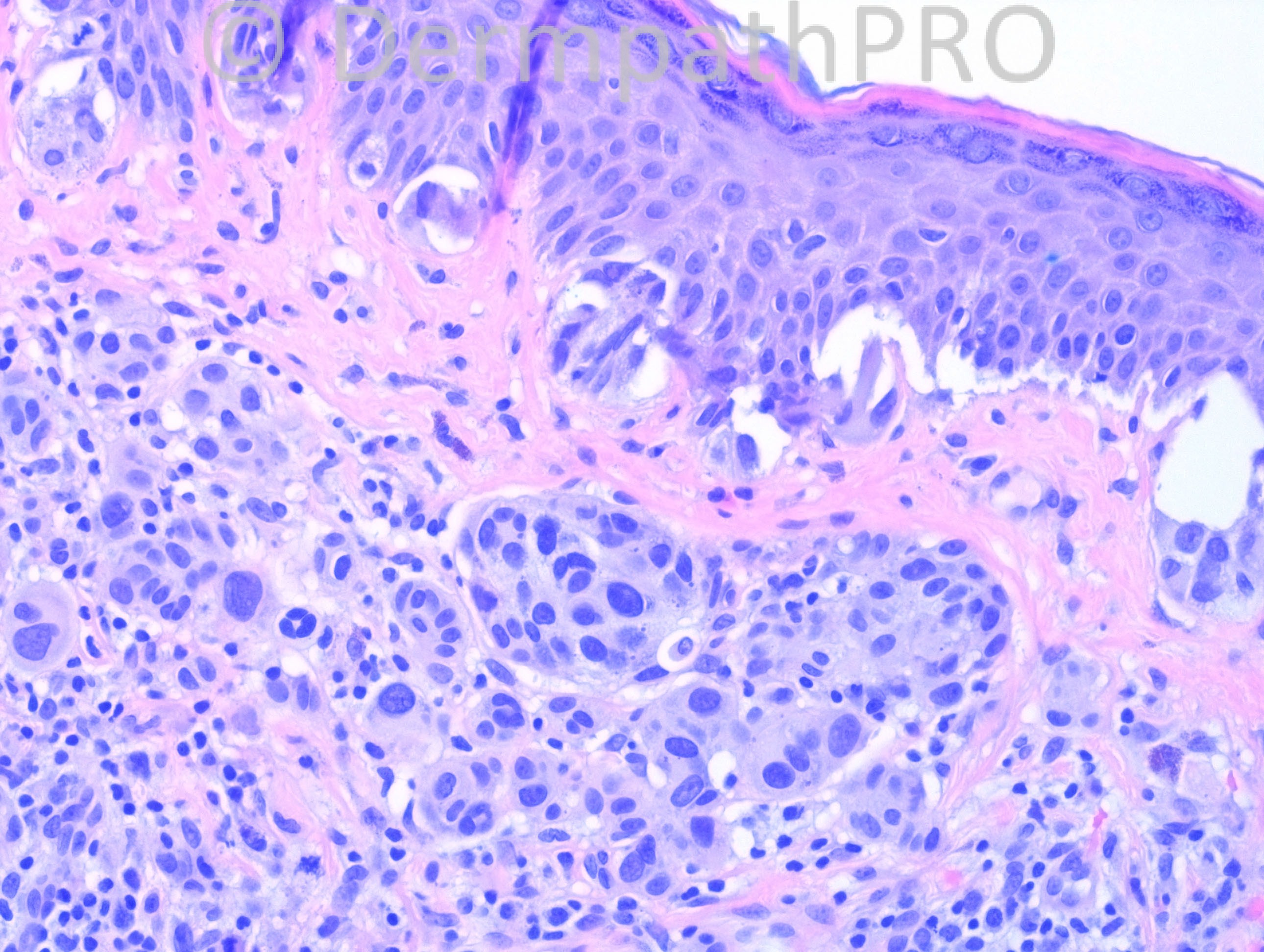
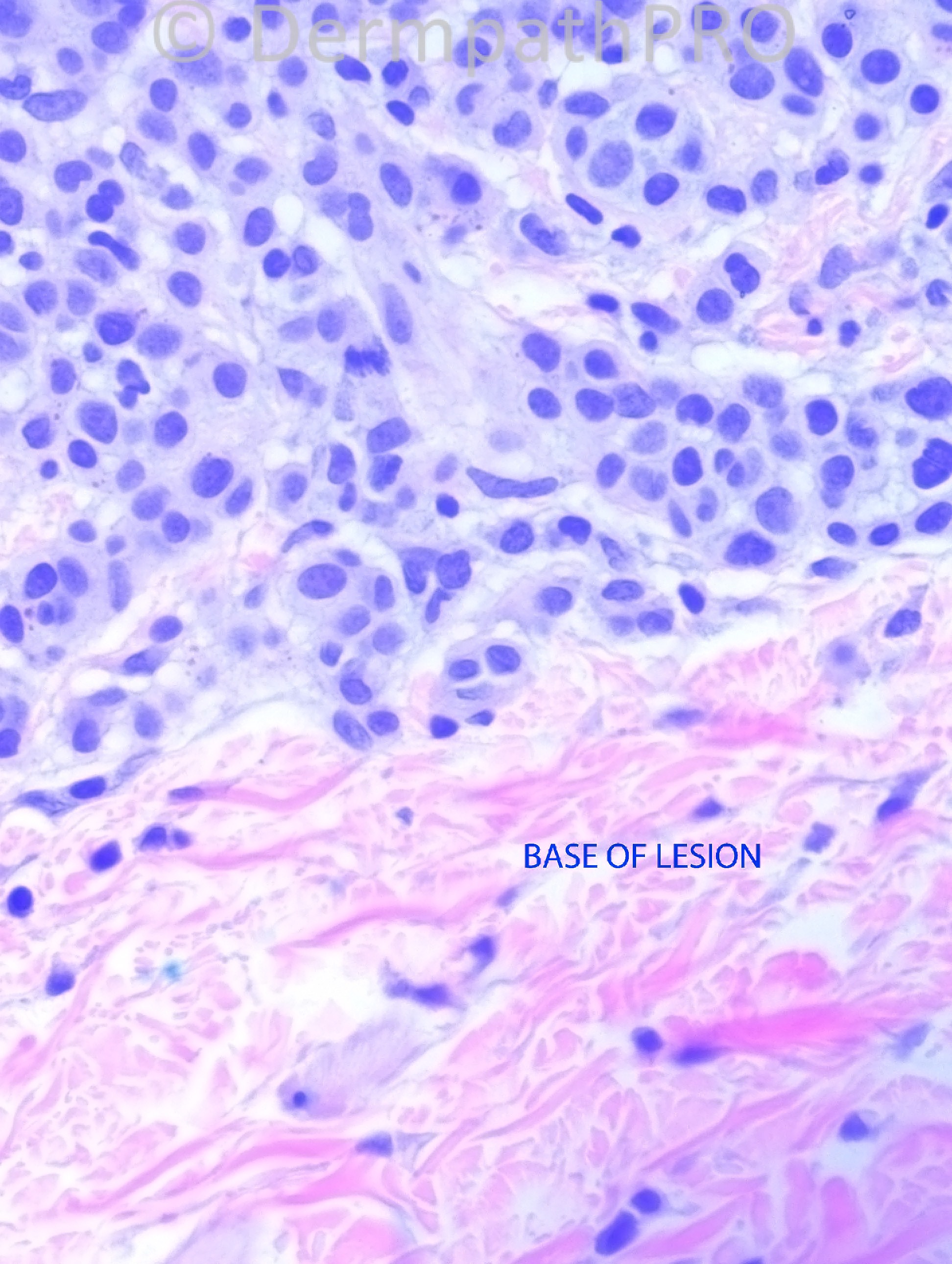
21 years-old female, with left upper back pigmented lesion. Clinical impression: nevus.

Case posted by Dr. Hafeez Diwan.

  Report Record

User Feedback


Dr. Richard Carr

Posted

"Odd" naevus some spitzoid features. A deep mitotic figure. We can't really assess silhouette but the compact nature of the base (as oppoased to the individual cells infiltrating reticular dermal collagen in a classic Spitz) is a bit concerning. That said DPN may have mitotic figures and lack maturation with depth. I would not call out right on these images but would like to see the glass. Could do CGH as differential includes MELTUMP. My diagnosis odd Spitzoid / naevoid lesion of uncertain malignant potential but favour benign behaviour (with everything crossed!). I suspect there may be a US v's rest of the world divide.

Share this comment


Link to comment
share_externally

Guest Romualdo C. L. Filho

Posted

I think it is melanoma, maybe a nevoid melanoma.

Share this comment


Link to comment
share_externally

Dr. Mona Abdel-Halim

Posted

I do not feel comfortable also with this lesion. Junctional nests show some atypia and pleomorphism. Also dermal component shows some pleomorphism. The sheet like arrangement of the dermal component with no enough stroma inbetween and the deep mitosis together with the lack of maturation, makes me consider nevoid melanoma. Will love to read more expert opinions.

Share this comment


Link to comment
share_externally

Guest Gustavo

Posted

Nevoid Melanoma

Share this comment


Link to comment
share_externally

Eman El-Nabarawy

Posted

Nevoid Melanoma.

Share this comment


Link to comment
share_externally

Robledo F. Rocha

Posted

There are some worrying features in this melanocytic proliferation that wouldn’t ensure me a good night’s sleep: a very small focus of pagetoid spread, nested atypical junctional component, small intradermal nests with pleomorphic cells clashing with surrounding cords of small cells, impaired maturation with depth, low pushing border and a mitotic figure in the deeper reaches.
Those findings really caught my eyes, so I favor nevoid melanoma.

Share this comment


Link to comment
share_externally

Guest Bansal_

Posted

My first impression is naevoid melanoma too.

Share this comment


Link to comment
share_externally

Guest Jim Davie MD

Posted

Nevoid melanoma with Spitz features, pending immunostains to confirm (Ki67, HMB45).

Agree that this is a case made for CGH, especially if the stains are less than helpful, using the newer panels that are more sensitive for Spitzoid melanoma.
Histologic features that caught my eye:
1. Two populations of dermal melanocytes: large, more atypical Spitzoid melanocytes with gray cytoplasm, and smaller melanocytes with amphophilic cytoplasm, nuclear pleomorphism with focal intranuclear inclusions, which appear to be irregularly intermixed.
2. Elevated mitotic activity, concentrated paradoxically in the smaller, less-Spitzoid cell component, in both the upper dermis [mitosis in bottom left corner, in mid-left photo], and the deep edge [middle of bottom left photo].
3. Asymmetric junctional component of Spitzoid melanocytes with sparse pagetoid scatter and focal confluent lentiginous nesting [right side of top left photo].
[i] It bears some resemblance to the Spitzoid nevoid melanoma in Difficult Opinions case#15 (DO015). [/i]

Share this comment


Link to comment
share_externally

Guest Rodrigo Restrepo

Posted

[color=#1C2837][font=arial, verdana, tahoma, sans-serif][size=4]Nevoid Melanoma[/size][/font][/color]

Share this comment


Link to comment
share_externally

Dr. Hafeez Diwan

Posted

I called this melanoma. Additional features were: patchy HMB45 positivity, without nice maturation, and focally more numerous Ki67 positivity, also without any obvious stratification.

Share this comment


Link to comment
share_externally


×
×
  • Create New...