Jump to content
In this section we have spot diagnoses posted on a daily basis since June 2010, now over 4000! You can review the archived cases and read the suggested diagnoses by users and the final comment by the contributors.
Case are uploaded each week day by 10 am UK time with the correct diagnosis will generally be posted at 8 pm UK time. Why not view the most recent spot diagnosis and proffer a diagnosis?

Case Number : Case 756 - 10th May Posted By: Guest

Please read the clinical history and view the images by clicking on them before you proffer your diagnosis.
Submitted Date :
   (0 reviews)

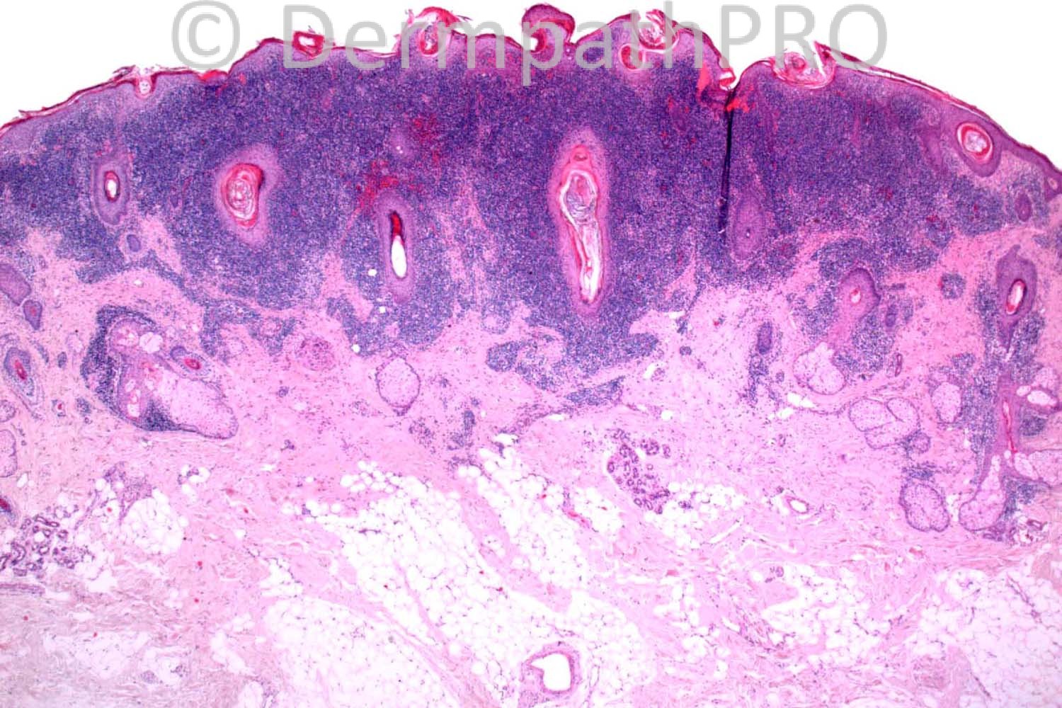
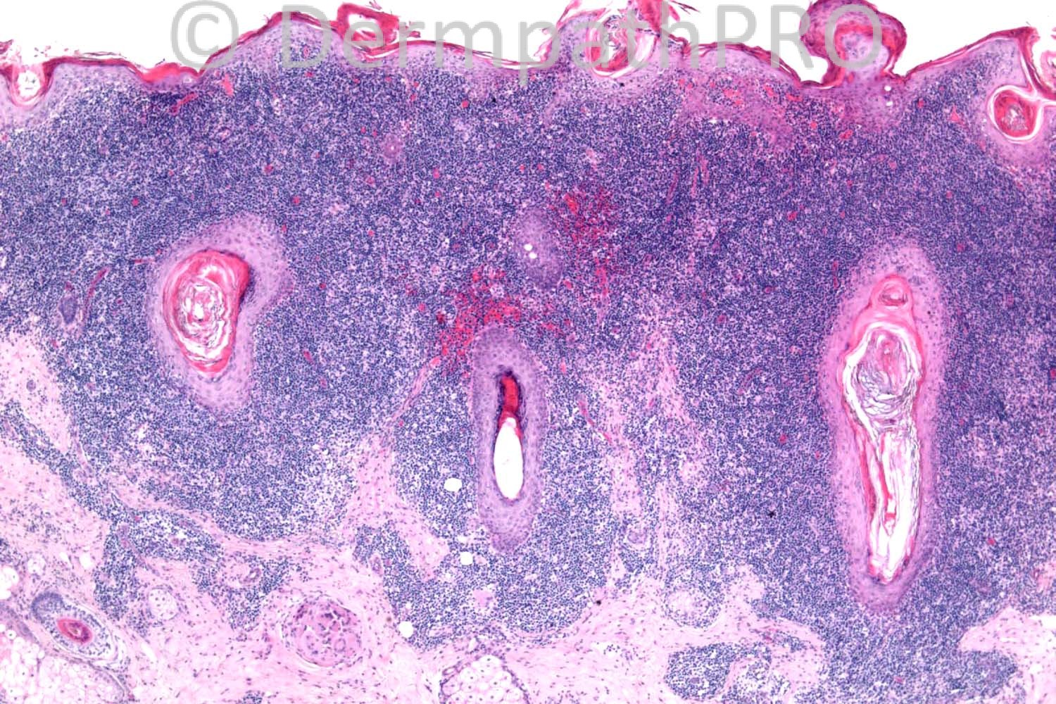
50 years-old female. Forehead lesion, nodular or cystic? ?AFX, ?DF

Case posted by Dr. Richard Carr, with thanks to Dr. Saleem Taibjee for sharing this case.


  Report Record

User Feedback


Dr. Mona Abdel-Halim

Posted

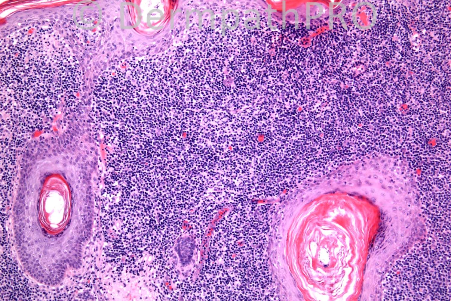
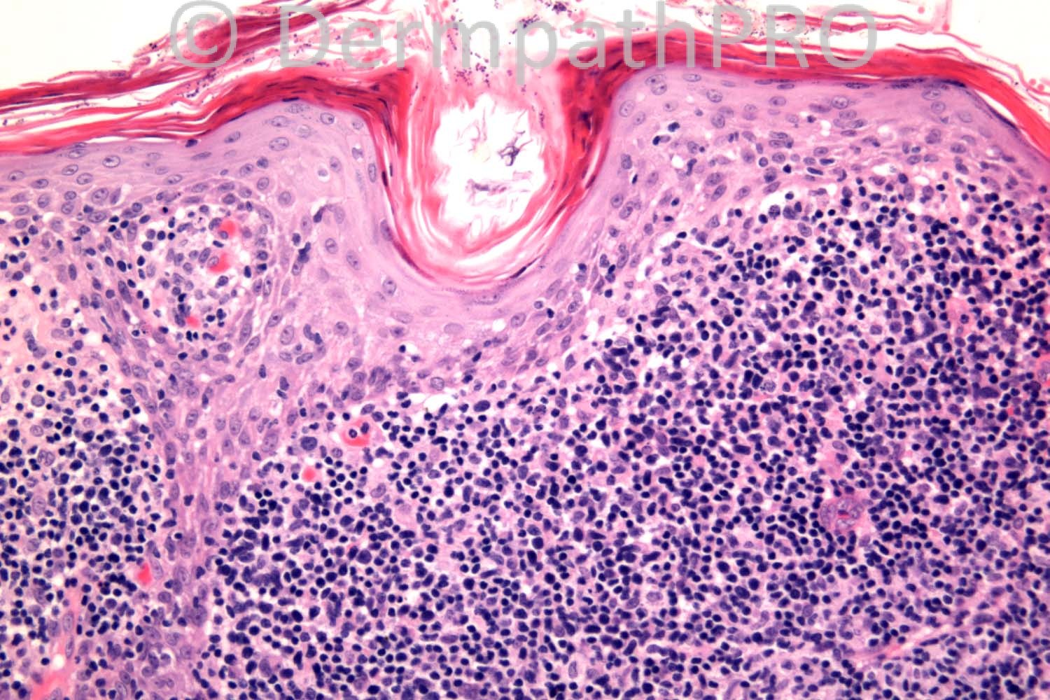
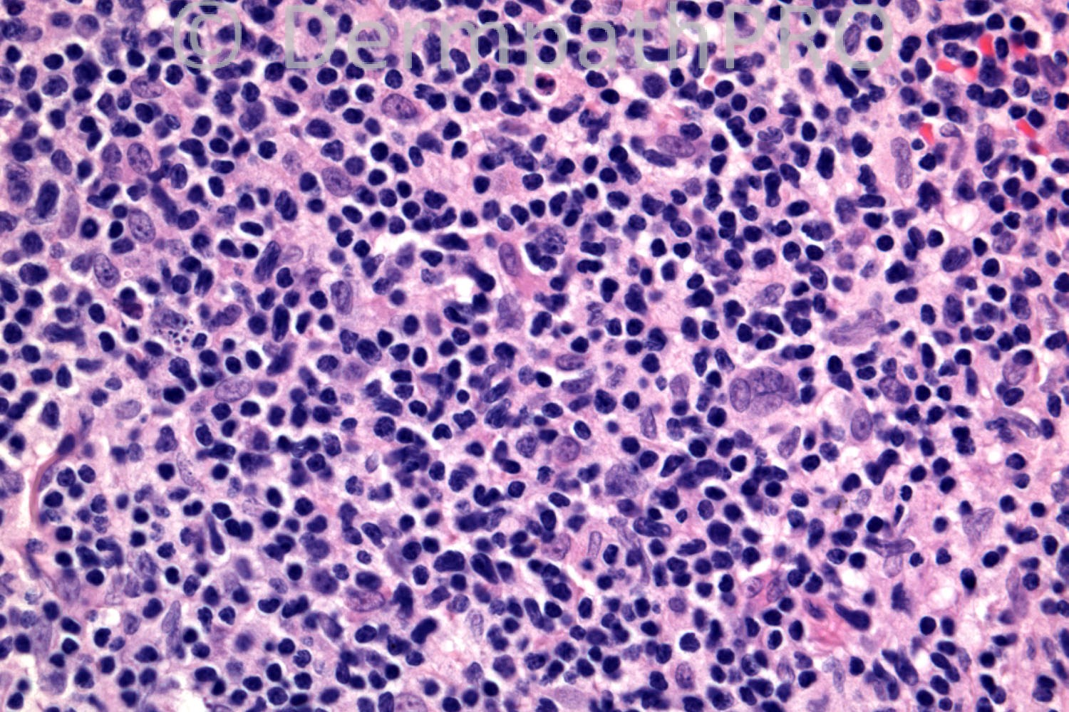
Lymphoid proliferation with a T cell pattern. Small to medium lymphocytic infiltrate with atypical irregular nuclei. Some epidermotropism. Could be primary cutaneous CD4 positive small/medium TCL (as lesions are usually solitary or localized) or T cell pseudolymphoma (lymphomatoid drug reaction). I think the idea of a single lesion of tumor stage MF without the clinical setting of patches and plaques else where is no more accepted. Will do: CD2, CD3, CD5, CD4 and CD8and TCR gene rearrangement studies.

Share this comment


Link to comment
share_externally

Guest Bansal_

Posted

? T cell lymphoma. Needs lymphoma panel.

Share this comment


Link to comment
share_externally

Guest Dr Engin Sezer

Posted

Folliculotrophic MF

Share this comment


Link to comment
share_externally

Sasi Attili

Posted

Solitary lesion with a top heavy dense infiltrate with some epidermotropism. Agree with Mona, but thinking this might be a pseudo!!

Share this comment


Link to comment
share_externally

Robledo F. Rocha

Posted

I think it's a pseudolymphoma. A top-heavy, folliculocentric and epidermotropic dense infiltrate of lymphocytes. I would expected, in a folliculotropic mycosis fungoides, mucinous expanded follicles or pilocentrism without epidermotropism. Also, intraepidermal lymphocyes are not larger than dermal lymphocytes.
This hypothesis is supported by the history case of a solitary nodule on the head, an uncommon presentation of a cutaneous lymphoma.

Share this comment


Link to comment
share_externally

Guest Romualdo C. L. Filho

Posted

Pseudolymphoma. I think it could be lupus erythematous with an uncommonly dense perifollicular lymphoid infiltrate or a pseudolymphomatous folliculitis.

Share this comment


Link to comment
share_externally

Dr. Richard Carr

Posted

It was a mixed infiltrate of B-cells, T-cells including CD4 and CD8 (no particular predominance). We did not peform PCR. The solitary nature, location and distribution of the cells might make you suggest a distinctive clinicopathological lesion in the literature (pretty uncommon).

Share this comment


Link to comment
share_externally

Eman El-Nabarawy

Posted

LPLK.

Share this comment


Link to comment
share_externally

Guest Jim Davie MD

Posted

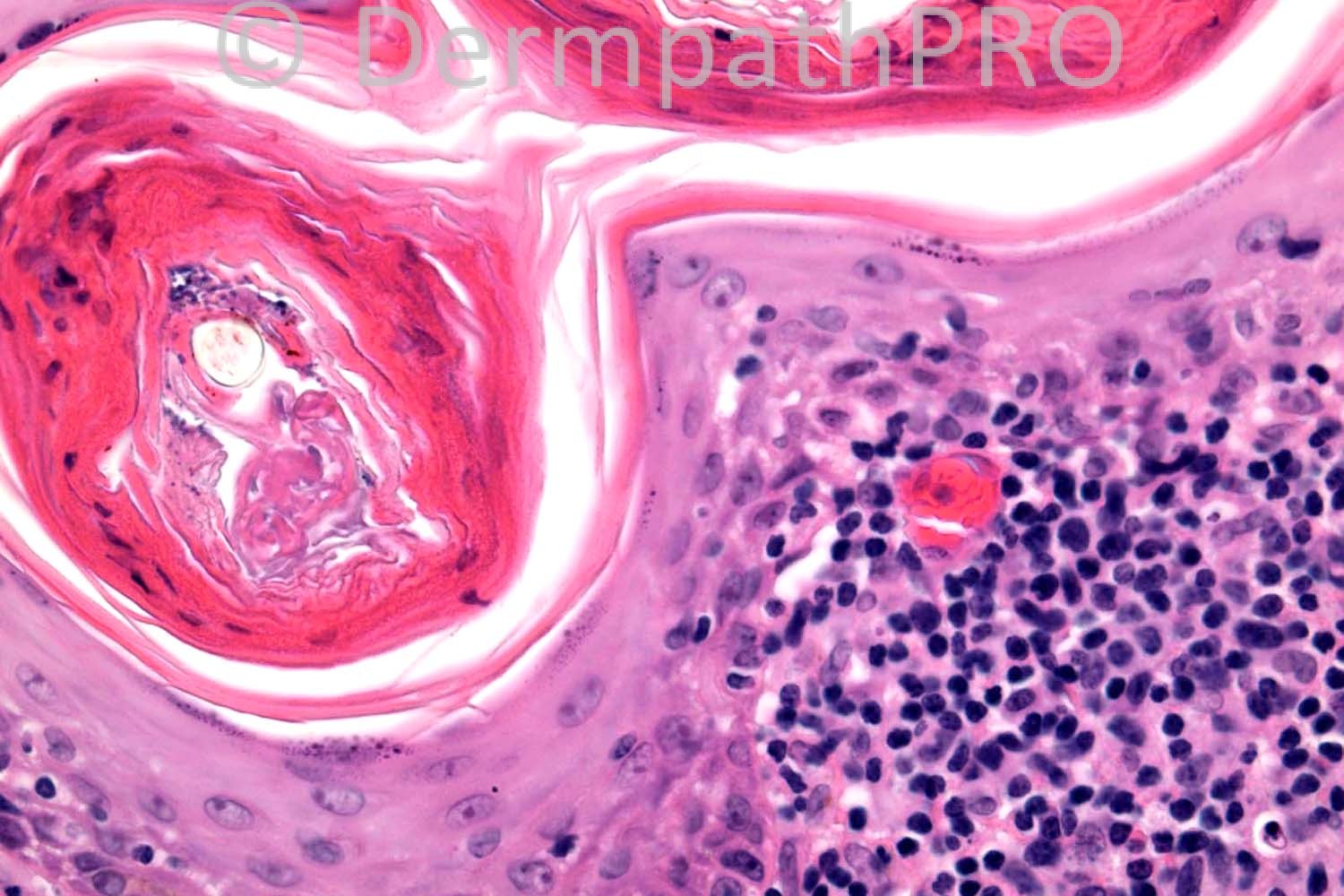
I agree this is most likely LPLK, with the differential including an unusual clinical presentation of lichen planopilaris.

- There is specific targeting of the epidermis and especially the follicular infundibula.
- Follicles show keratin plugging and hypergranulosis with lichen-planus like epidermal changes.
- No perieccrine or sebaceous adnexal infiltrate. No perivascular infiltrate. (these would be expected in lupus).
- There is a foreign body granuloma adjacent to the bottom-left edge of the infiltrate.

Share this comment


Link to comment
share_externally

nick turnbull

Posted


Primary cutaneous follicle center lymphoma?

Share this comment


Link to comment
share_externally

Guest Rodrigo Restrepo

Posted

Majocchi´s granuloma

Share this comment


Link to comment
share_externally

Dr. Mona Abdel-Halim

Posted

Given the immunophenotyping results, the mixed T and B cell population, I would suggest: Pseudolymphomatous folliculitis.

Share this comment


Link to comment
share_externally

Dr. Richard Carr

Posted

Well done Mona!! although I think we had some very good alternate suggestions and discussion.

We also favoured pseudolymphoma and Dr Saleem Taibjee specifically raised the possibility of pseudolymphomatous folliculitis. We shared the case with Dr Dmitry Kazakov who agreed and speculated that the numerous fungal spores in the follicular infundibula (on PAS, and in the last image) may be relevant. A pseudolymphomatous variant of LPLK would certainly be a good alternate suggestion. I put this case in to draw your attention to pseudolymphomatous folliculitis.

References
Arai E, Okubo H, Tsuchida T, Kitamura K, Katayama I. Pseudolymphomatous folliculitis: a clinicopathologic study of 15 cases of cutaneous pseudolymphoma with follicular invasion. Am J Surg Pathol. 1999 Nov;23(11):1313-9.

Kazakov DV, Belousova IE, Kacerovska D, Sima R, Vanecek T, Vazmitel M, Pizinger K, Michal M. Hyperplasia of hair follicles and other adnexal structures in cutaneous lymphoproliferative disorders: a study of 53 cases, including so-called pseudolymphomatous folliculitis and overt lymphomas. Am J Surg Pathol. 2008 Oct;32(10):1468-78.

Share this comment


Link to comment
share_externally

Guest Dr Engin Sezer

Posted

Great case, many thanks Dr. Carr and Dr. Taibjee

Share this comment


Link to comment
share_externally


×
×
  • Create New...