Jump to content
In this section we have spot diagnoses posted on a daily basis since June 2010, now over 4000! You can review the archived cases and read the suggested diagnoses by users and the final comment by the contributors.
Case are uploaded each week day by 10 am UK time with the correct diagnosis will generally be posted at 8 pm UK time. Why not view the most recent spot diagnosis and proffer a diagnosis?

Case Number : Case 757 - 13th May Posted By: Guest

Please read the clinical history and view the images by clicking on them before you proffer your diagnosis.
Submitted Date :
   (0 reviews)

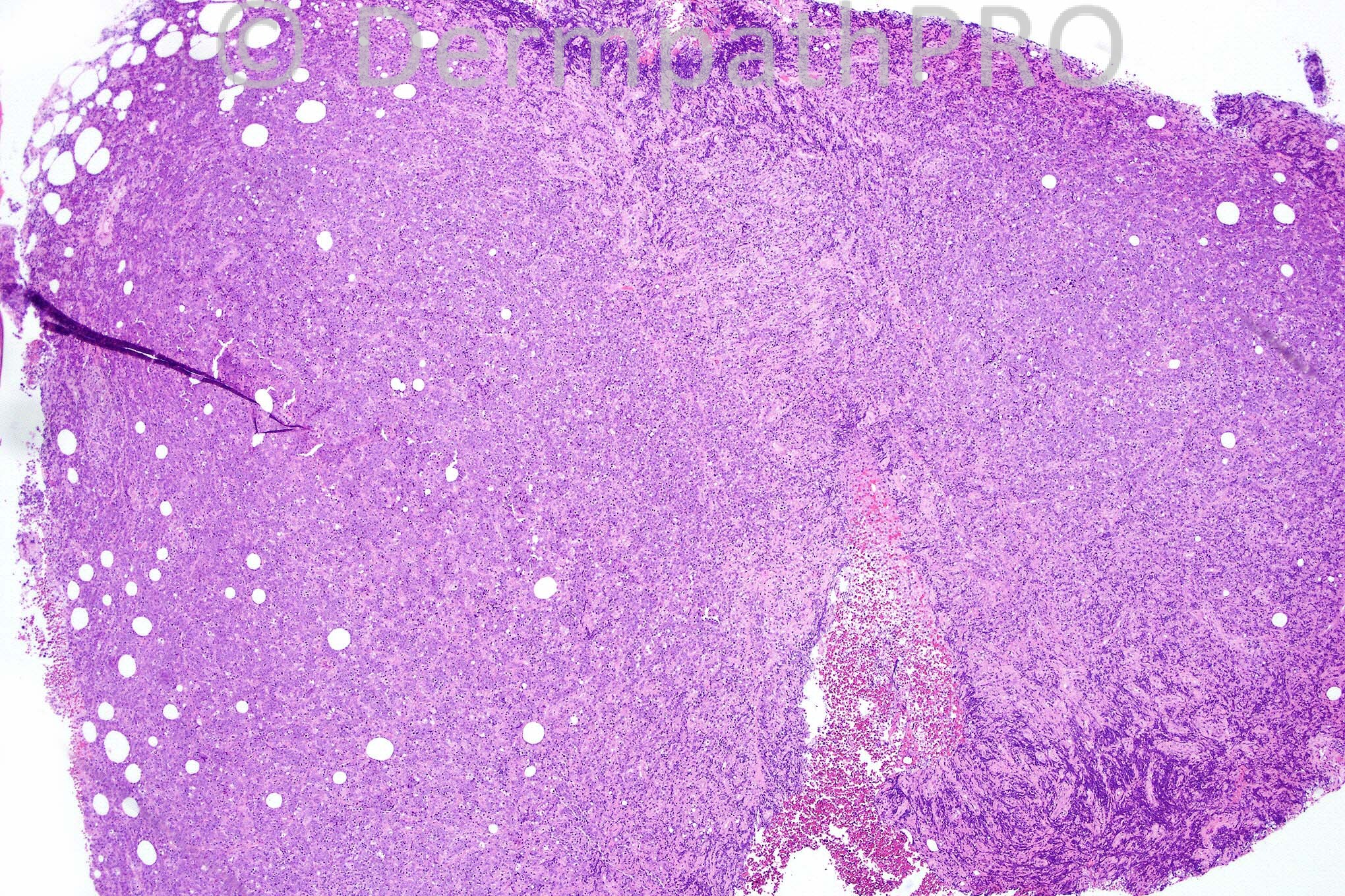
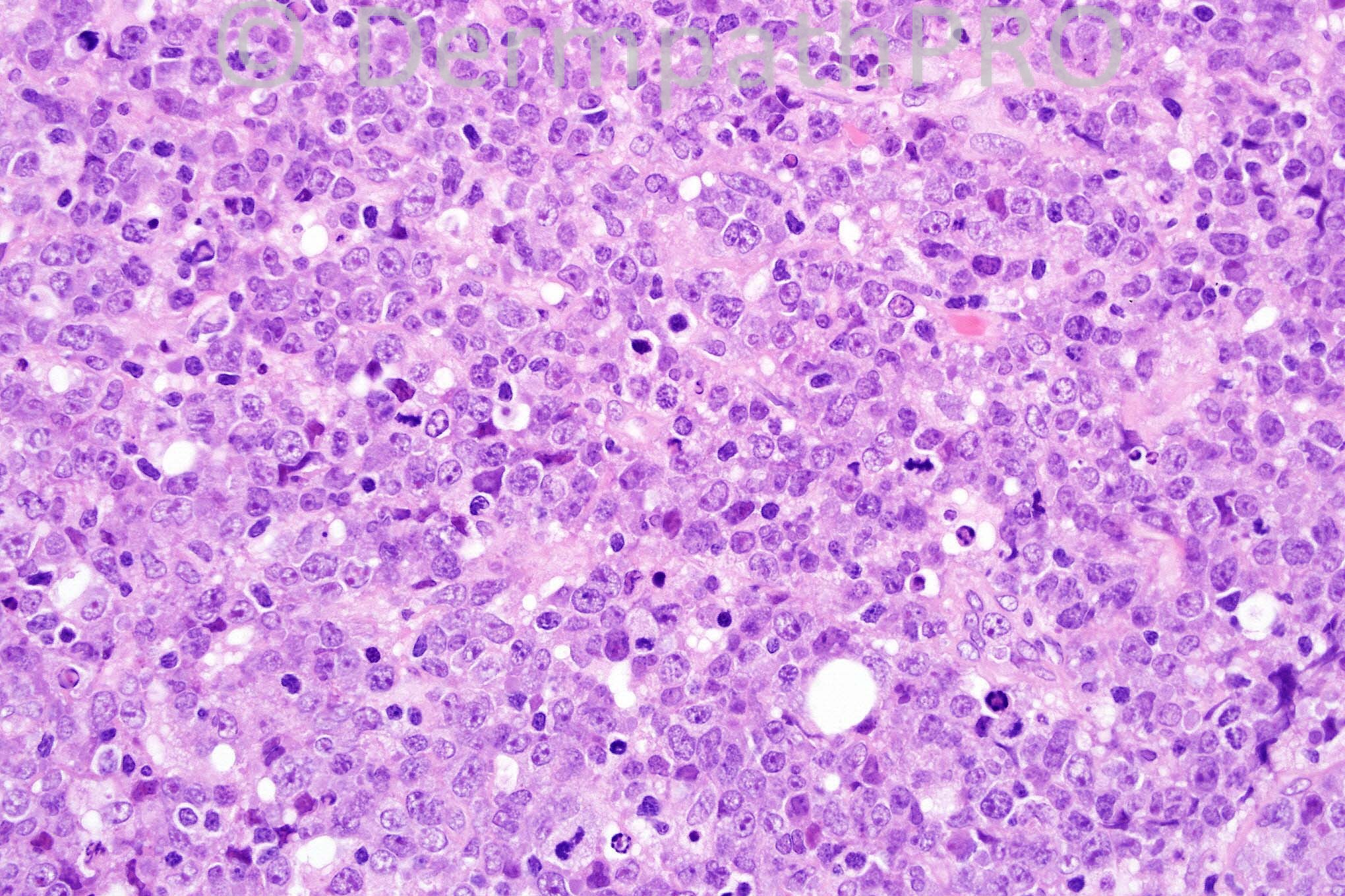
70 years-old male with a history of nodules and plaques on the abdomen.


  Report Record

User Feedback


Guest Dr Engin Sezer

Posted

Epithelioid mailignant peripheral nerve sheath tumor (based on palisading pattern in Fig. 1 )

Share this comment


Link to comment
share_externally

Guest Dr Engin Sezer

Posted

History of NF??

Share this comment


Link to comment
share_externally

Merkel cell carcinoma vs lymphoma

Share this comment


Link to comment
share_externally

Sasi Attili

Posted

Would favour a high grade lymphoma, but need immunos to rule out poorly diff melanoma/ carcinoma's/ sarcoma

Share this comment


Link to comment
share_externally

Guest Zena Slim (SpR Wessex)

Posted

A dense atypical lymphoid population with numerous blasts, apoptotic bodies and mitoses. I would favour tumour-stage Mycosis Fungoides. The possibilty of transformation to an anaplastic large cell lymphoma should also be considered.

Share this comment


Link to comment
share_externally

Guest Bansal_

Posted

DD: high grade lymphoma (?DLBCL), melanoma, undiff carcinoma, sarcoma. On morphology, favour lymphoma.

Share this comment


Link to comment
share_externally

Eman El-Nabarawy

Posted

Favor B cell lymphoma.

Share this comment


Link to comment
share_externally

Guest Marcia

Posted

Favor lymphoma, diffuse large B cell lymphoma. Leg type?

Share this comment


Link to comment
share_externally

Guest Rodrigo Restrepo

Posted

Large B cell lymphoma

Share this comment


Link to comment
share_externally

Robledo F. Rocha

Posted

Sheets of cohesive large atypical cells extending into the subcutis comprising cells with irregularly indented nuclei, dispersed chromatin and prominent nucleoli. These microscopic findings and a case history of lesions confined to one anatomic region in an old man make me think of primary cutaneous anaplastic large cell lymphoma.
Immunophenotype study is vital to distinguish this hypothesis from other diffuse large cell lymphomas, like cutaneous follicle centre cell lymphoma with a diffuse growth pattern and tumor stage of mycosis fungoides with large cell transformation, and from diffuse large cell nonlymphoid neoplasms.

Share this comment


Link to comment
share_externally

Dr. Mona Abdel-Halim

Posted

Thought of lymphoma: Anaplastic large TCL is my first impression. Considering the clincal localization to one region makes transformed tumor stage MF less likely. Definitely diffuse large BCL is a possibility. Immunophenotyping is mandatory.

Share this comment


Link to comment
share_externally

Guest Romualdo C. L. Filho

Posted

Large cell lymphoma. Diffuse large B cell lymphoma is my first possibility.

Share this comment


Link to comment
share_externally

Iskander H. Chaudhry

Posted

Hello everyone! Thanks for your contributions today, the diagnosis is diffuse large B-cell lymphoma.

Share this comment


Link to comment
share_externally

Guest Hazem Hamed

Posted

Interesting case. Was EBER performed on this case? I would add EBER to the immuno panel to exclude DLBCL of the elderly.

Share this comment


Link to comment
share_externally


×
×
  • Create New...