Jump to content
In this section we have spot diagnoses posted on a daily basis since June 2010, now over 4000! You can review the archived cases and read the suggested diagnoses by users and the final comment by the contributors.
Case are uploaded each week day by 10 am UK time with the correct diagnosis will generally be posted at 8 pm UK time. Why not view the most recent spot diagnosis and proffer a diagnosis?

Case Number : Case 758 - 14th May Posted By: Guest

Please read the clinical history and view the images by clicking on them before you proffer your diagnosis.
Submitted Date :
   (0 reviews)

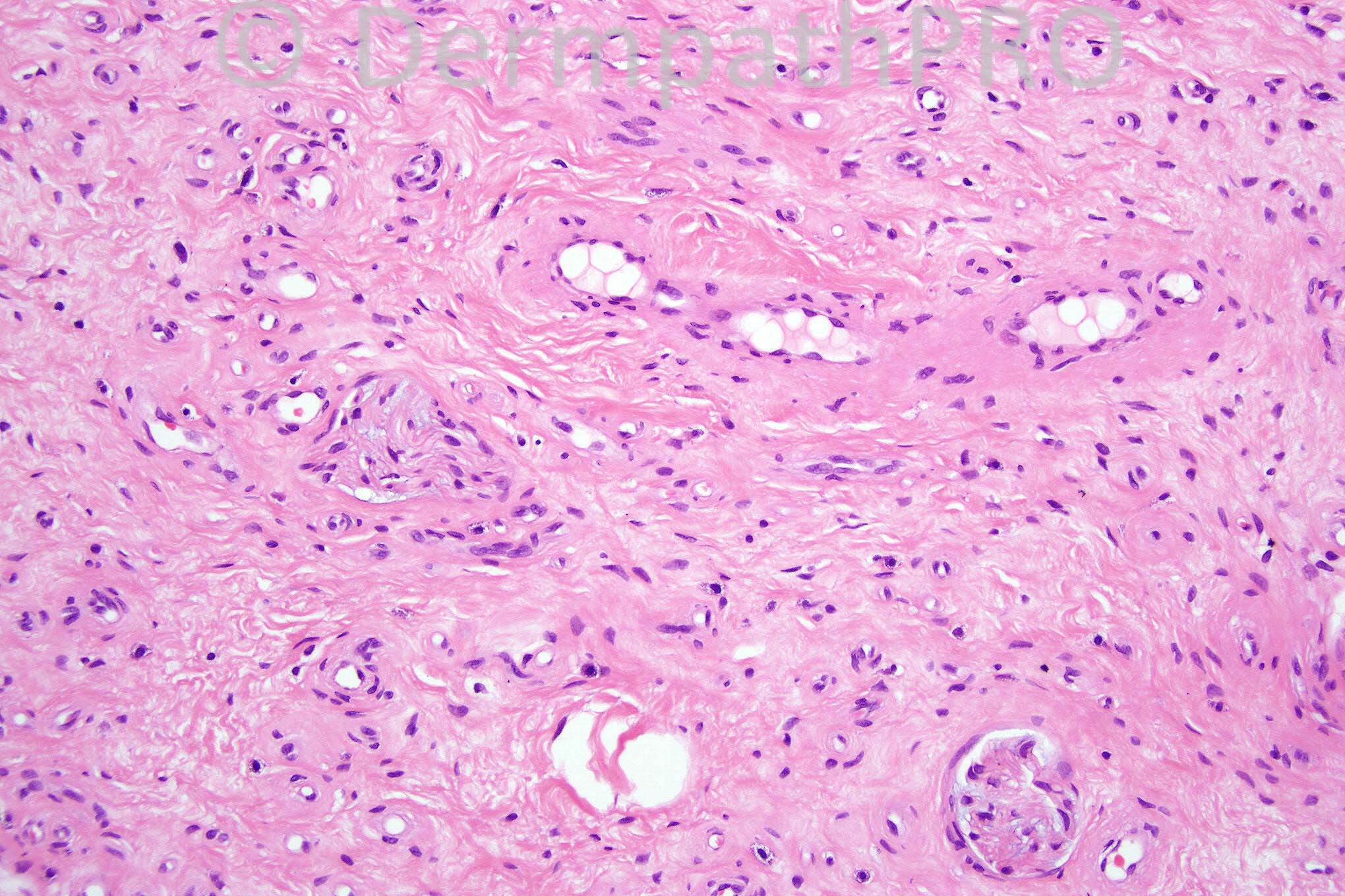
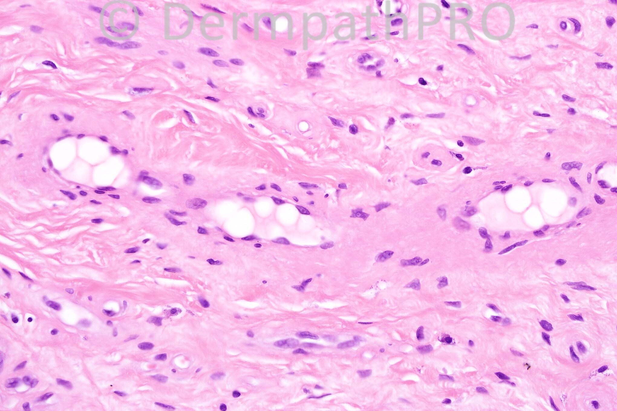
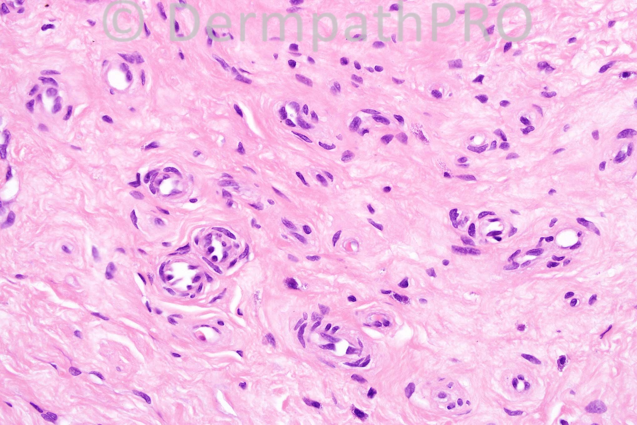
3 years-old female with a vascular lesion on the face.


  Report Record

User Feedback


Guest Zena Slim (SpR Wessex)

Posted

This lesion is within the capillary malformation / portwine stain (nevus flammeus)category including 'salmon patch. Is there any evidence of this lesion being related to a congenital clinical syndrome such as Sturge Webber, or Klipper-Trenaunay?

Share this comment


Link to comment
share_externally

Guest Romualdo C. L. Filho

Posted

The absence of a lobular pattern militates against a diagnosis of capillary or infantile hemangioma. The involvement of deep dermis and subcutaneous tissue does not favor a salmon patch. I think it is a vascular hamartoma, more in keeping with a port-wine stain.

Share this comment


Link to comment
share_externally

Guest Dr Engin Sezer

Posted

Microvenular hemangima

Share this comment


Link to comment
share_externally

Sasi Attili

Posted

The hyalinised stroma and the thin walled capillaries are probably more in keeping with a microvenular haemangioma (though not seeing the infiltration of arrector pili, typical of this entity).

Share this comment


Link to comment
share_externally

Robledo F. Rocha

Posted

Thick-walled and thin-walled blood vessels with round or oval contours evenly distributed throughout a fibrotic dermis and extending into subcutaneous tissue without clear limits. These features suggest a high-flow vascular malformation with direct arteriovenous shuntings or fistulas.
I think it is arteriovenous malformation. Maybe there are other clinical signs in addition to the visual ones, like pulsation, murmur and warmness, but it seems to be in quiescent stage.

Share this comment


Link to comment
share_externally

Guest Jaime A Mejia MD

Posted

This is killing me. Ducking instead of answering the challenge I say that vascular lesions in infancy are disproportionately benign and localized in nature, plaques or segmental lesions are of certain risk and mutifocally cutaneous lesions associated with internal hemangiomas as well. If this is a segmental stuff, [b]PHACE [/b]syndrome should be considered in the workup. Having said that, my ignoramus perception is that this lesion is not well lobulated, rather diffuse, with primitive vessels and hyalinized ones, fat and myfibroblastoid whorls semi-glomeruloid funny structures. Help me GLUT-1! and vascular hamartoma of some kind are in my list. Certainly not a capillary hemangioma, not so sure about infantile hemangio(endothelio)ma (GLUT-1?). Kassabach-Merrit, nop, I do not think so. Microvenular hemangioma, never seen one in a child, can be. You can tell, I am dumbfounded on this. Wait to see the results.

Share this comment


Link to comment
share_externally

Dr. Mona Abdel-Halim

Posted

I think this does not belong to vascular malformations because there is actual vascular proliferation. The pattern of the proliferation, the hyalinized stroma and the plump nuclei are making me consider microvenular hemangioma however it is more likely to be present in young adults.

Share this comment


Link to comment
share_externally

Iskander H. Chaudhry

Posted

This was a difficult one, the diagnosis is involuting infantile hemangioma.

Share this comment


Link to comment
share_externally

Guest Jaime A Mejia MD

Posted

Thanks.

Share this comment


Link to comment
share_externally

Robledo F. Rocha

Posted

What microscopic features is one supposed to pursue in order to diagnose an infantile hemangioma as an involuting or as an involuted lesion?

Share this comment


Link to comment
share_externally


×
×
  • Create New...