Jump to content
In this section we have spot diagnoses posted on a daily basis since June 2010, now over 4000! You can review the archived cases and read the suggested diagnoses by users and the final comment by the contributors.
Case are uploaded each week day by 10 am UK time with the correct diagnosis will generally be posted at 8 pm UK time. Why not view the most recent spot diagnosis and proffer a diagnosis?

Case Number : Case 764 - 22nd May Posted By: Guest

Please read the clinical history and view the images by clicking on them before you proffer your diagnosis.
Submitted Date :
   (0 reviews)

60 years-old female with a right calf lesion. Clinical impression: dysplastic nevus vs. melanoma.

Case posted by Dr. Hafeez Diwan.


  Report Record

User Feedback


Guest Dr Engin Sezer

Posted

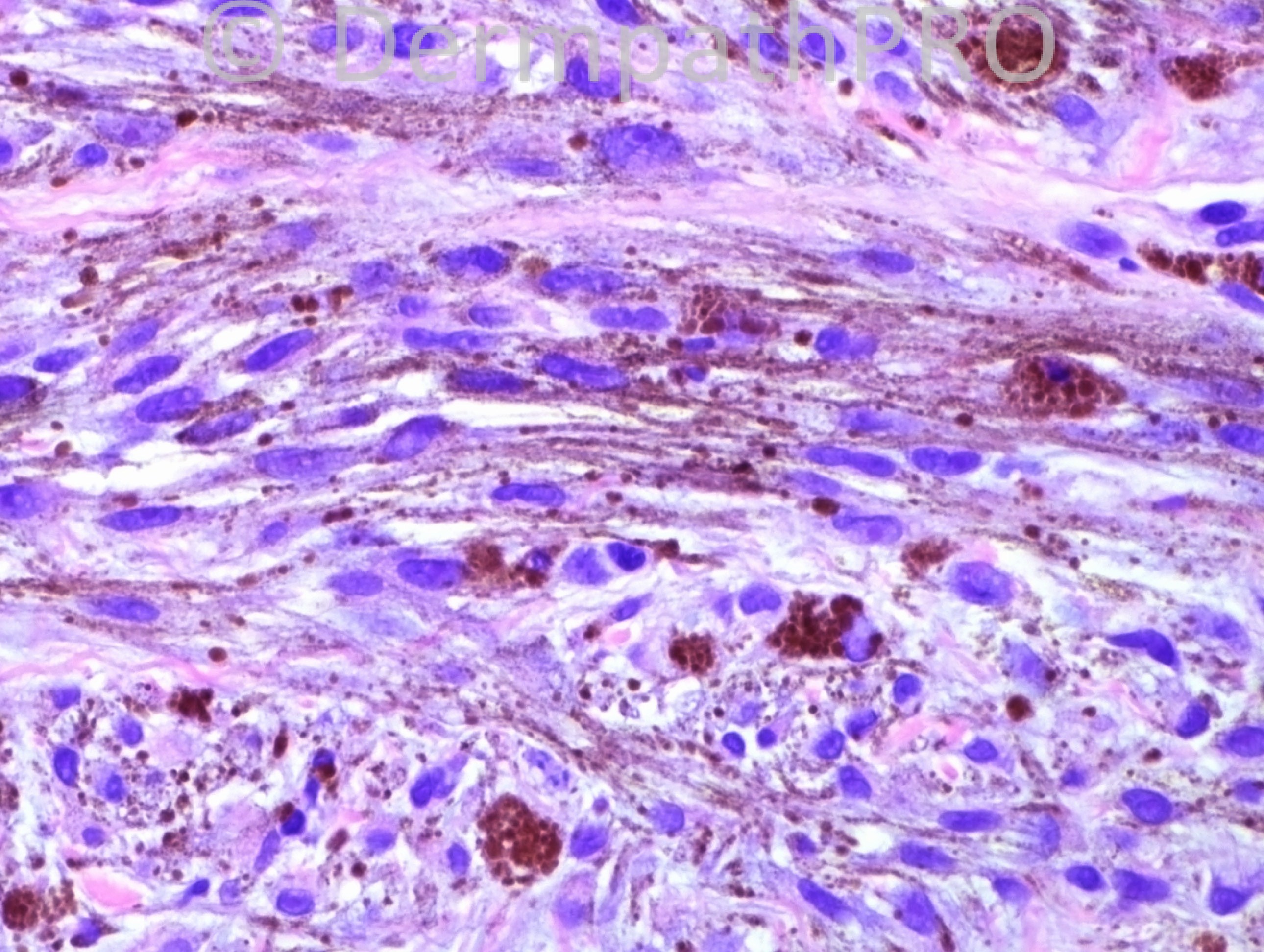
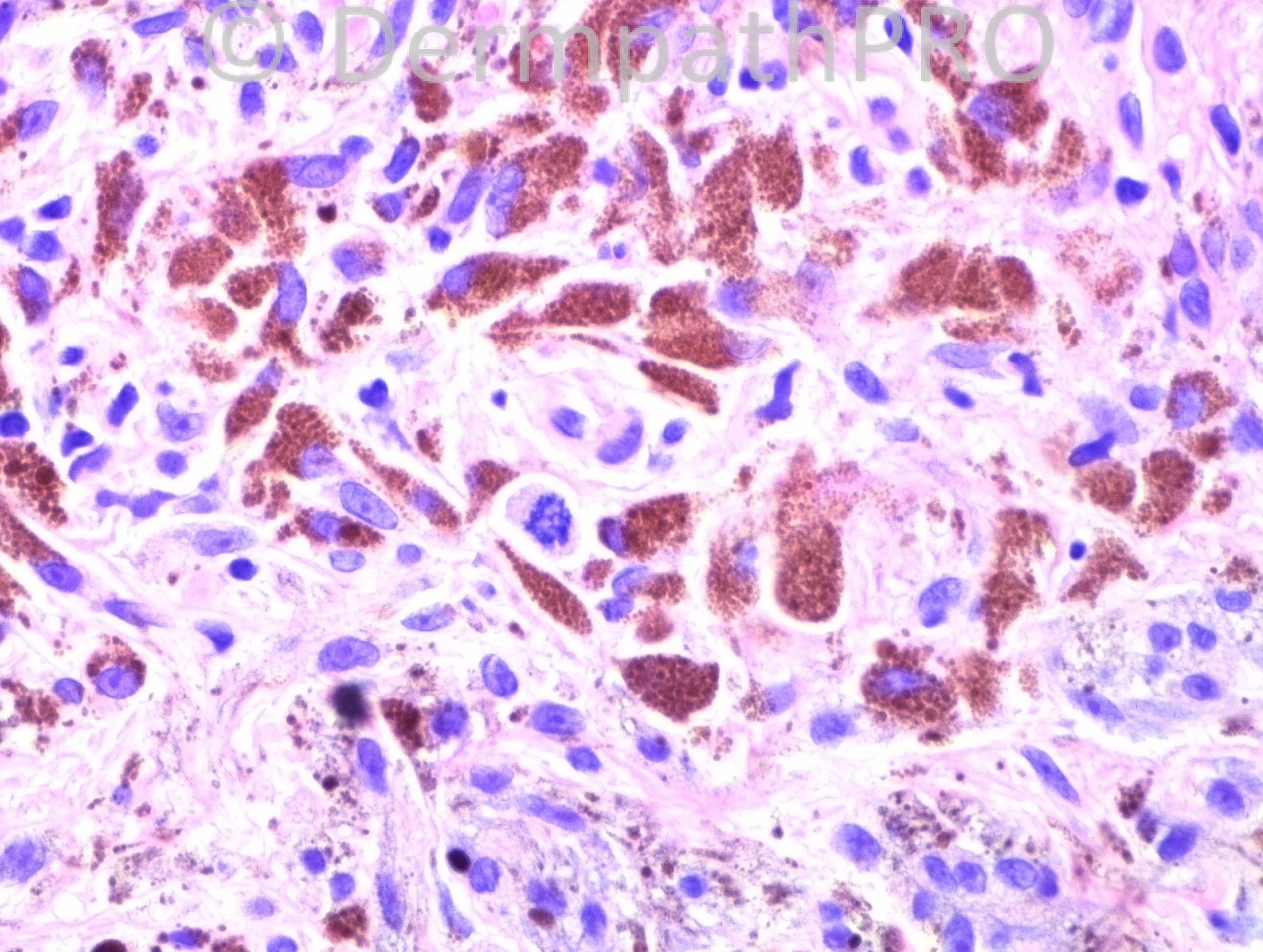
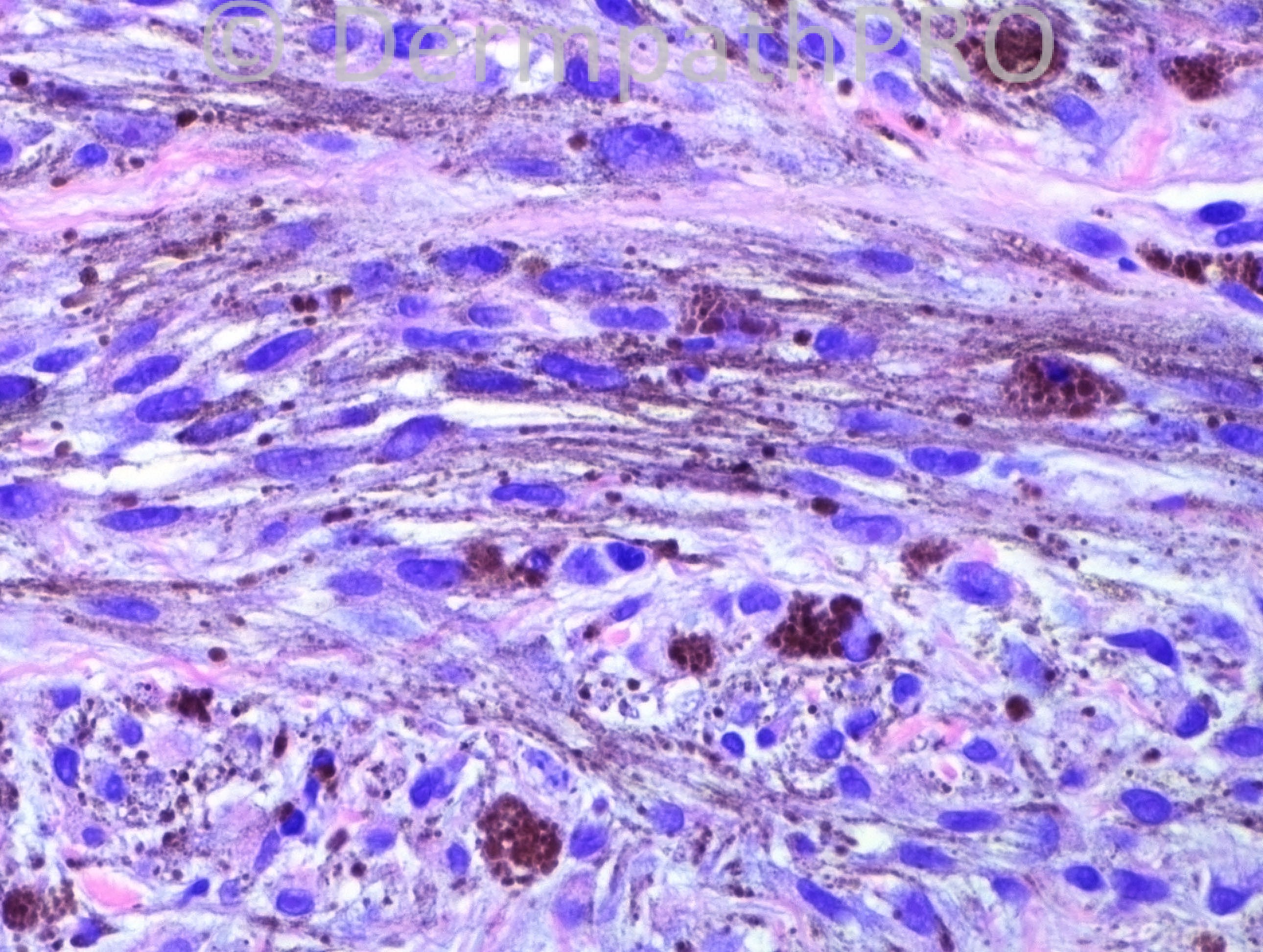
Melanocytic lesion with high grade pleomorphism and atypical mitoses. Spindle cell/blue nevus-like melanoma

Share this comment


Link to comment
share_externally

Guest Marcia

Posted

Melanoma.

Share this comment


Link to comment
share_externally

Guest Bansal_

Posted

? melanoma, ? atypical blue naevus.

Share this comment


Link to comment
share_externally

IgorSC

Posted

It is difficult to diagnose based only in high power pictures, but i will try: spindle and heavily pigmented melanocytes with atypia and some mitotic figures. As we know, it is difficult to differentiate malignant blue nevus from atypical blue nevus and some cellular blue nevus. Many histologic and cytologic features are necessary for this differentiation, including size of the lesion, borders, mitotic count, atypia and others. Based only on these high power images it could be a Malignant blue nevus x Atypical blue nevus.

Share this comment


Link to comment
share_externally

Robledo F. Rocha

Posted

A pigmented lesion on a man’s back or on a woman’s calf always concerns me, mainly if there are atypical melanocytes and mitotic activity, like this case.
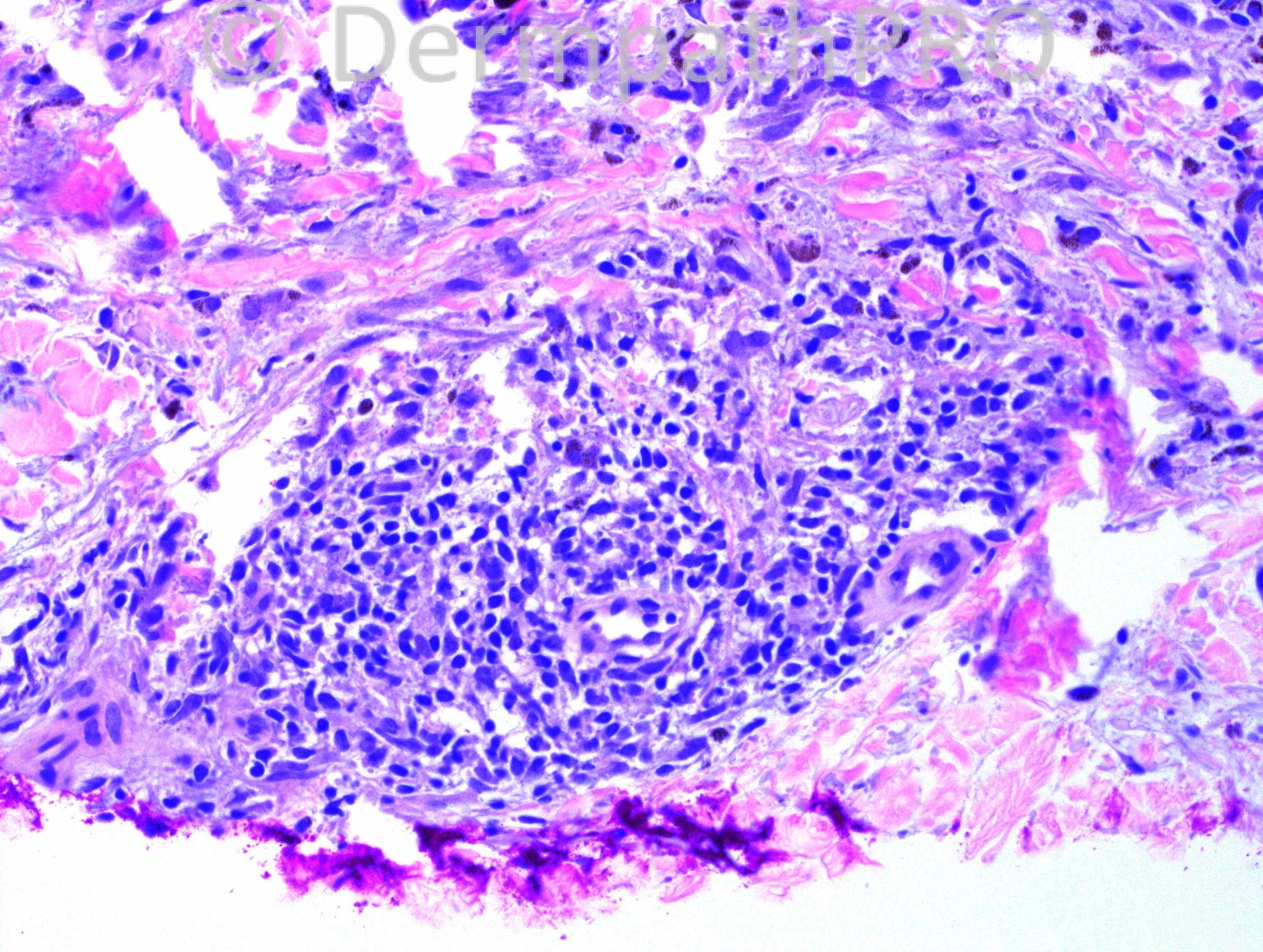
My first impression is malignant blue nevus (pictures #1 and #3) arising in a preexisting benign cellular blue nevus (picture #2).
On the other hand, there is only one discernible mitotic figure in the midst of blander cells, and cytologic atypia may be confined to the same microscopic field shown by both picture #1 and picture #3. Since I can’t access the features that I would take in count to distinguish the differential diagnosis, I can’t dispel doubt between malignant blue nevus X cellular blue nevus.
I prefer not to use controversial expressions like “melanocytic tumor of uncertain malignant potential (MELTUMP)” and “atypical cellular blue nevus” because they are not diagnosis at all, but a more sophisticated way for saying “I don’t know”!

Share this comment


Link to comment
share_externally

Dr. Mona Abdel-Halim

Posted

I think it is blue nevus like melanoma

Share this comment


Link to comment
share_externally

Guest Dr. Wilber Martínez

Posted

Blue nevus-like melanoma (malignant blue nevus)

Share this comment


Link to comment
share_externally

Guest Guillermo Solis

Posted

Melanoma (blue nevus like), or metastatic melanoma.

Share this comment


Link to comment
share_externally

Guest Rodrigo Restrepo

Posted

[color=#1C2837][font=arial, verdana, tahoma, sans-serif][size=4]Melanoma[/size][/font][/color]

Share this comment


Link to comment
share_externally

Dr. Hafeez Diwan

Posted

I ended up calling this melanoma (blue nevus like). At our consensus conference, the atypical blue nevus discussion was brought up.

Share this comment


Link to comment
share_externally


×
×
  • Create New...