Jump to content
In this section we have spot diagnoses posted on a daily basis since June 2010, now over 4000! You can review the archived cases and read the suggested diagnoses by users and the final comment by the contributors.
Case are uploaded each week day by 10 am UK time with the correct diagnosis will generally be posted at 8 pm UK time. Why not view the most recent spot diagnosis and proffer a diagnosis?

Case Number : Case 765 - 23rd May Posted By: Guest

Please read the clinical history and view the images by clicking on them before you proffer your diagnosis.
Submitted Date :
   (0 reviews)

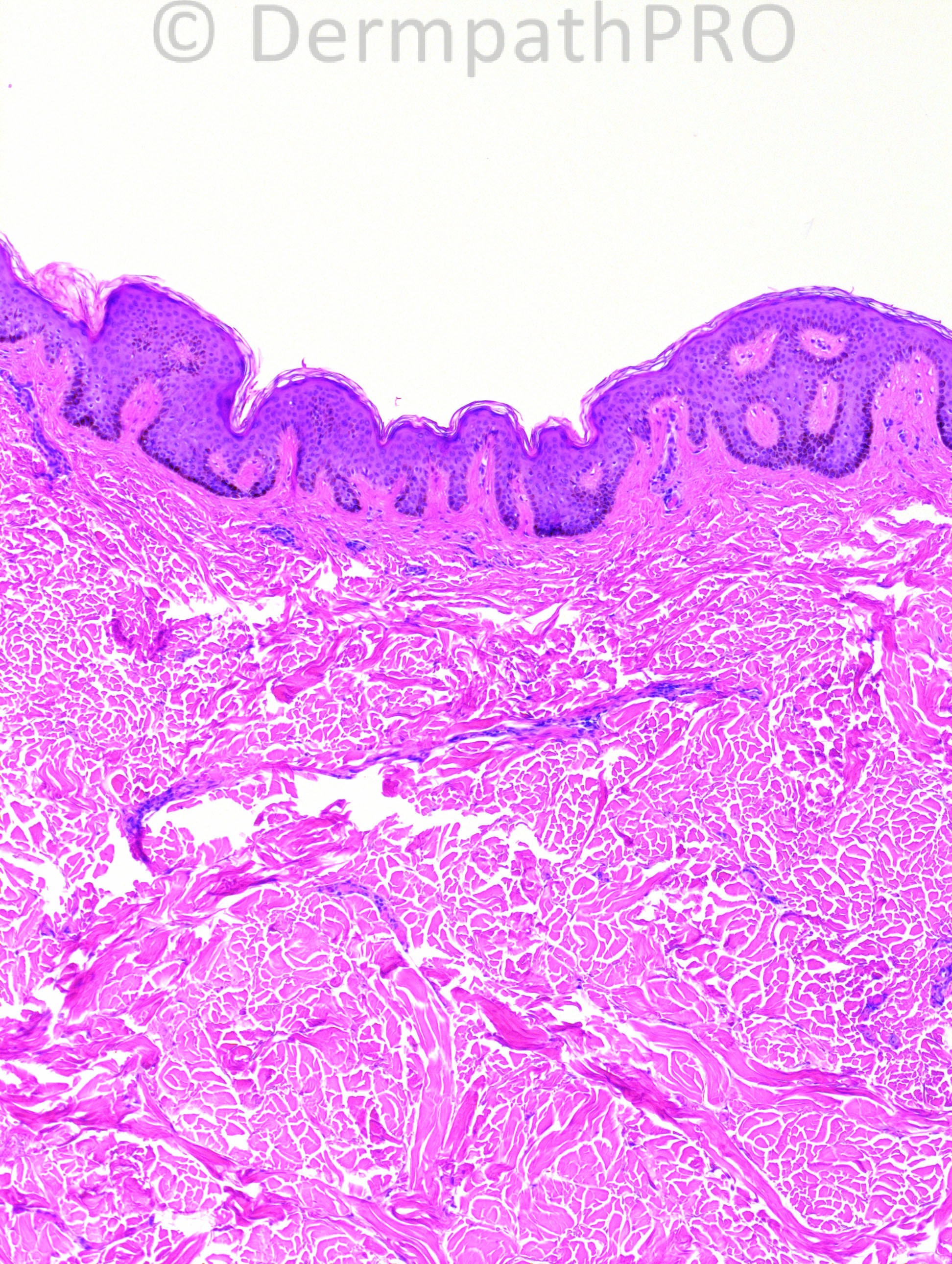
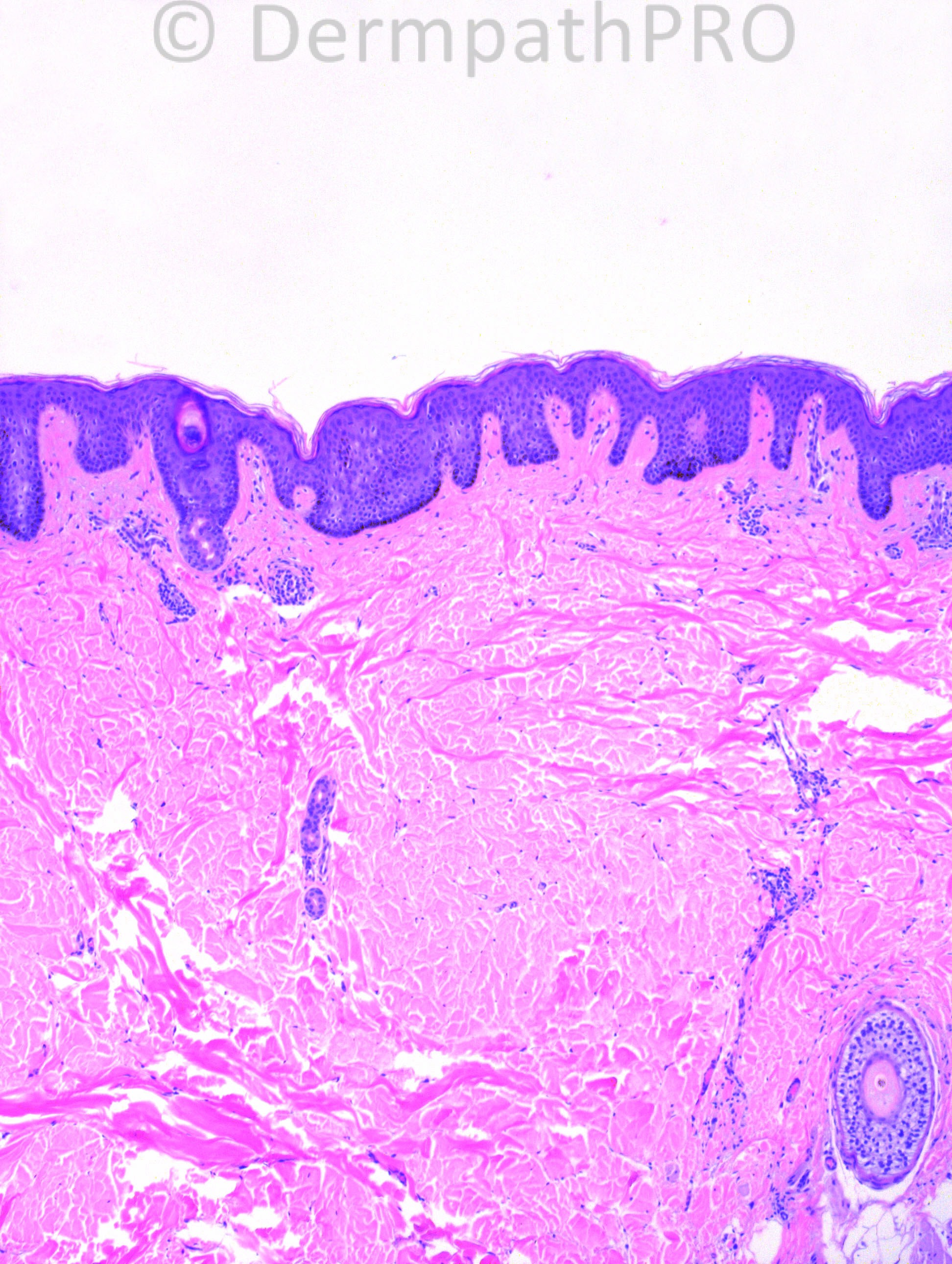
52 years-old female with left back lesion: a dimpled, soft plaque.


Case posted by Dr. Hafeez Diwan.


  Report Record

User Feedback


Guest Marcia

Posted

Collagenoma?

Share this comment


Link to comment
share_externally

Dr. Mona Abdel-Halim

Posted

Collagenoma with fragmented elastic fibres.

Share this comment


Link to comment
share_externally

nick turnbull

Posted

Pseudoxanthoma elasticum?

Share this comment


Link to comment
share_externally

Guest Guillermo Solis

Posted

Anetoderma.

Share this comment


Link to comment
share_externally

Dr. Mona Abdel-Halim

Posted

Lovely suggestion of anetoderma!!!! Fits with the clinical

Share this comment


Link to comment
share_externally

IgorSC

Posted

Elastofibroma dorsi (better known as only "Elastofibroma" today because it can appear at other locations). The bottom rigth figure shows fragmented elastic fibers. Localization is also typical.

Share this comment


Link to comment
share_externally

Guest Maria george

Posted

Some people may argue for elastofibroma but you may call it connective tissue nevus.

Share this comment


Link to comment
share_externally

Sasi Attili

Posted

[color=#1C2837][font=arial, verdana, tahoma, sans-serif][size=4]Elastofibroma dorsi[/size][/font][/color]

Share this comment


Link to comment
share_externally

Guest Jim Davie MD

Posted

I favor Connective tissue nevus. [list]
[*]The elastic fibers appear mildly increased and thinned, with normal expected artifactual fracturing and not diffuse globular beading.
[*]There is dermatofibroma-like hyperpigmentation and square psoriasiform hyperplasia of epidermis.
[*]Dermis is slightly thickened (limited by scope of photos).
[*]Location is superficial.
[/list]

Share this comment


Link to comment
share_externally

Robledo F. Rocha

Posted

I don't think it is collagenoma (aka connective tissue nevus) or elastofibroma. Both diseases manifest clinically as a mass. Anetoderma fits well on the case history of a depressed plaque.
Microscopic features also favor anetoderma. There are widened spaces between collagen fibers and, after elastic stain, one can see fragmentation and loss of elastic fibers.
In elastofibroma I'd expected to find streaks of fat and numerous coarse elastic fibers.

Share this comment


Link to comment
share_externally

Guest Rodrigo Restrepo

Posted

[color=#1C2837][font=arial, verdana, tahoma, sans-serif][size=4]Anetoderma[/size][/font][/color]

Share this comment


Link to comment
share_externally

Dr. Hafeez Diwan

Posted

I called this anetoderma. This is what they were thinking clinically as well.

Share this comment


Link to comment
share_externally


×
×
  • Create New...