Jump to content
In this section we have spot diagnoses posted on a daily basis since June 2010, now over 4000! You can review the archived cases and read the suggested diagnoses by users and the final comment by the contributors.
Case are uploaded each week day by 10 am UK time with the correct diagnosis will generally be posted at 8 pm UK time. Why not view the most recent spot diagnosis and proffer a diagnosis?

Case Number : Case 766 - 24th May Posted By: Guest

Please read the clinical history and view the images by clicking on them before you proffer your diagnosis.
Submitted Date :
   (0 reviews)

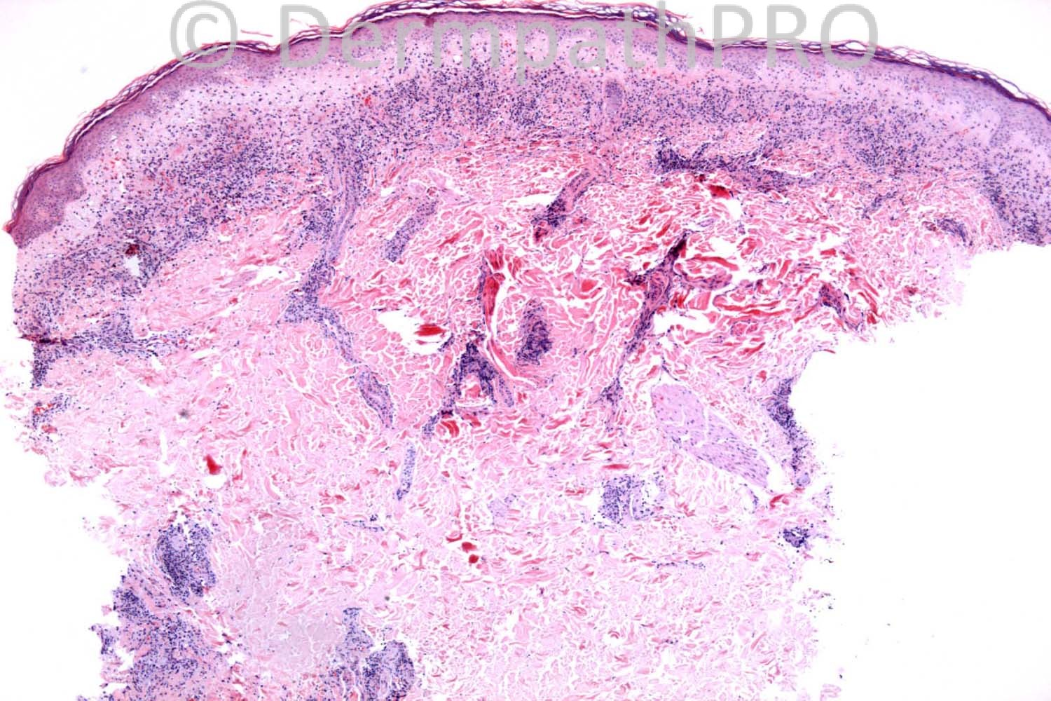
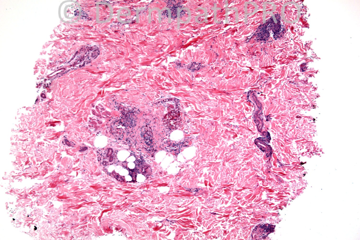
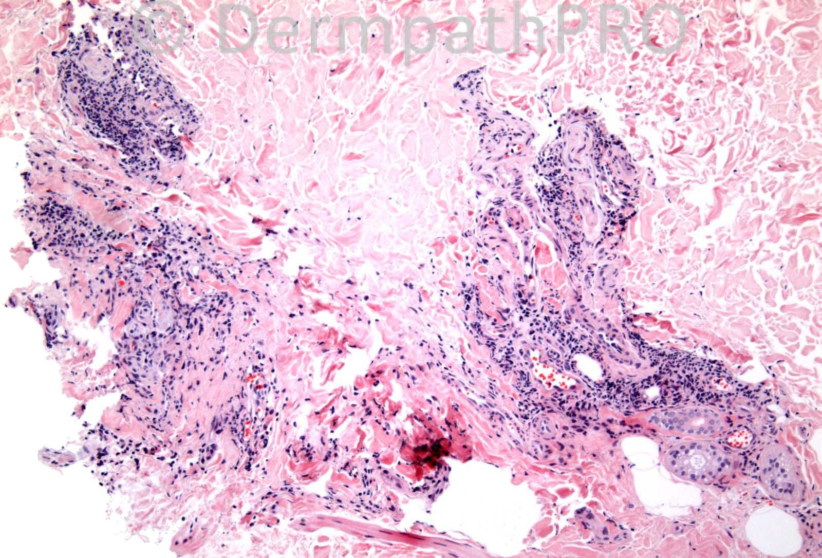
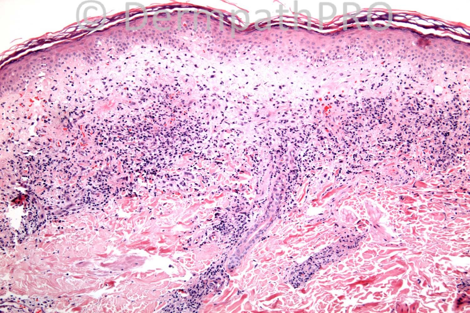
55 years-old male. Diabetic. Rash on right buttock. ?Necrobiosis lipoidica.

Case posted by Dr. Richard Carr.

  Report Record

User Feedback


Sasi Attili

Posted

SCLE? Band like lichenoid infiltrate with papillary dermal edema associated with a superficial and deep perivascular (and perineural) lymphocytic infiltrate.

Share this comment


Link to comment
share_externally

Guest Guillermo Solis

Posted

Fixed drug eruption.

Share this comment


Link to comment
share_externally

Guest Maria george

Posted

Things to be discussed here are lupus, perioniosis, pigmented purpura (lichen aurus), and lichen sclerosus.

Share this comment


Link to comment
share_externally

Dr. Mona Abdel-Halim

Posted

Drug reaction vs Lupus

Share this comment


Link to comment
share_externally

Guest Dr Gonzalo de Toro

Posted

Lupus

Share this comment


Link to comment
share_externally

Guest Bansal_

Posted

DD: Lupus, drug eruption.

Share this comment


Link to comment
share_externally

Robledo F. Rocha

Posted

It can be lupus erythematosus, fixed drug eruption or Mucha-Habermann disease, all of them in early phase. I favor Mucha-Habermann disease because the lesion is not localized in a sun-exposed site, what argues against lupus erythematosus, and it lacks neutrophils and eosinophils, as would be expected on fixed drug eruption.

Share this comment


Link to comment
share_externally

Guest Marcia

Posted

I like the drug eruption hypothesis

Share this comment


Link to comment
share_externally

Guest Jim Davie MD

Posted

Lupus-like Drug reaction / Fixed Drug Eruption, less favor Lupus.

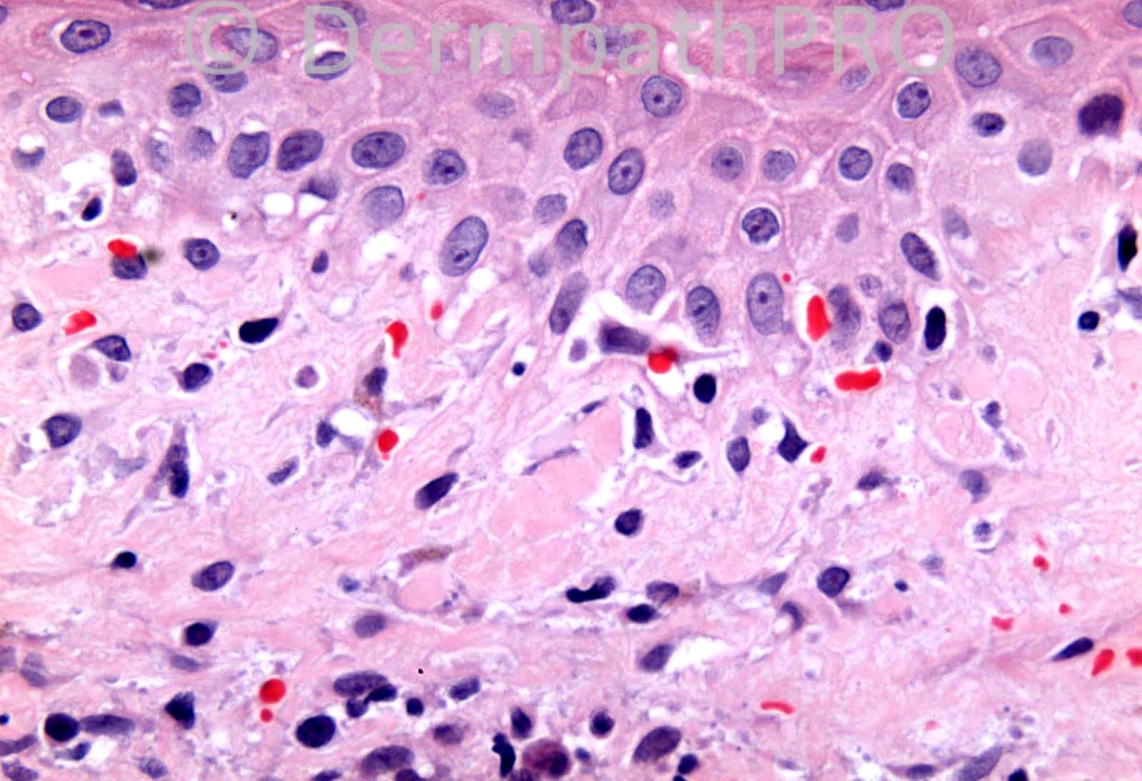
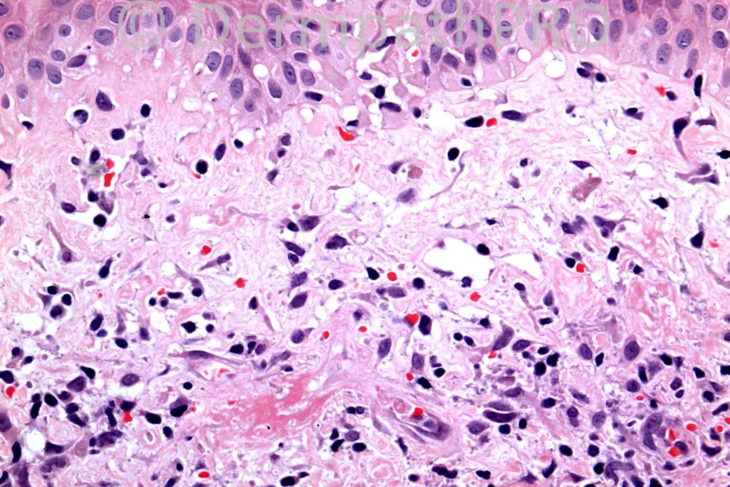
- Dermis shows lichenoid superficial and deep perivascular dermatitis. Papillary dermis shows numerous, focally aggregated colloid bodies, extravasated red cells, melanophages, and impressive edema.
- Epidermis shows lichen-planus-like reactive changes, with subtle sawtooth attenuation of rete, interface vacuolar change with junctional necrotic keratinocytes, mildly to markedly thickened basement membrane.
(I'd want DIF or connective-tissue/anti-histone serologies if the clinical context wasn't sufficient.)

Share this comment


Link to comment
share_externally

Dr. Richard Carr

Posted

I will give you all a little more time but only one person has mentioned the final clinicopathological diagnosis. The history supplied was submitted by a general practioner and not a dermatologist. Unfortunately I suggested lupus which it clearly was not (solitary lesion on the buttock of a male). I did however recommend referral to a dermatologist. The vacuolar change I believe is a red herring as it is not mentioned in the books for the final clinpath diagnosis (suggested by typical clinical appearance of a slightly bronze/purple plaque limited to the buttock.

Share this comment


Link to comment
share_externally

Dr. Mona Abdel-Halim

Posted

Lichen aureus???

Share this comment


Link to comment
share_externally

Guest Guillermo Solis

Posted

Abscence of neutrophils or eosinophils does not rule out drug reactions.

Share this comment


Link to comment
share_externally

Guest Jim Davie MD

Posted

Idiopathic perniosis might have interface vacuolar changes and necrotic keratinocytes, and the exaggerated superficial edema. But couldn't account for the aggregated lichen-amyloidosus-like colloid bodies in the papillary dermis (bottom-right photo).

Share this comment


Link to comment
share_externally

Guest F  Vermander

Posted

pernionis/ cold panniculitis ? Vacuolar change usually not mentioned but possible

Share this comment


Link to comment
share_externally

Guest Irina

Posted

lichenoid purpura of Gougerot-Blum/lichen aureus
the fact that it can have vacuolar alteration is mentioned in Bernie Ackerman's book.

Share this comment


Link to comment
share_externally

Eman El-Nabarawy

Posted

Lichen aureus.

Share this comment


Link to comment
share_externally

Guest Rodrigo Restrepo

Posted

Perniosis

Share this comment


Link to comment
share_externally

Sasi Attili

Posted

Hmmm....I am trying to think of conditions where vacuolar change hasn't been described. As far as I am aware lichen aureus can have vacuolar/ lichenoid change but is really the best fit, given the clinical.

Share this comment


Link to comment
share_externally

Robledo F. Rocha

Posted

After the last detailed clinical description, I go with lichenoid purpura of Gougerot-Blum, even though in this present case the infiltrate uncharacteristically can be found also around the deep vascular plexus.

Share this comment


Link to comment
share_externally

Robledo F. Rocha

Posted

[quote name='Guillermo Solis' timestamp='1369425423']
Abscence of neutrophils or eosinophils does not rule out drug reactions.
[/quote]
I really agree with your statment, Dr. Solis. However the abscence of those cells in the infitrate makes less likely the diagnosis of drug eruption, and compels the pathologist to consider other hypothesis which might fit on the clinicopathological picture.

Share this comment


Link to comment
share_externally

Guest Romualdo

Posted

I think superficial and deep perivascular lymphocytic infiltrate, lymphocytic vasculitis, peri-eccrine infiltrate, subepidermal edema and interface changes in concert with the clinical findings are consistent with the diagnosis of perniosis.

Share this comment


Link to comment
share_externally

Dr. Richard Carr

Posted

Well done to Maria George. Wow you guys really are committed to the site - I was hoping for one or two more posts! The clinical diagnosis was typical of lichen aureus / pigmented purpuric dermatosis (PPD). We had mentioned lupus and cold injury in the initial report and I accept fixed drug reaction as a good differential diagnosis. However on reviewing the case with the clinical images the red cell extravasation, lymphocytic vasculitis pattern with fibrin in the wall of a superficial vessel (lower left image) scattered iron (on Perl's) in both the superficial band-like and deep peri-vascular and periadnexal component supported the final diagnosis of lichen aureus. Apologies for my post as Dr Akerman is clearly correct I suppose I should have said usually inconspicuous vacuolar / lichenoid change (it is the first time I have seen it so dramatic myself with obvious colloid bodies). I do not see basement membrane thickening. What I did not know is that lichen aureus / PPD can involve the deep plexus (mentioned in Weedon) and can be such a very good histological mimic of both lupus and cold injury / perniosis (I have not seen such prominent lichenoid change in perniosis so far). So a nice case to highlight that interface / lichenoid reaction pattern with superficial band, deep plexus and periadnexal involvement consider lichen aureus/PPD. Regards to all and enjoy the rest of your weekends - and take a break!!

Share this comment


Link to comment
share_externally


×
×
  • Create New...