Jump to content
In this section we have spot diagnoses posted on a daily basis since June 2010, now over 4000! You can review the archived cases and read the suggested diagnoses by users and the final comment by the contributors.
Case are uploaded each week day by 10 am UK time with the correct diagnosis will generally be posted at 8 pm UK time. Why not view the most recent spot diagnosis and proffer a diagnosis?

Case Number : Case 767 - 27th May Posted By: Guest

Please read the clinical history and view the images by clicking on them before you proffer your diagnosis.
Submitted Date :
   (0 reviews)

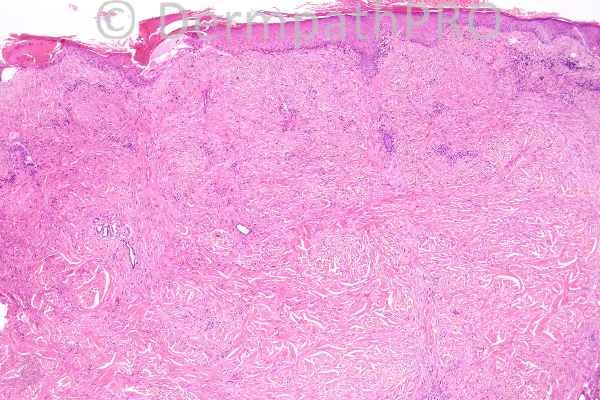
69 years-old female with a sclerotic plaque on the cheek.


  Report Record

User Feedback


Guest Marcia

Posted

Desmoplastic melanoma?

Share this comment


Link to comment
share_externally

IgorSC

Posted

I am always concerned with clinical sclerotic plaques on the face and cheek accompanied by dermal sclerosis and chronic inflammatory infiltrate. Desmoplastic melanoma x cicatricial fibrosis. S-100 and Sox-10 are good options for immunohistochemical evaluation.

Share this comment


Link to comment
share_externally

Dr. Mona Abdel-Halim

Posted

I have the same concern, will consider this first possibility.

Share this comment


Link to comment
share_externally

Guest Bostjan

Posted

Desmoplastic melanoma

Share this comment


Link to comment
share_externally

Guest Dr Engin Sezer

Posted

Agree

Share this comment


Link to comment
share_externally

Robledo F. Rocha

Posted

Like my colleagues, I think desmoplastic melanoma must be considered every time one deals with a scar-like lesion on chronically sun-damaged skin of head and neck, mainly in eldery patients.
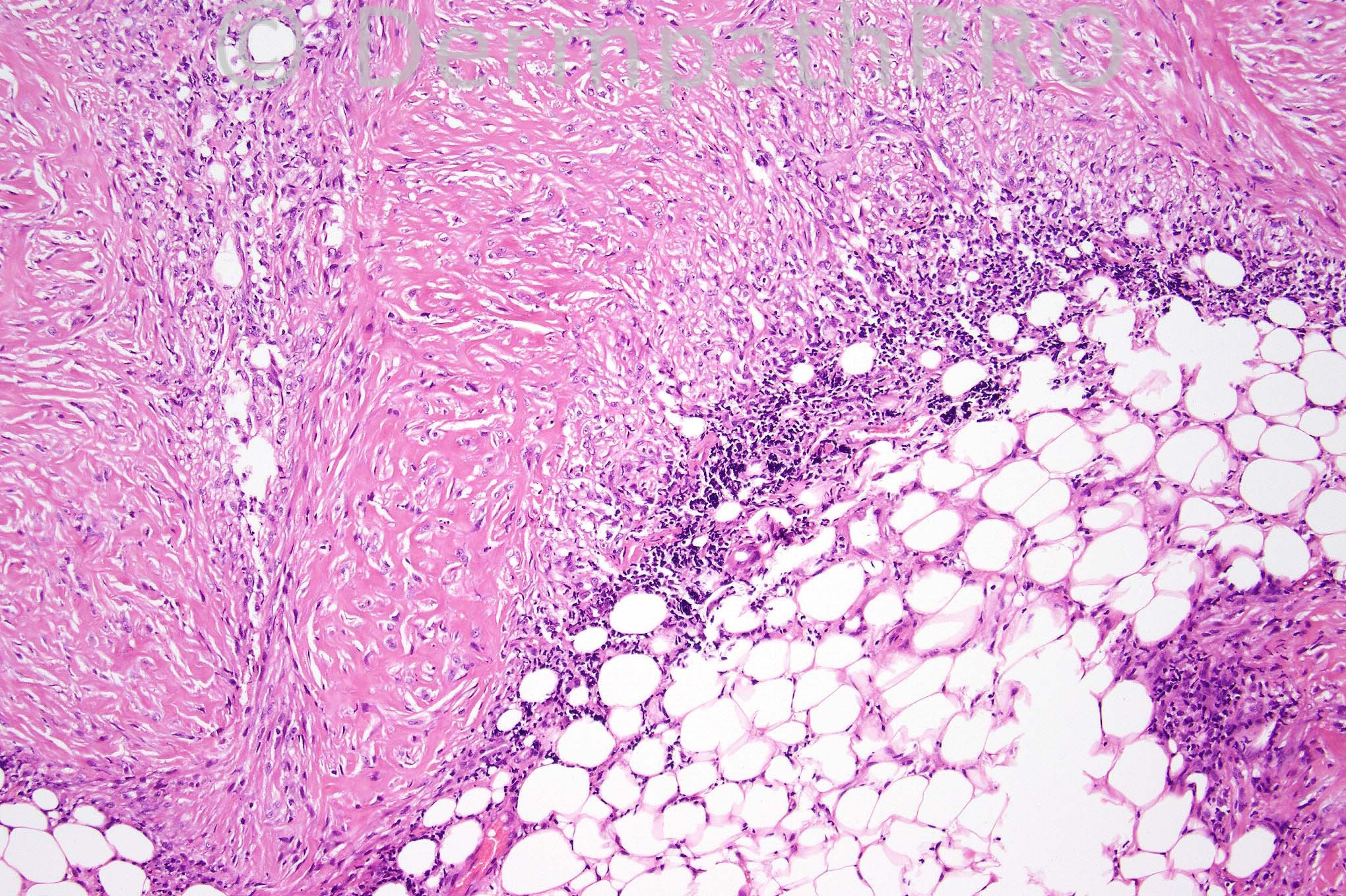
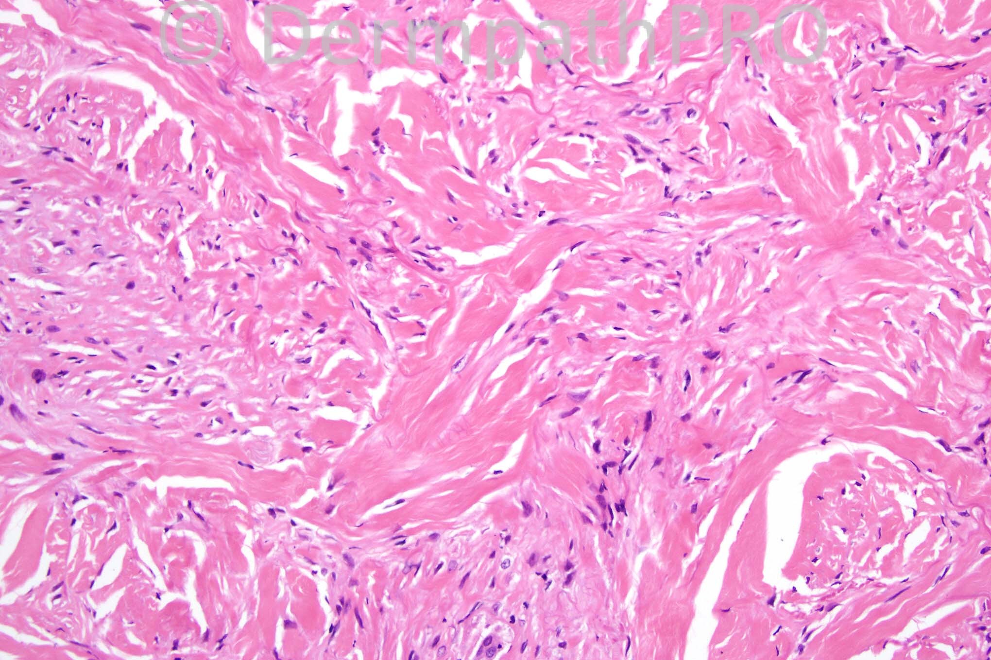
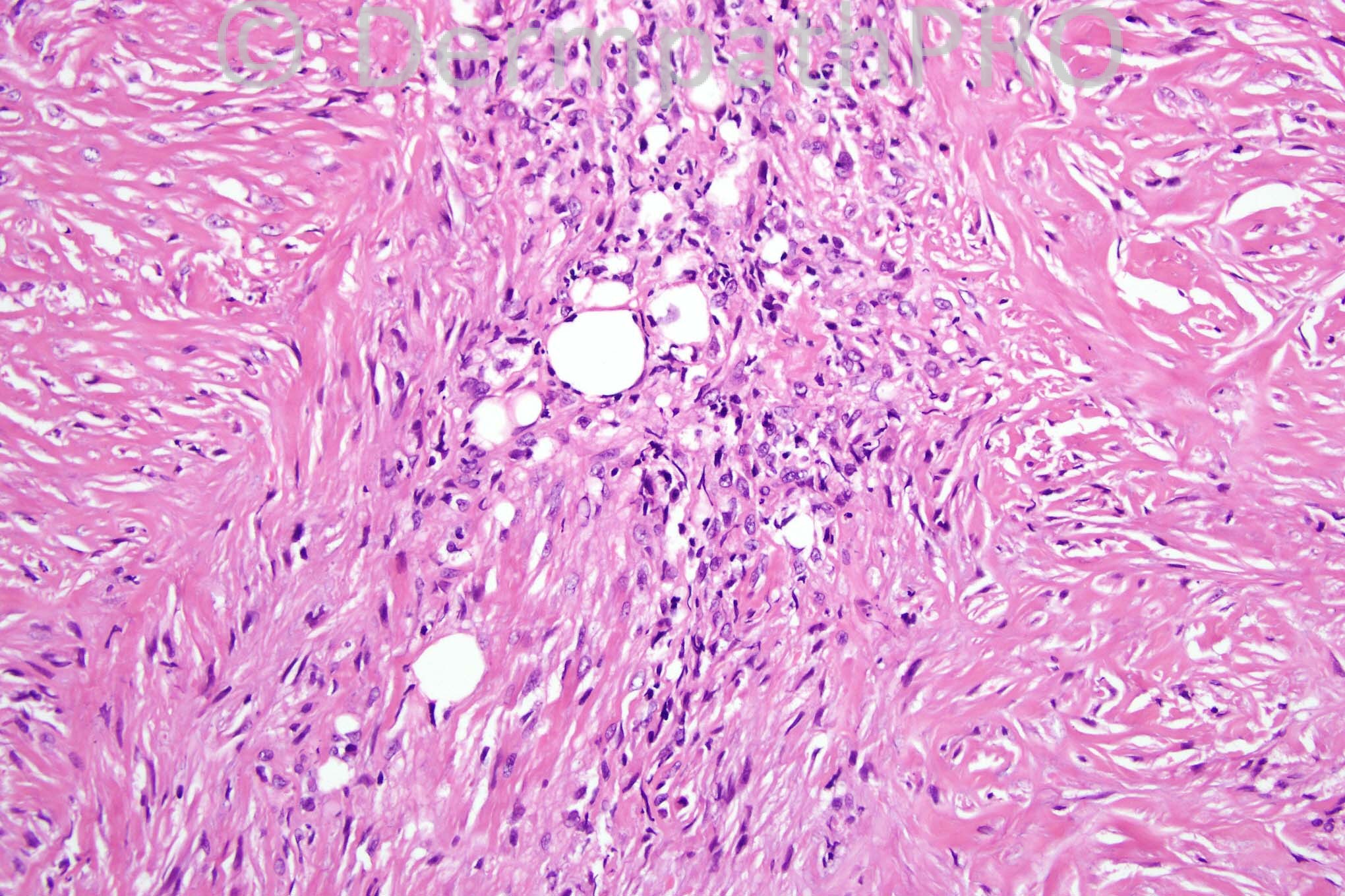
In the pictures shown here there are some microscopic features that favor desmoplastic melanoma over its differential diagnosis:
● fascicles of bland amelanotic spindle cells embedded in a somewhat myxoid stroma;
● spindle cells permeate throughout the dense collagenized dermis and extend into the subcutaneous fat;
● some nodular patchy lymphoid infiltrates;
● scattered pleomorphic cells close to fat cells.
Last but not least, I'd ask the attending physician if there's a history of previous surgical intervention on the biopsied site. It could be another type of melanoma locally recurring with desmoplastic appearance.

Share this comment


Link to comment
share_externally

Guest Maria george

Posted

Like my colleagues, thinking of [color=#1C2837][font=arial, verdana, tahoma, sans-serif][size=4]Desmoplastic melanoma.Lymphoid infiltrates are a good clue.[/size][/font][/color]

Share this comment


Link to comment
share_externally

Guest Rodrigo Restrepo

Posted

[color=#1C2837][font=arial, verdana, tahoma, sans-serif][size=4]Desmoplastic melanoma[/size][/font][/color]

Share this comment


Link to comment
share_externally

Guest Bansal_

Posted

Desmoplastic melanoma

Share this comment


Link to comment
share_externally

Guest Iskander

Posted

Dear All. If you would like to consider another diagnosis as there was no cytological atypia. Iskander

Share this comment


Link to comment
share_externally

Sasi Attili

Posted

[quote name='Guest' timestamp='1369736347']
Dear All. If you would like to consider another diagnosis as there was no cytological atypia. Iskander
[/quote]
My first impression was actually inflammatory morphoea. But since everyone was unanimous about desmoplastic melanoma, chose to keep quiet !! The differential would include an old scar/ ruptured & healed cyst (though no cyst lining seen). There is some palisading of inflammatory cells around irregularly distributed nodules of scar tissue, though I couldn't convince myself of a frank granuloma's or necrobiosis (considering atypical facial necrobiosis).

Share this comment


Link to comment
share_externally

Guest Bansal_

Posted

Presumably it is morphoea then.

Share this comment


Link to comment
share_externally

Dr. Mona Abdel-Halim

Posted

Do not feel it is inflammatory, more with a spindle cell tumor with dense sclerotic collagenous stroma. Could it be hypopigmented blue nevus. it is said in McKee's book that it overlaps and may be identical to sclerosing (desmoplastic) blue nevus. Also it said that it may show concentric or laminated sclerosis resembling storiform collagenoma which I can perceive vaguely in some areas... Hope I am correct.

Share this comment


Link to comment
share_externally

Guest Dr Engin Sezer

Posted

Sclerosing perineuroma

Share this comment


Link to comment
share_externally

Dr. Mona Abdel-Halim

Posted

This was my first impression after excluding desmoplastic melanoma Dr Engin but the site is so atypical... But it could be!!!

Share this comment


Link to comment
share_externally

Iskander H. Chaudhry

Posted

Thanks for the additional comments everyone, the diagnosis for this was morphea, well done to Sasi!

Share this comment


Link to comment
share_externally

Dr. Mona Abdel-Halim

Posted

Well done Dr Sasi :-)

Share this comment


Link to comment
share_externally


×
×
  • Create New...