Jump to content
In this section we have spot diagnoses posted on a daily basis since June 2010, now over 4000! You can review the archived cases and read the suggested diagnoses by users and the final comment by the contributors.
Case are uploaded each week day by 10 am UK time with the correct diagnosis will generally be posted at 8 pm UK time. Why not view the most recent spot diagnosis and proffer a diagnosis?

Case Number : Case 768 - 28th May Posted By: Guest

Please read the clinical history and view the images by clicking on them before you proffer your diagnosis.
Submitted Date :
   (0 reviews)

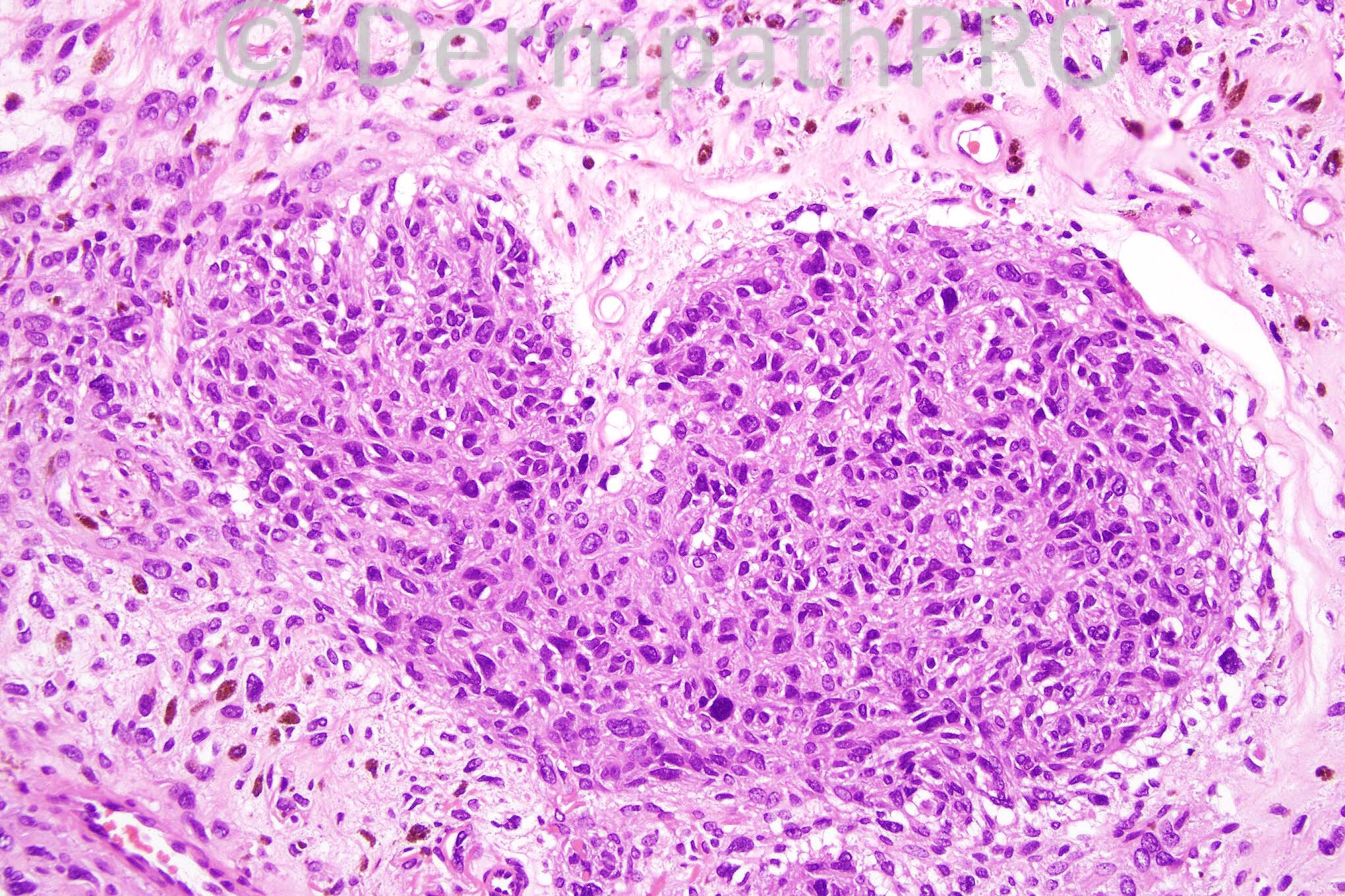
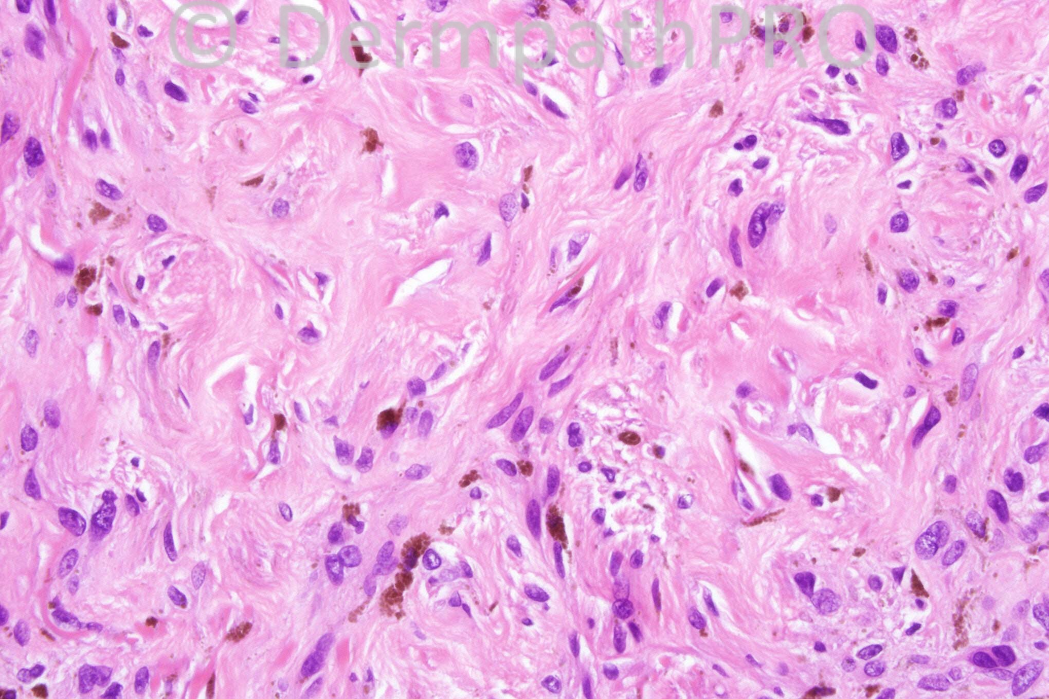
74 years-old male with a long-standing lesion on the buttock.


  Report Record

User Feedback


Guest Iskander

Posted

Dear All - please have another look at yesterday's post diagnosis as there were no correct answers :(. Iskander

Share this comment


Link to comment
share_externally

Guest Dr Engin Sezer

Posted

MFH

Share this comment


Link to comment
share_externally

Guest Maria george

Posted

Iskandar,this is phyohyphomycosis and previous one is heterotopic neural coriostoma!if not epitheliod sarcoma.

Share this comment


Link to comment
share_externally

Sasi Attili

Posted

I am worried by the pigment production and the irregular nests of epithelioid cells some of which do display pleomorphism but can't see obvious mitoses. Some of the nests seem to be inside vessels! I would like to see the silhouette but suspecting that this is a melanoma.

Share this comment


Link to comment
share_externally

Guest Maria george

Posted

The previous one is metastatic breast carcinoma this one possibly deep fungal infection.

Share this comment


Link to comment
share_externally

Guest Maria george

Posted

Both could be metastases.The previous from breast and this from a melanoma.

Share this comment


Link to comment
share_externally

Dr. Mona Abdel-Halim

Posted

Metastatic blue nevus like melanoma

Share this comment


Link to comment
share_externally

Guest Bansal_

Posted

DD: Melanoma, Metastatic Carcinoma. Needs immuno panel.

Share this comment


Link to comment
share_externally

Dr. Richard Carr

Posted

I did not understand most of the suggestions above. Yesterday's was amusing too - shows that diagnosing on a few images can be treacherous! What about a blue naevus?

Share this comment


Link to comment
share_externally

Guest Bansal_

Posted

Yes Richard, possibly why EQAs based on a few photomicrographs will never work!

Share this comment


Link to comment
share_externally

Iskander H. Chaudhry

Posted

Thanks for everyone for going back to yesterday's case, the diagnosis is now on so do check it out!

It seems today's was no easier! This was an atrophic cellular blue nevus, many thanks Richard for the intervention! You're right, it can be tricky judging a lesion by a few photos alone, and we are all here to learn :)

Share this comment


Link to comment
share_externally


×
×
  • Create New...