Jump to content
In this section we have spot diagnoses posted on a daily basis since June 2010, now over 4000! You can review the archived cases and read the suggested diagnoses by users and the final comment by the contributors.
Case are uploaded each week day by 10 am UK time with the correct diagnosis will generally be posted at 8 pm UK time. Why not view the most recent spot diagnosis and proffer a diagnosis?

Case Number : Case 769 - 29th May Posted By: Guest

Please read the clinical history and view the images by clicking on them before you proffer your diagnosis.
Submitted Date :
   (0 reviews)

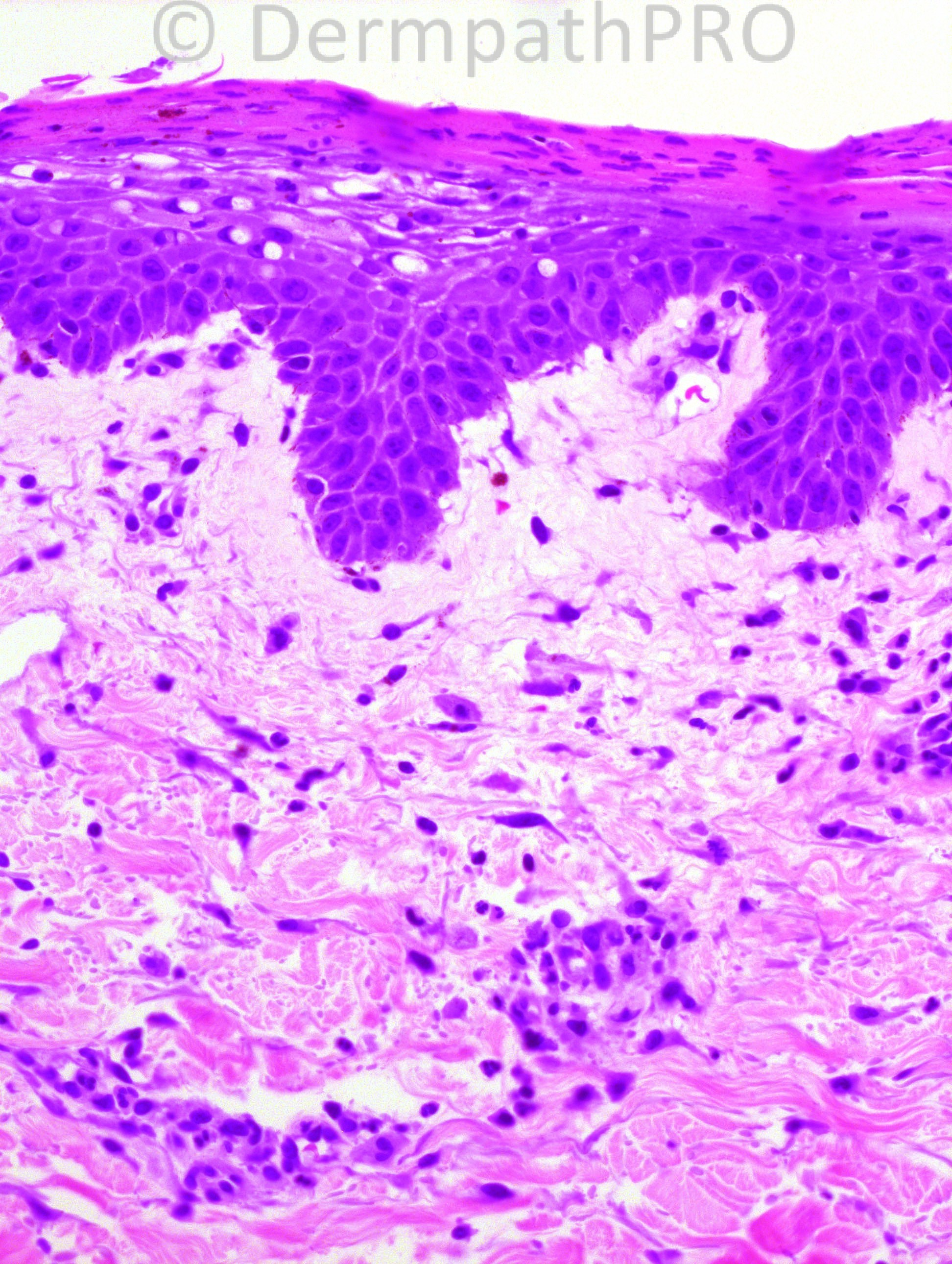
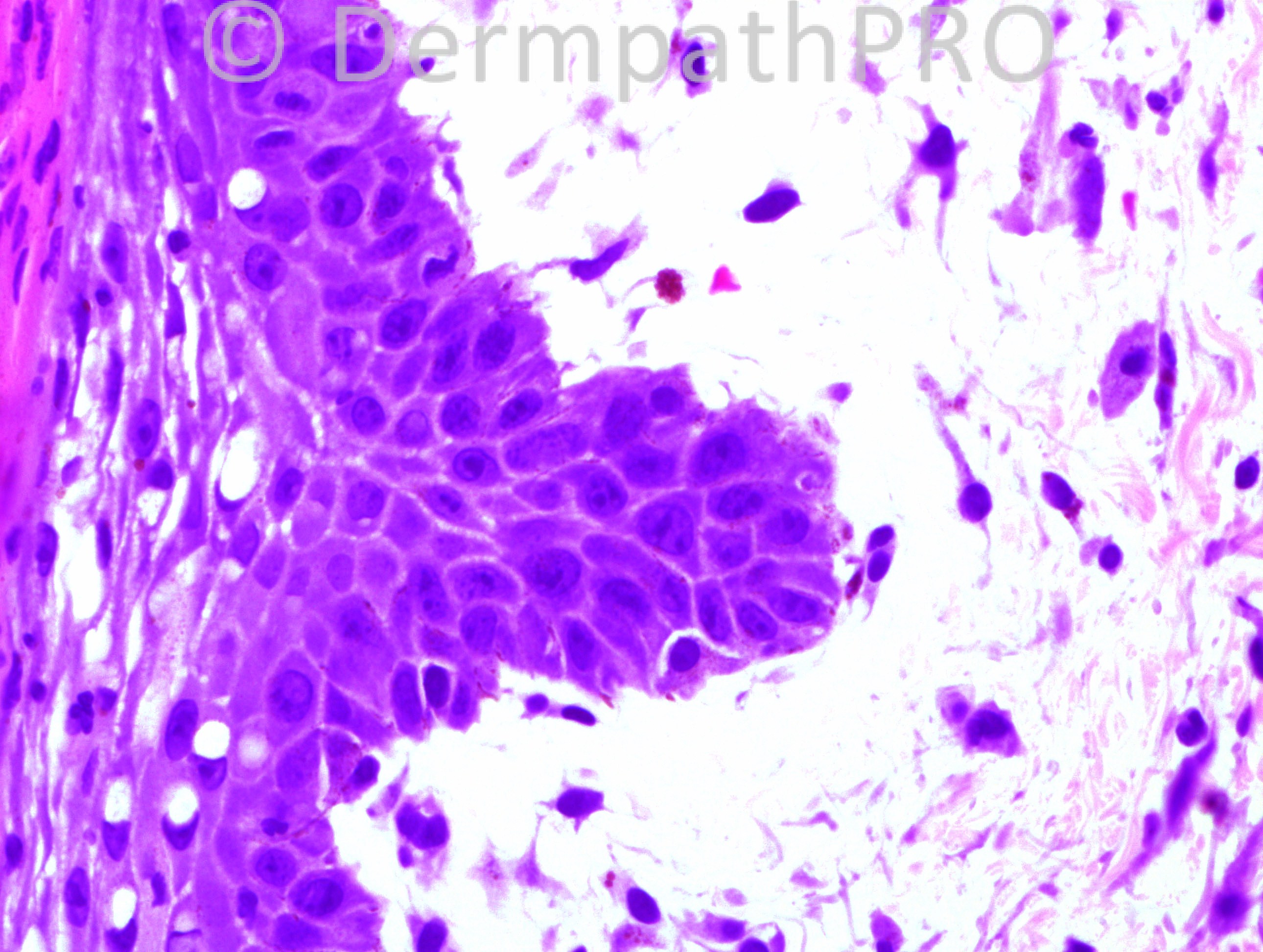
50 years-old male with HIV and failure to thrive for 3 months, with diffuse desquamating rash, and edema. The patient has very low protein and albumin levels.

Case posted by Dr. Hafeez Diwan.


  Report Record

User Feedback


Guest Romualdo

Posted

Mastocytoma.

Share this comment


Link to comment
share_externally

Guest Guillermo Solis

Posted

Agree with solitary mastocytoma

Share this comment


Link to comment
share_externally

Guest Dr Engin Sezer

Posted

Agree

Share this comment


Link to comment
share_externally

Robledo F. Rocha

Posted

Increased density of "fried egg" cells with perivascular accentuation in an edematous papillary dermis.
Agree with early solitary mastocytoma.
But is an early disease enough to produce a mass, as described on case history?

Share this comment


Link to comment
share_externally

Guest Bansal_

Posted

Probable early mastocytoma. Would corroborate with Tol Blue / MGG.

Share this comment


Link to comment
share_externally

nick turnbull

Posted

The epidermis shows parakeratosis and i see vacuolar changes and maybe slight paleness also. The significant mast cell population is almost UP like, but that wouldnt fit the clinical. Would mastocytoma be more compact mast cells filling the dermis? Early mastocytoma? Im not sure about the epidermal changes. Im just a few months into my dermpath training.

Share this comment


Link to comment
share_externally

IgorSC

Posted

Mastocytoma, but it is difficult to imagine these histologic pictures making an ear mass. Maybe that´s why "early" mastocytoma.

Share this comment


Link to comment
share_externally

Guest Dr Wilber J. Martínez

Posted

Mastocytoma

Share this comment


Link to comment
share_externally

Sasi Attili

Posted

Even though there are prominent mast cells in this section, I think they are just associated with the prominent edema which I suspect is partly mucin in the upper dermis. Would like an Alcian blue to confirm. I agree with Nick that mast cells are much more prominent and in aggregates filling the dermis, in a mastocytoma. History is not right for PMLE or any other inflammatory dermatosis. Is this a variant of a mucinous nevus? Can't explain the secondary changes in the epidermis though!

Share this comment


Link to comment
share_externally

Guest Guillermo Solis

Posted

Some times i can not distinguish between oedema of papillary dermis or mucin deposits, mucinosis is a nice option, altough remember that children often strokes lesions and evokes Darier's sign with edema and degranulation of mast cells and in a few days this inflammation lead to epidrmal changes.

Share this comment


Link to comment
share_externally

Eman El-Nabarawy

Posted

Mastocytoma.

Share this comment


Link to comment
share_externally

Guest Maria george

Posted

Mucinosis?

Share this comment


Link to comment
share_externally

Dr. Hafeez Diwan

Posted

There has been some kind of glitch and in fact this happens to be a biopsy from an HIV patient with a diffuse rash, marked edema, and involvement of the intertriginous areas. He has very low protein and albumin levels. So this is a case of Kwashiorkor, showing the dermal edema and the pallor of the superficial dermis. I apologize for the error. We will try to put the 12 year-old's case tomorrow.

Share this comment


Link to comment
share_externally

Dr. Richard Carr

Posted

This is a classic!! I had tried to invent some story like systemic mastocytosis and severe diarrhoea causing nutritional deficiency! Nick had hinted at this in his comments. I decided to keep my powder dry because I could not fit the histology with the clinical information supplied. But really we could have all done better. On first looking at a biopsy always say to yourself what is the site of the biopsy (blind of clinical information of course) then you will have a better chance of picking up errors (that do occur from time to time in all labs and institutions and it appears teaching interactive forums). Ask yourself does this look like ear skin? Answer no! This was also very clearly not a mastocytoma. What an interesting few days we are having.

Share this comment


Link to comment
share_externally


×
×
  • Create New...