Jump to content
In this section we have spot diagnoses posted on a daily basis since June 2010, now over 4000! You can review the archived cases and read the suggested diagnoses by users and the final comment by the contributors.
Case are uploaded each week day by 10 am UK time with the correct diagnosis will generally be posted at 8 pm UK time. Why not view the most recent spot diagnosis and proffer a diagnosis?

Case Number : Case 771 - 31st May Posted By: Guest

Please read the clinical history and view the images by clicking on them before you proffer your diagnosis.
Submitted Date :
   (0 reviews)

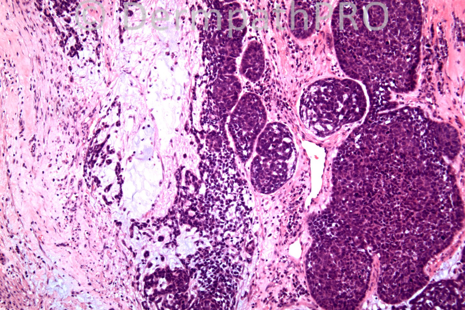
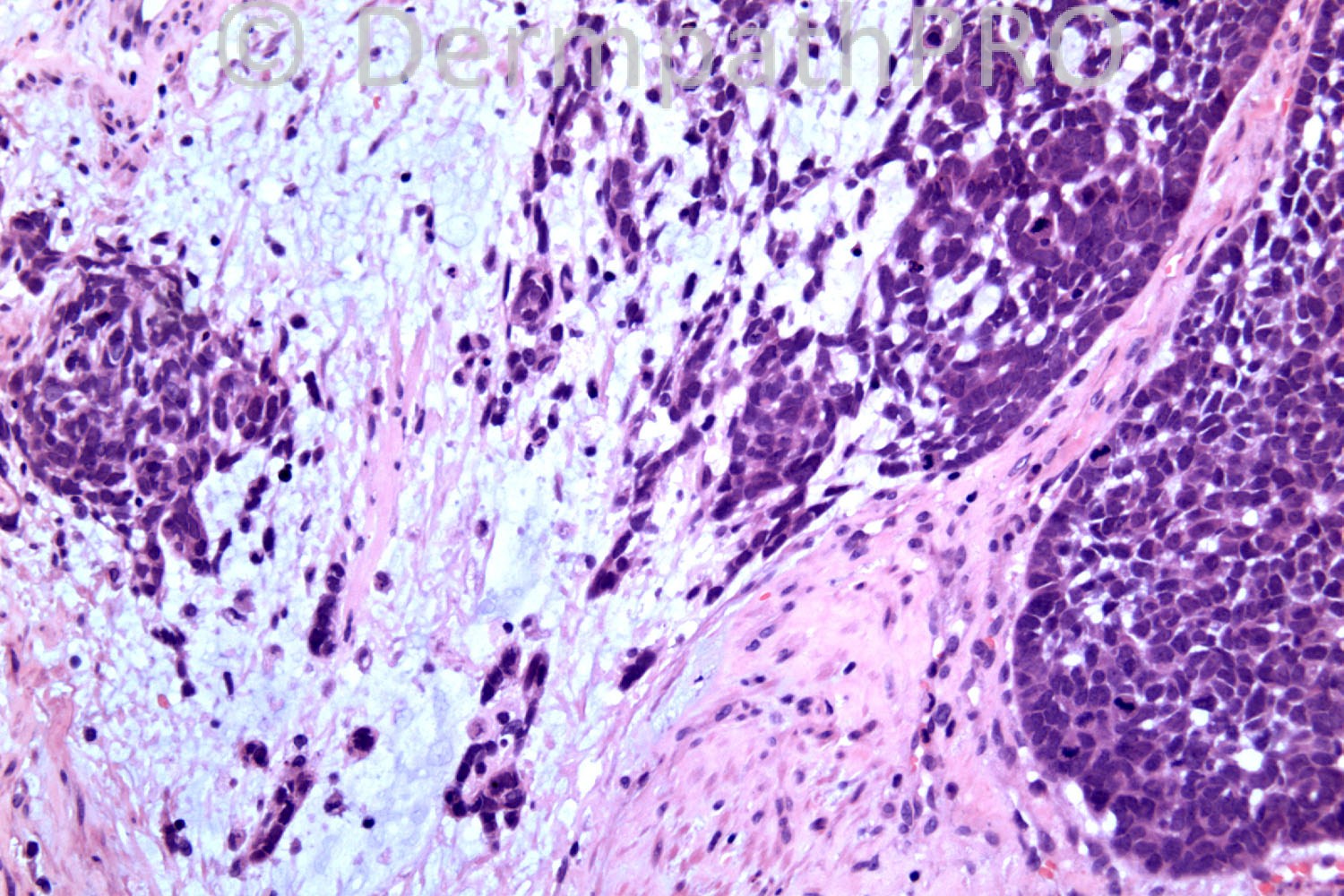
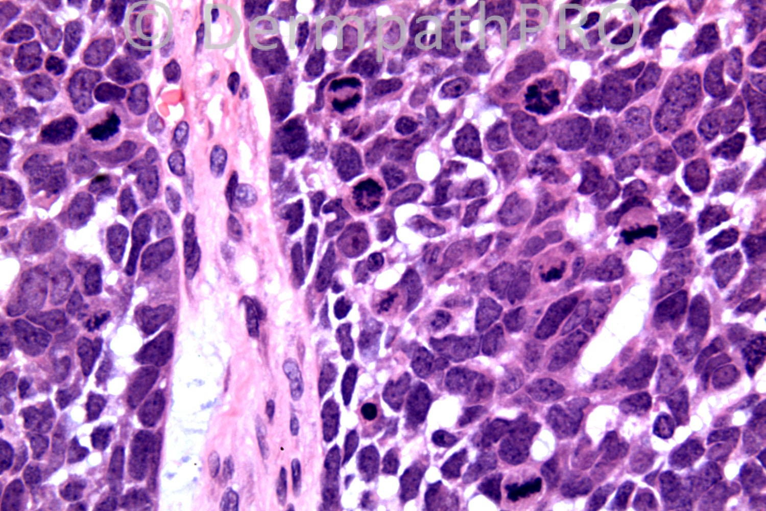
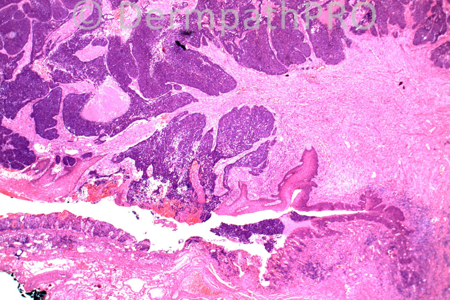
84 years-old female. Painful perianal lump excised.

Case posted by Dr. Richard Carr.

  Report Record

User Feedback


basaloid squamous cell carcinoma of the anal region

Share this comment


Link to comment
share_externally

Guest Romualdo

Posted

Basaloid squamous carcinoma.

Share this comment


Link to comment
share_externally

Dr. Mona Abdel-Halim

Posted

Basaloid Squamous cell carcinoma

Share this comment


Link to comment
share_externally

IgorSC

Posted

I agree, basaloid squamous cell carcinoma. There´s an "in situ" area and also comedo-like necrosis.

Share this comment


Link to comment
share_externally

Robledo F. Rocha

Posted

Basaloid squamous cell carcinoma, probably associated with HPV infection.

Share this comment


Link to comment
share_externally

Dr. Richard Carr

Posted

Thanks for all the responses. Yes we also thought quite neuroendocrine-like but a wide panel of neuroendocrine markers were all negative (full panel below). Additional immunoprofile as follows;
BerEP4 - diffuse strong +ve
EMA - peripheral tumour cells only +ve
CK5 - 30% of tumour cells +ve
CAM 5.2 - 20% of tumour cells +ve
P63 focal +ve
S-100, CK7, CK20, TTF-1, CDX-2 all -ve
Unusually the lesion was only focally positive for keratins and p63 (usually basaloid anal squamous cell carcinoma are diffuse). But p16 was positive in the high grade anal intra-epithelial neoplasia (well done IgorSC) and also diffusely in the tumour. BCC might be a consideration with diffuse BerEP4 and the stromal mucin but the lesion lack palisading and the lesion was also focally EMA positive in the basaloid epithelium (we never see that in a BCC).
In our opinion final diagnosis; poorly differentiated basaloid anal carcinoma
(regarded clinically as a variant of squamous carcinoma)

Share this comment


Link to comment
share_externally

Sasi Attili

Posted

I find this quite interesting Richard. Let us play devil's advocate for a moment:

Ignoring the histology if someone told you that a particular skin tumour is strongly Ber-EP4 +ve and focally EMA +ve, you would favour a BCC wouldn't you? Why is this not a metatypical BCC on that grounds, as strong Ber-EP4 +vetivity is virtually never seen in SCC's!

Share this comment


Link to comment
share_externally

Dr. Richard Carr

Posted

Thanks Sasi yes you are right, as a general rule, but I would always double check the focal EMA staining was in squamoid areas (not peripheral basaloid areas). In every "metaypical/basosquamous/malignant squamous component" BCC there are generally areas typical on morphology and IHC of a typical BCC (if not it is a basaloid SCC/basosquamous carcinoma NOS). A BCC with malignant squamous component is currently considered to carry a similar metastatic rate to SCC so a little academic. However there are no papers of large series of metastatic BCC that supported the diagnosis with IHC and in my experience cutaneous basaloid SCC/basosquamous carcinoma NOS can very closely mimic BCC. So there is scope for a good paper on metastatic BCC and basosquamous carcinomas! If you read the original papers on BerEP4 it was always reported to be positive in a proportion of systemic SCC (lung, oesophagus, cervix, ENT cases etc ) as opposed to cutaneous SCC so I am not surprised to see it in anal carcinoma (a mucosal carcinoma). EMA was only focal but definitely positive in the basaloid epithelium (we still have not seen that in BCC) and this was important in the distinction here. So a completely negative EMA in the basaloid epithelium (with strong control staining in sebaceous glands and plasma cells) is even more characteristic of BCC than diffuse BerEP4. Incidentally we do have a series of basaloid cutaneous SCC (unpublished) wtih about 50% showing moderate to widespread BerEP4 expression in >10% of the epithelium (but never totally diffuse and strong), EMA is at least focally positive in the basaloid epithelium in all cases and we also published (abstract only unfortunately) similar findings in basaloid bowen's. Remember BerEP4 also stains widely diffuse in neuroendocrine carcinomas (including Merkel) and some adenocarcinomas (rarely in cutaneous adenocarcinomas). So diffuse strong BerEP4 while being highly characteristic of BCC is not entirely specific. I like the suggestion of Sox-2 (I have no experience) and I have also heard that CD44 can help in distinction. I will send Mark some spares. I do think this was a very difficult case and has a very unusual IHC profile because up till now anal basaloid carcinomas have also always been diffusely p63 positive (does not help in distinction with BCC that are also diffuse for p63). Incidentally on p16 in our experience (and to quote Dr Kutzner) it is for Bowen's what BerEP4 is for BCC. It seems to light up basaloid bowen's from all anatomic locations and I don't know if this is independant of HPV or not. We know that basaloid ano-genital carcinomas and p16 correlate with a high incidence of high risk HPV types. Incidently there are reports of p16 also staining BCC. Because of the in situ tumour here (and that was very focal actually) we plumped for basaloid anal SCC. The tumours are preferentially treated with radiotherapy so it is an important distinction. Hope this helps!

Share this comment


Link to comment
share_externally


×
×
  • Create New...