Jump to content
In this section we have spot diagnoses posted on a daily basis since June 2010, now over 4000! You can review the archived cases and read the suggested diagnoses by users and the final comment by the contributors.
Case are uploaded each week day by 10 am UK time with the correct diagnosis will generally be posted at 8 pm UK time. Why not view the most recent spot diagnosis and proffer a diagnosis?

Case Number : Case 880 - 1st November Posted By: Guest

Please read the clinical history and view the images by clicking on them before you proffer your diagnosis.
Submitted Date :
   (0 reviews)

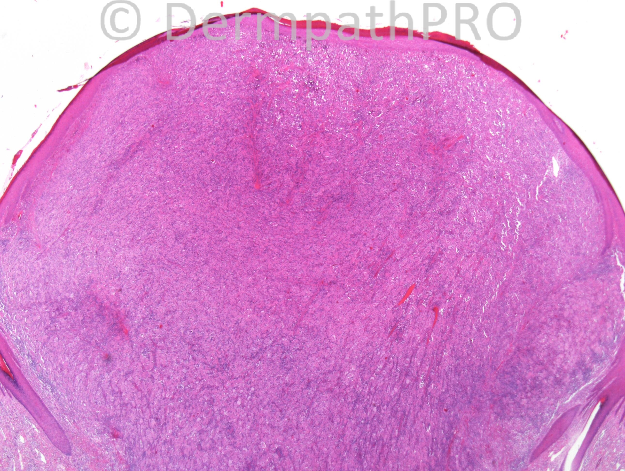
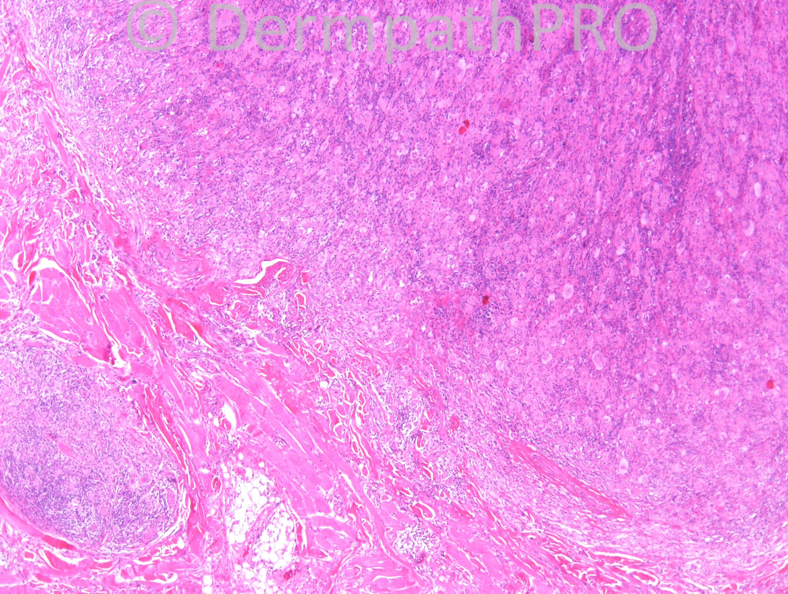
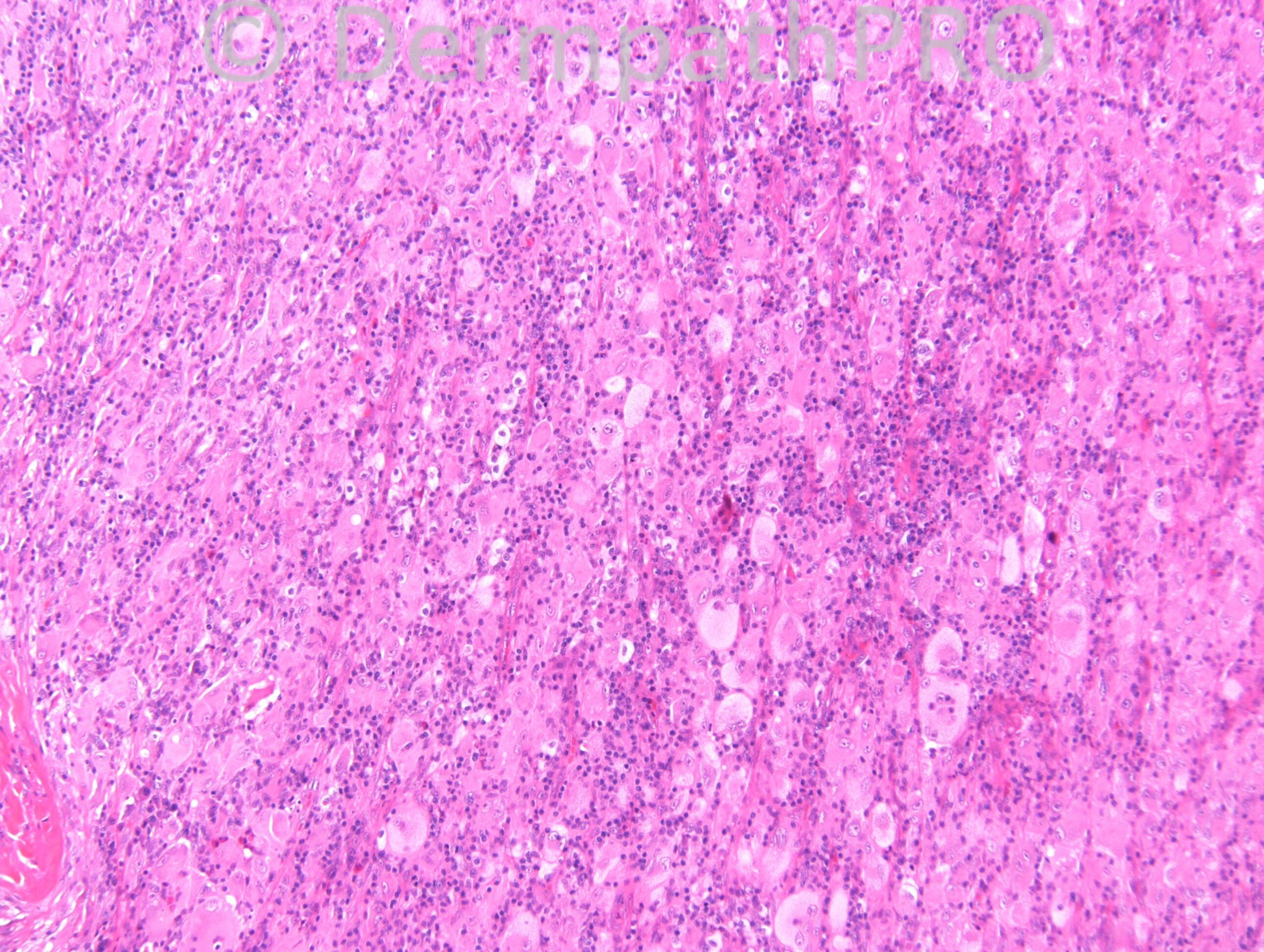
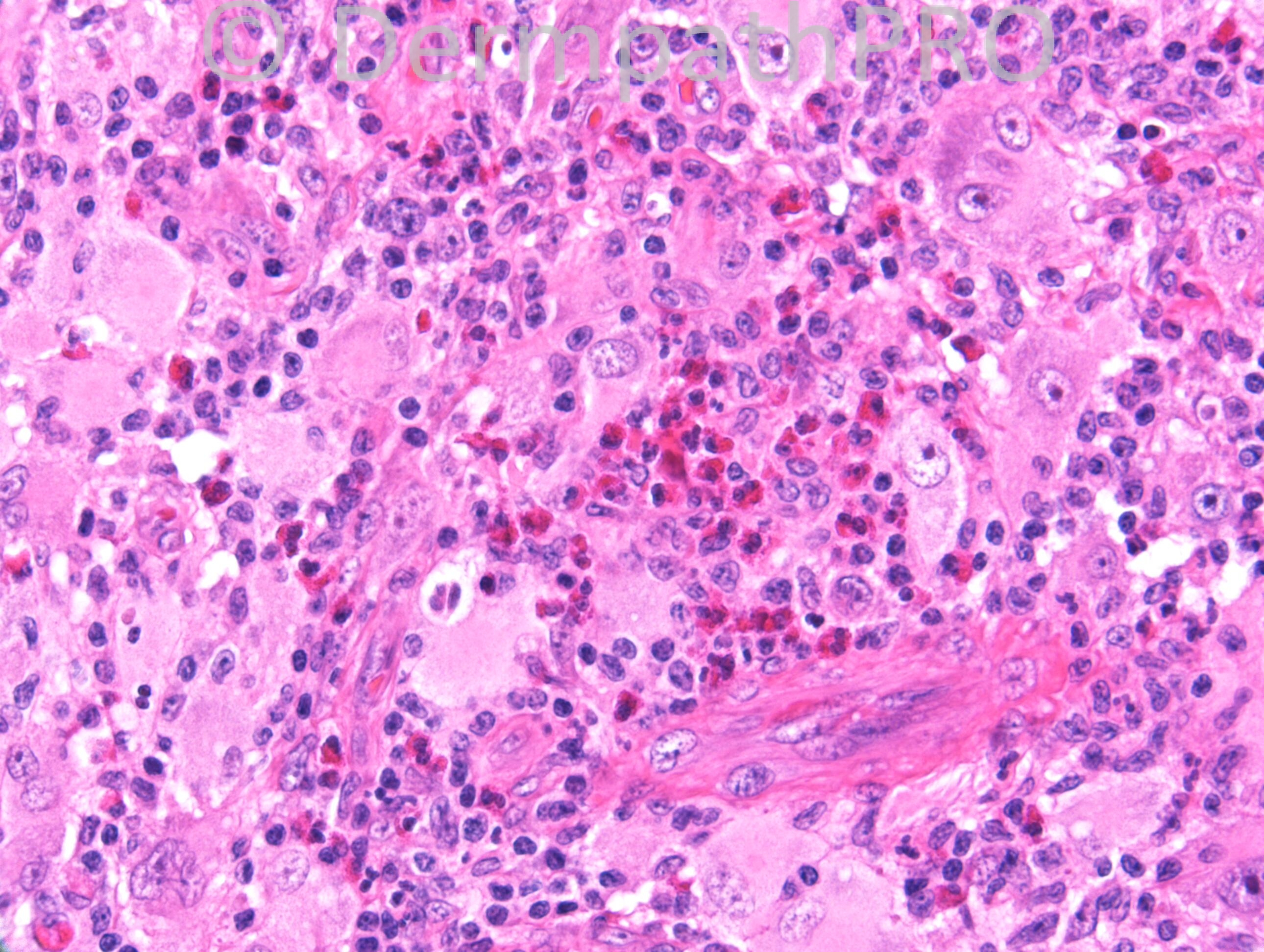
47 years old male. Upper chin. Grew to 1cm in 5 weeks. Surgeon queries epithelialized granuloma or KA.

Case posted by Dr. Richard Carr.

  Report Record

User Feedback


Guest Romualdo

Posted

I think the apparent presence of emperipolesis takes precedence over other uncommon findings and agree with Mona, although the presence of eosinophils and Touton-like giant cells brings to mind the diagnosis of xanthogranuloma.

Share this comment


Link to comment
share_externally

Guest Rodrigo Restrepo

Posted

Agree, cutaneous Rosai Dorfman Disease.

Share this comment


Link to comment
share_externally

Robledo F. Rocha

Posted

There are more eosinophils than would make me feel confortable with the hypothesis of Rosai-Dorfman disease. Thus, I’m tending towards an alternative diagnosis like solitary reticulohistiocytoma. Touton-type giant cells, emperipolesis, and inflammatory infiltrate with numerous eosinophils can occur in solitary reticulohistiocytoma.

Share this comment


Link to comment
share_externally

Sasi Attili

Posted

This is a common conundrum we face and a routine exam case where people are marked on their ability to spell the right name. I favour this to be in the solitary RH spectrum, but I don't think the histiocytes have the typical ground-glass cytoplasm. I agree that lesion has quite a few features as those described in cutaneous RDD and immunos are necessary (CRDD is S100 +ve while RH is -ve). However, I don't really believe that this is an entity distinct from a solitary RH- apart from the S100 +vity and obvious emperipolesis. CRDD I believe is within the spectrum of RH, as it is usually does not progress to systemic RDD and is distinct from systemic RDD.. So the name CRDD is actually confusing, unless we agree to call all lesions with Emperipolesis and S100 +vity cutaneous RDD and that is the definition of it! But I am not sure to what purpose it serves to split similar entities and create confusion?

I believe both are reactive phenomenon (like cutaneous pseudolymphoma.... but with a different cellular/ immuno profile). Both are derived from Histiocytes. One is S100 +ve and the other isn't! In fact even a Xanthogranuloma being histiocyte derived, is in the same spectrum, but with a different immuno profile. [i]The prognosis/ treatment is the same in all these solitary entities related to proliferation of histiocytes.[/i]

I think it all depends on whether you are a lumper or a splitter. As a clinician, all that I am interested is that whether this is benign or not (probably is- in this case). Maybe 'Benign Reactive Histiocytic proliferation' to encompass all these benign, reactive entities, is more apt? However it gives us great pleasure as diagnosticians to be able to make a 'diagnosis' when actually we are just giving a name to a simple reactive phenomenon associated with histiocytic proliferation of a particular phenotype. :)

Share this comment


Link to comment
share_externally

xanthogranuloma ,
no enough plasma cells to call it Rosai Dorfman
immunos are needed

Share this comment


Link to comment
share_externally

Dr. Richard Carr

Posted

I called it an adult type eruptive xanthogranuloma. S100 was weakly positive in the histiocytes but the control was super strong so I disregarded it, CD68 was impressively positive. There is emperipolesis (rather focal) and this has been described. The lesion has quite a number of perfect Touton giant cells (I did not highlight these in the images just to create a bit more difficulty) although I appreactiate morphologically there is an overlap with reticulohistiocytoma. I don't recall seeing this number of esoinophils in CRDD myself. What struck me in this case were some quite huge nuclei in the histiocytes with very prominent nucleoli that in isolation could be quite concerning - not sure I photographed the best examples of that though. Hope you like my new camera!!
Enjoy your weekends.

Share this comment


Link to comment
share_externally


Join the conversation

You can post now and register later. If you have an account, sign in now to post with your account.

Guest
Add a comment...

×   Pasted as rich text.   Paste as plain text instead

  Only 75 emoji are allowed.

×   Your link has been automatically embedded.   Display as a link instead

×   Your previous content has been restored.   Clear editor

×   You cannot paste images directly. Upload or insert images from URL.

Loading...

×
×
  • Create New...