Jump to content
In this section we have spot diagnoses posted on a daily basis since June 2010, now over 4000! You can review the archived cases and read the suggested diagnoses by users and the final comment by the contributors.
Case are uploaded each week day by 10 am UK time with the correct diagnosis will generally be posted at 8 pm UK time. Why not view the most recent spot diagnosis and proffer a diagnosis?

Case Number : Case 882 - 5th November Posted By: Admin_Dermpath

Please read the clinical history and view the images by clicking on them before you proffer your diagnosis.
Submitted Date :
   (0 reviews)

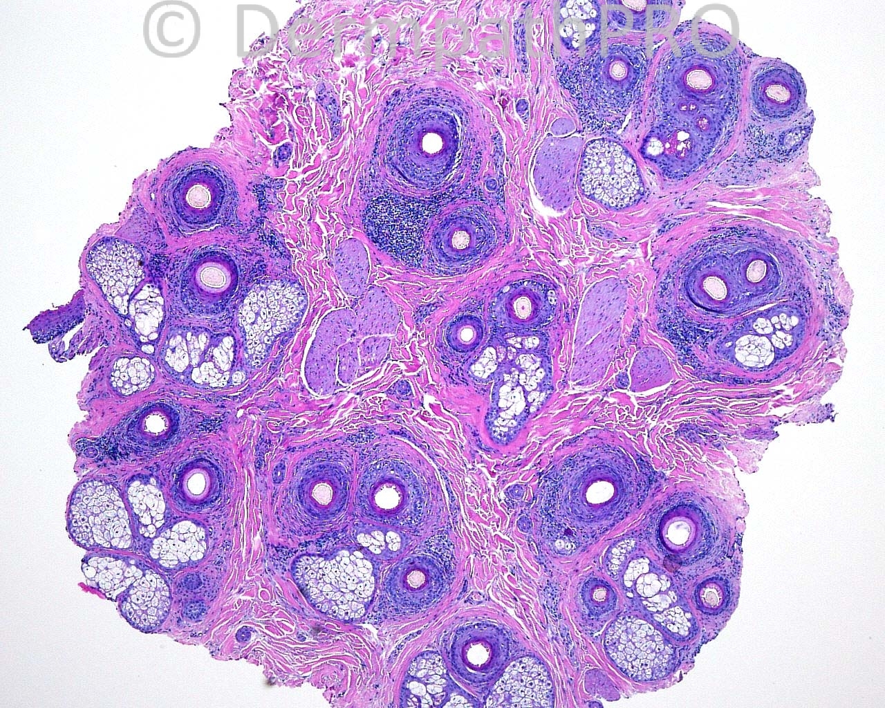
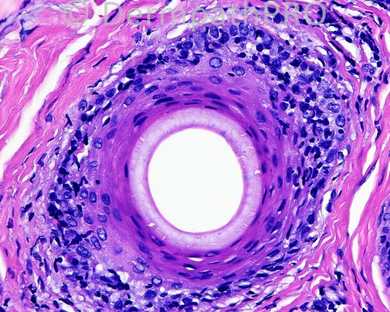
The patient is a 46 year old white woman with a regressing frontal hairline with clinical perifollicular inflammation.

Case posted by Dr. Mark Hurt


  Report Record

User Feedback


Guest Maria George

Posted

Most likely Frontal fibosing alopecia.
Things to be discussed are lichenplanopilaris, follicular MF, Lupus,...

Share this comment


Link to comment
share_externally

Sasi Attili

Posted

perifollicular lichenoid inflammation + mild/ early perifollicular fibrosis + history, in keeping with LPP= FFP.

Share this comment


Link to comment
share_externally

Guest Marwa Fawzy

Posted

[color=#1C2837][font=arial, verdana, tahoma, sans-serif][size=4]Frontal fibosing alopecia.[/size][/font][/color]

Share this comment


Link to comment
share_externally

Guest Romualdo

Posted

I agree with my colleagues: frontal fibrosing alopecia.

Share this comment


Link to comment
share_externally

Dr. Mona Abdel-Halim

Posted

The clinical presentation is of frontal fibrosing alopecia, pathological features are those of lichen planopilaris. It is said that frontal fibrosing alopecia is a form of lichen planopilaris affecting females mostly menopausal.

Share this comment


Link to comment
share_externally

Nice case. The clinical description is of a FFA.
The histopathologic pictures shows nice horizontal sections at the isthmus level with perifollicular inflamation and fibrosis composed basically of lymphocites. There´s no interfollicular fibrosis or neutrophils.
There´s lichenoid inflamation of the outer root sheath with some apoptotic bodies.

The first picture shows some follicular drop out. It is compatible with Lymphocytic fibrosing alopecia and the hole case is Frontal fibrosing alopecia.

Share this comment


Link to comment
share_externally

Guest Juan Carlos Garcés, Ecuador

Posted

Agree with frontal fibrosing alopecia

Share this comment


Link to comment
share_externally

Guest Saleem Taibjee

Posted

Agree, FFA. Worth inquiring if there are clinical features of Graham-Little syndrome.
I tend to give an indication of the degree of active inflammation versus burnt out follicular fibrosis in such cases to guide the clinician as to the merits of immunosuppressive treatment for individual cases.

Share this comment


Link to comment
share_externally

Guest Rodrigo Restrepo

Posted

F[color=#1C2837][font=arial, verdana, tahoma, sans-serif][size=4]rontal fibrosing alopecia[/size][/font][/color]

Share this comment


Link to comment
share_externally

Robledo F. Rocha

Posted

Interface lymphocytic scarring alopecia consistent with lichen planopilaris. Case history of a regressing frontal hairline indicates frontal fibrosing alopecia variant. Maybe the eyebrows are also involved.

Share this comment


Link to comment
share_externally

Guest Julio Salas

Posted

Great and very illustrative LPP FFA case.

Share this comment


Link to comment
share_externally


Join the conversation

You can post now and register later. If you have an account, sign in now to post with your account.

Guest
Add a comment...

×   Pasted as rich text.   Paste as plain text instead

  Only 75 emoji are allowed.

×   Your link has been automatically embedded.   Display as a link instead

×   Your previous content has been restored.   Clear editor

×   You cannot paste images directly. Upload or insert images from URL.

Loading...

×
×
  • Create New...