Jump to content
In this section we have spot diagnoses posted on a daily basis since June 2010, now over 4000! You can review the archived cases and read the suggested diagnoses by users and the final comment by the contributors.
Case are uploaded each week day by 10 am UK time with the correct diagnosis will generally be posted at 8 pm UK time. Why not view the most recent spot diagnosis and proffer a diagnosis?

Case Number : Case 886 - 11th November Posted By: Guest

Please read the clinical history and view the images by clicking on them before you proffer your diagnosis.
Submitted Date :
   (0 reviews)

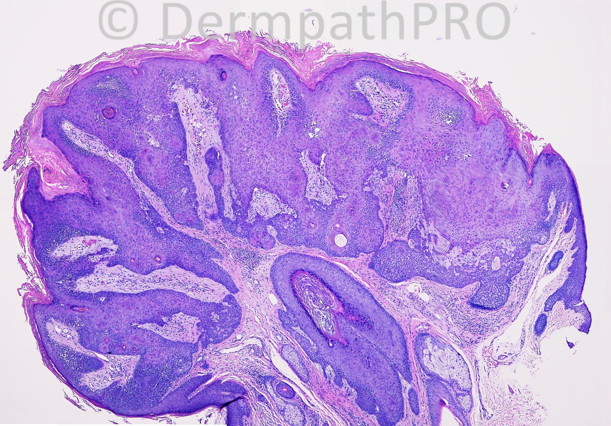
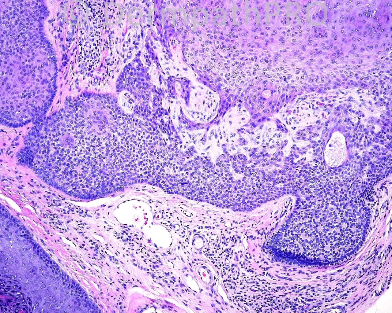
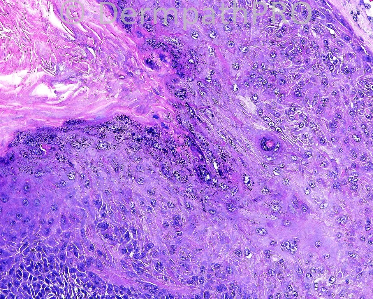
The patient is a 64 year old white man with an excision of a 5 mm, elevated, verrucous skin lesion taken from the left side of the chin.

Case posted by Dr. Mark Hurt


  Report Record

User Feedback


Desmoplastic tricholemmoma (verruca vulgaris + tricholemmal differentiation).

Share this comment


Link to comment
share_externally

Guest Mazen charaf iddin

Posted

agree with dr: Nidal, inverted follicular keratosis

Share this comment


Link to comment
share_externally

Arti Bakshi

Posted

Desmoplastic trichelemmoma (esp in the 3rd image).

Share this comment


Link to comment
share_externally

Sasi Attili

Posted

Trichilemmoma with focal desmoplastic change- I believe this might be viral in origin. There are prominent keratohyalin granules seen...

Share this comment


Link to comment
share_externally

Dr. Mona Abdel-Halim

Posted

Desmoplastic trichilemmoma related to viral origin

Share this comment


Link to comment
share_externally

Guest Marwa Fawzy

Posted

[color=#1C2837][font=arial, verdana, tahoma, sans-serif][size=4]Desmoplastic trichilemmoma[/size][/font][/color]

Share this comment


Link to comment
share_externally

Robledo F. Rocha

Posted

Tricholemmoma.
About the HPV-related changes, are they delusive, pure coincidence or a confirmation of an old hypothesis?

Share this comment


Link to comment
share_externally

Dr. Richard Carr

Posted

I thought partly basaloid, focally desmoplastic tricholemmoma with inverted follicular keratosis-like areas. CD34 would be helpful to confirm the diagnosis (positive in all tricholemmoma in our experience although can be quite focal). I have not been convinced by the suggestion these lesions are viral aetiology and seen no direct evidence in these images.

Share this comment


Link to comment
share_externally

Eman El-Nabarawy

Posted

I can't see the characteristic clear cytoplasm of trichilemmoma except in the area of focal desmoplastic change. I suggest collision of inverted follicular keratosis with desmoplastic trichilemmoma.

Share this comment


Link to comment
share_externally

Guest Tiberiu Tebeica

Posted

warty lesion with features of inverted follicular keratosis and desmoplastic trichilemmoma. both fit into the trichilemmal verruca category.

Share this comment


Link to comment
share_externally

Guest Romualdo

Posted

Squamous eddies similar to those seen in irritated seborrheic keratoses, papillomatosis and prominent granular layer resembling a verruca vulgaris are possible in trichilemmomas, the first in the keratinizing variety. So, my diagnosis is trichilemmoma, desmoplastic and keratinizing.

Share this comment


Link to comment
share_externally


Join the conversation

You can post now and register later. If you have an account, sign in now to post with your account.

Guest
Add a comment...

×   Pasted as rich text.   Paste as plain text instead

  Only 75 emoji are allowed.

×   Your link has been automatically embedded.   Display as a link instead

×   Your previous content has been restored.   Clear editor

×   You cannot paste images directly. Upload or insert images from URL.

Loading...

×
×
  • Create New...