Jump to content
In this section we have spot diagnoses posted on a daily basis since June 2010, now over 4000! You can review the archived cases and read the suggested diagnoses by users and the final comment by the contributors.
Case are uploaded each week day by 10 am UK time with the correct diagnosis will generally be posted at 8 pm UK time. Why not view the most recent spot diagnosis and proffer a diagnosis?

Case Number : Case 887 - 12th November Posted By: Guest

Please read the clinical history and view the images by clicking on them before you proffer your diagnosis.
Submitted Date :
   (0 reviews)

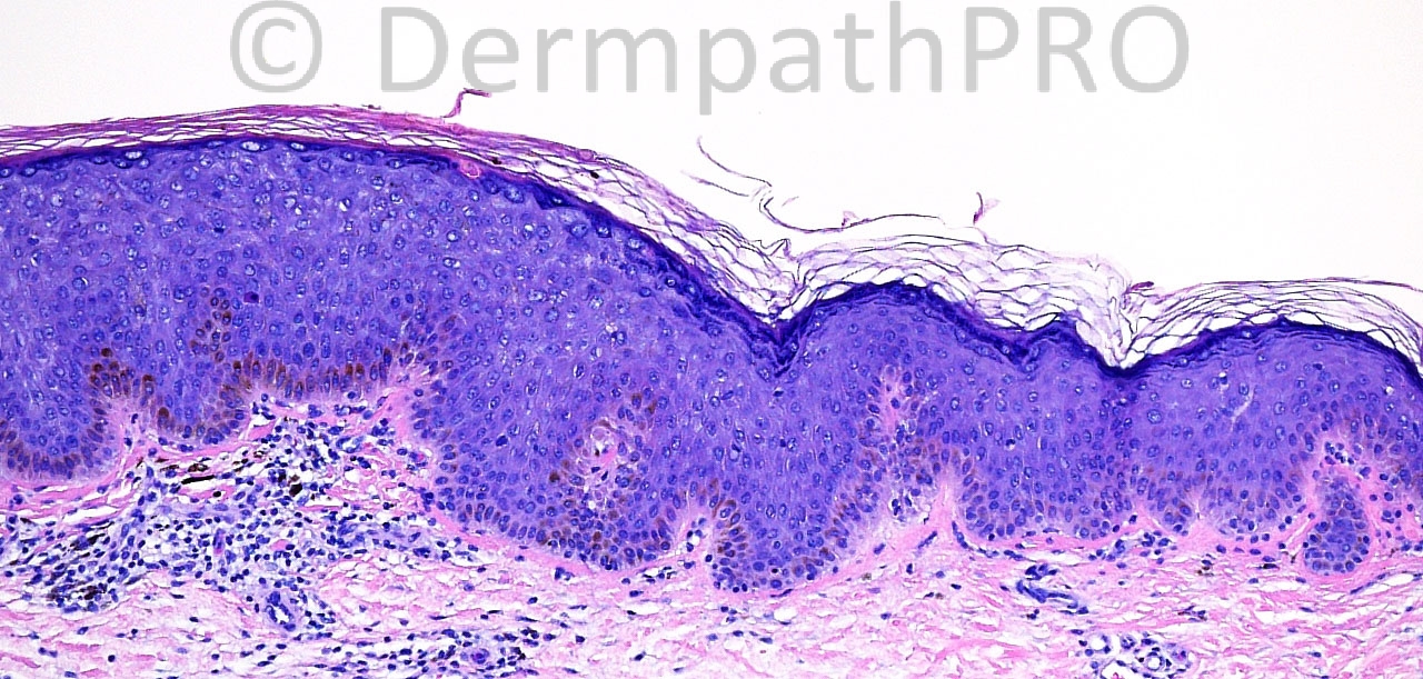
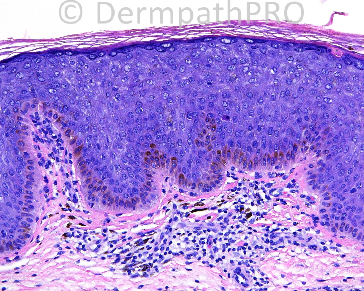
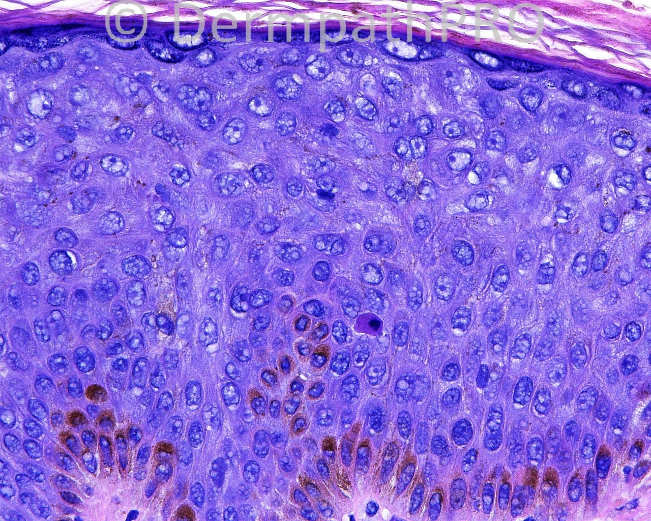
The patient is a 67 year old man with a history of malignant melanoma. The patient now has an excision taken from the left, upper aspect of the chest.

Case posted by Dr. Mark Hurt


  Report Record

User Feedback


Guest Romualdo

Posted

The nuclei on the left side of figure # 2 (lesion) are slightly enlarged in relation to the nuclei on the the right side (normal epidermis). I think this is large cell acanthoma.

Share this comment


Link to comment
share_externally

Robledo F. Rocha

Posted

A well demarcated thickening of the epidermis due to proliferation of enlarged keratinocytes without nuclear atypia. There's also hyperpigmentation of the basal layer. I go with large cell acanthoma.

Share this comment


Link to comment
share_externally

Guest Rodrigo Restrepo

Posted

[color=#1C2837][font=arial, verdana, tahoma, sans-serif][size=4]Large cell acanthoma[/size][/font][/color]

Share this comment


Link to comment
share_externally

Guest Jim Davie MD

Posted

Very interesting case. Without stains, I would hesitate to call it Bowen's disease. However, I don't think a large cell acanthoma should spare the basal layer as seen here. (See earlier LCA: [url="https://dermpathpro.com/spot-diagnosis-1/_/2013-spot-diagnoses/september-2013/case-838-3rd-september-r865"]case[/url]). I think it's a clonal SK, with differential less favoring Bowen's disease. CK10 / Bcl2 may be helpful.[list]
[*]Low grade atypia with dyskeratosis, mild pleomorphism/enlargement. The vescicular nuclear artefact obscures the presence of slightly enlarged and multiple nucleoli within the enlarged suprabasal keratinocyte population.
[*]The Ki-67 shows basal sparing and full-thickness increase in staining, with absence of any maturation. Large cell acanthomas and melanoacanthomas should have a low Ki67 index, with low-level distribution.
[*]Pagetoid melanocytes are low level, and likely incidental to disruptive effects.
[/list]

Share this comment


Link to comment
share_externally

pigmented Bowen's vs melanoma in situ ( Melan A shqowed some pagetoid spread of melanocystes)
I don't see nests to call it metastatic intraepidermal melanoma

Share this comment


Link to comment
share_externally

Is there a history of topical treatment which may have induced epidermal proliferation?

Share this comment


Link to comment
share_externally

Robledo F. Rocha

Posted

Immunohistochemistry was crucial to the correct diagnosis. In HE pictures alone, I didn't suspect all that cellular proliferation in the full-thickness of the epidermis as highlighted by Ki-67.
Thanks, Dr. Hurt, to share this interesting case.

Share this comment


Link to comment
share_externally

Sasi Attili

Posted

I have trouble with the diagnosis Bowens just on the basis of Ki67. Is there enough scientific evidence to say that high Ki67 activity is 100% specific for malignancy? Would be interested to know.

Share this comment


Link to comment
share_externally

Guest Jim Davie MD

Posted

[quote name='Sasi Attili' timestamp='1384296889']
I have trouble with the diagnosis Bowens just on the basis of Ki67. Is there enough scientific evidence to say that high Ki67 activity is 100% specific for malignancy? Would be interested to know.
[/quote]

I agree Ki67 is useful, but not sufficient in itself for malignancy.

A recent JCP article [ [url="http://onlinelibrary.wiley.com/doi/10.1111/j.1600-0560.2011.01825.x/abstract"]ABSTRACT link[/url]] studied histologic and immunohistochemical markers to help reliably distinguish clonal SK and Bowen's. Of the IPEX markers tested -- Ki-67, bcl-2, cytokeratin 7 and cytokeratin 10 -- they suggested CK10 and Bcl-2 as most useful. (Ki67 didn't make the cut).

[size=4][b][color=#000000]Cytokeratin 10-negative nested pattern enables sure distinction of clonal se[/color][/b][color=#000000][b]borrheic keratosis from pagetoid Bowen's disease. [/b][/color][/size][color=#000000]Böer-Auer, A., Jones, M. and Lyasnichaya, O. V. [/color]
[size=4][color=#000000]Journal of Cutaneous Pathology, 39: 225–233. (2012) [/color][/size]

Share this comment


Link to comment
share_externally

Dr. Richard Carr

Posted

There is a saying don't dismiss your first impression. My first thought was pigmented subtle Bowen's but I was not brave enough to contradict all those (seemingly good) responses - I had not considered large cell acanthoma but it seemed reasonable. The photos are a little blue overall and I assumed this was a colour balance issue! Showing a high mitotic figure would have been helpful though.

Share this comment


Link to comment
share_externally


Join the conversation

You can post now and register later. If you have an account, sign in now to post with your account.

Guest
Add a comment...

×   Pasted as rich text.   Paste as plain text instead

  Only 75 emoji are allowed.

×   Your link has been automatically embedded.   Display as a link instead

×   Your previous content has been restored.   Clear editor

×   You cannot paste images directly. Upload or insert images from URL.

Loading...

×
×
  • Create New...