Jump to content
In this section we have spot diagnoses posted on a daily basis since June 2010, now over 4000! You can review the archived cases and read the suggested diagnoses by users and the final comment by the contributors.
Case are uploaded each week day by 10 am UK time with the correct diagnosis will generally be posted at 8 pm UK time. Why not view the most recent spot diagnosis and proffer a diagnosis?

Case Number : Case 890 - 15th November Posted By: Guest

Please read the clinical history and view the images by clicking on them before you proffer your diagnosis.
Submitted Date :
   (0 reviews)

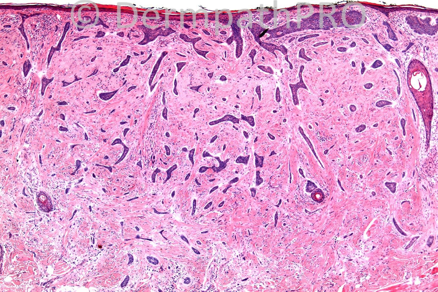
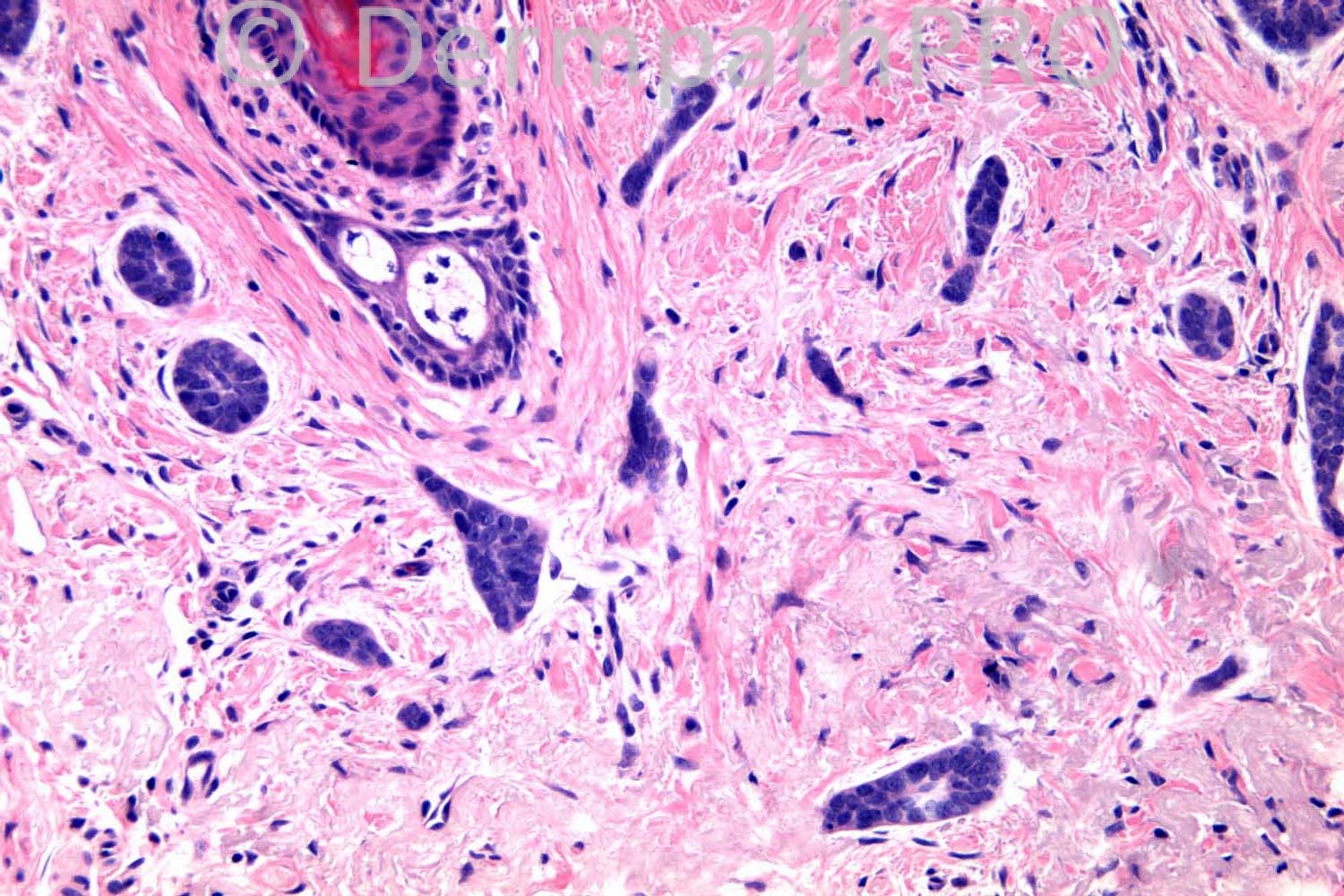
75 years old female. ?BCC on upper lip.

Case posted by Dr. Richard Carr.


  Report Record

User Feedback


Robledo F. Rocha

Posted

I favor columnar trichoblastoma over morpheaform basal cell carcinoma because this well-circunscribed lesion has a depression in the center of its surface. Also I didn't find solar elastosis below the lesion.

Share this comment


Link to comment
share_externally

I favour an infiltrative basal cell carcinoma. There is peripheral palisading (the first photo), retraction artifact and absence of horn cysts (although there is a foreign-body giant cell in the third photo). What does CK20 show?

Share this comment


Link to comment
share_externally

Infiltrattive BCC
CK20 is usually positive in TE and not BCC
BCL-2 and CD10 ARE usually positive in THE BASALOID cells OF BCC and not TE( only the basaloid cells of the periphery of TE will BE cd10 POSITIVE

Share this comment


Link to comment
share_externally

Guest Saleem Taibjee

Posted

Nice tricky case.
It is going to come down to a vote of infiltrative BCC versus DTE.
I favour DTE here based on criteria including overlying dell, quality of stromal reaction, granulomatous reaction. However, there does seem to be increased mucin, and immunos might sway me either way i.e. Merkel cells or otherwise.

Share this comment


Link to comment
share_externally

Sasi Attili

Posted

on first impression I thought DTE. However looking at the pics closely, there does seem to be a retraction artefact around the nests favouring a BCC. Also in the first pic towards the top right edge, I think Richard might be hiding the superficial BCC component of the tumor!. It is suspicious of that anyway. However, I agree that immunos would be helpful.

Having said that, I believe that BCC and DTE are children of the same mother! They might have a different father though :)

Share this comment


Link to comment
share_externally

Dr. Richard Carr

Posted

I have asked the dermpathpro team to post the IHC at 4pm GMT. I am the father of this case by the way. Another father (of dermatopathology) once said the diagnosis of DTE is one of the most difficult in dermatopathology.

Share this comment


Link to comment
share_externally

Sasi Attili

Posted

[quote name='Dr. Richard Carr' timestamp='1384517030']
I have asked the dermpathpro team to post the IHC at 4pm GMT. I am the father of this case by the way. Another father (of dermatopathology) once said the diagnosis of DTE is one of the most difficult in dermatopathology.
[/quote] who is the mother? :P

Share this comment


Link to comment
share_externally

Iskander H. Chaudhry

Posted

Dear All, please view the case of the week if you have a moment - it is a new feature of the site and we hope it will generate some discussion.

Share this comment


Link to comment
share_externally

Sasi Attili

Posted

[quote name='Iskander H. Chaudhry' timestamp='1384519240']
Dear All, please view the case of the week if you have a moment - it is a new feature of the site and we hope it will generate some discussion.
[/quote]
Please post the link and include the link in the blog menu/ home page Iskander. Can't find it!

Share this comment


Link to comment
share_externally

Dr. Mona Abdel-Halim

Posted

Favour infiltrative BCC,,,, waiting for the immunos!!

Share this comment


Link to comment
share_externally

Guest Romualdo

Posted

The presence of large epitelial islands, considerable nuclear atypia in some foci, retraction artifact and lack of concentric bands of collagen enveloping elongated epitelial strands favour morpheaform BCC.

Share this comment


Link to comment
share_externally

Iskander H. Chaudhry

Posted

Dear All, you can access the case of the week from the home page or by clicking on the link below:

[url="https://dermpathpro.com/case_of_the_week.html/_/case-1-r3"]https://dermpathpro.com/case_of_the_week.html/_/case-1-r3[/url]

Share this comment


Link to comment
share_externally

Dr. Richard Carr

Posted

[img]https://dermpathpro.com/uploads/spot_diagnosis_comment_img/CASE890_RAC6317x10_CK20_MR.jpg[/img]


[img]https://dermpathpro.com/uploads/spot_diagnosis_comment_img/CASE890_RAC6316x5_BerEP4_MR.jpg[/img]


[img]https://dermpathpro.com/uploads/spot_diagnosis_comment_img/CASE890_RAC6316x20b_Ki67_MR.jpg[/img]


[img]https://dermpathpro.com/uploads/spot_diagnosis_comment_img/CASE890_RAC6316x10b_Bcl2_MR.jpg[/img]


[img]https://dermpathpro.com/uploads/spot_diagnosis_comment_img/CASE890_RAC6316x10_CD34_MR.jpg[/img]


[img]https://dermpathpro.com/uploads/spot_diagnosis_comment_img/CASE890_RAC6316x10_CD10_MR.jpg[/img]

Share this comment


Link to comment
share_externally

Dr. Mona Abdel-Halim

Posted

Waaaaaaaaaaao !!!!!!
As I expected,,,,, beautifully demonstrated immunos....
I love this :-))

Share this comment


Link to comment
share_externally

Guest Tiberiu Tebeica

Posted

Because there is nuclear pleomorphism, some nuclear palisading and retraction artifact, I favor infiltrative BCC. Of course, the classic differential includes DTE, but the stroma and the cytologic features don't look great for it, and MAC, for which I don't see the classic stratification, with keratocysts on top. I would still perform IHC to rule these two out.

Share this comment


Link to comment
share_externally

Robledo F. Rocha

Posted

Oops, immunohistochemistry changed my mind. It's basal cell carcinoma.

Share this comment


Link to comment
share_externally

Dr. Richard Carr

Posted

Mona you just made my day! Yes it is a basal cell
[quote name='Romualdo' timestamp='1384527431']
The presence of large epitelial islands, considerable nuclear atypia in some foci, retraction artifact and lack of concentric bands of collagen enveloping elongated epitelial strands favour morpheaform BCC.
[/quote]
Says it all - IHC is helpful if you have any doubt. Merkel cells limited to trapped follicles (and perifollicular dermis). Ki67 is nearly 50% (should be ~5% in DTE). BerEP4 just highlights the abnormal larger irregular foci not allowed in DTE. Remember DTE has the nice curvilinear cords only 2 to 3 cells in width although this case does have some cords mimicking the pattern closely. Dr Bernard Akerman was the father of dermatopathology who said DTE can be one of the most difficult diagnoses in dermatopathology.

Share this comment


Link to comment
share_externally

Guest Jim Davie MD

Posted

On H&E, additional features that suggested infiltrative BCC was the definitive mucinous stroma surrounding nests in the lowest high-power photo, and presence of mitotic activity and/or hyperchromatism in the same.

Share this comment


Link to comment
share_externally

Guest Tiberiu Tebeica

Posted

The immunostains in this case are neat and nicely light up the diagnosis. Another marker I got to work with these days is TDAG51, a follicular stem cell marker that reliably differentiates between trichoepithelioma and BCC. For similar cases, usually a panel made of TDAG51 and BerEP4 is enough to put one on the right track.

Share this comment


Link to comment
share_externally


Join the conversation

You can post now and register later. If you have an account, sign in now to post with your account.

Guest
Add a comment...

×   Pasted as rich text.   Paste as plain text instead

  Only 75 emoji are allowed.

×   Your link has been automatically embedded.   Display as a link instead

×   Your previous content has been restored.   Clear editor

×   You cannot paste images directly. Upload or insert images from URL.

Loading...

×
×
  • Create New...