Jump to content
In this section we have spot diagnoses posted on a daily basis since June 2010, now over 4000! You can review the archived cases and read the suggested diagnoses by users and the final comment by the contributors.
Case are uploaded each week day by 10 am UK time with the correct diagnosis will generally be posted at 8 pm UK time. Why not view the most recent spot diagnosis and proffer a diagnosis?

Case Number : Case 895 - 22nd November Posted By: Guest

Please read the clinical history and view the images by clicking on them before you proffer your diagnosis.
Submitted Date :
   (0 reviews)

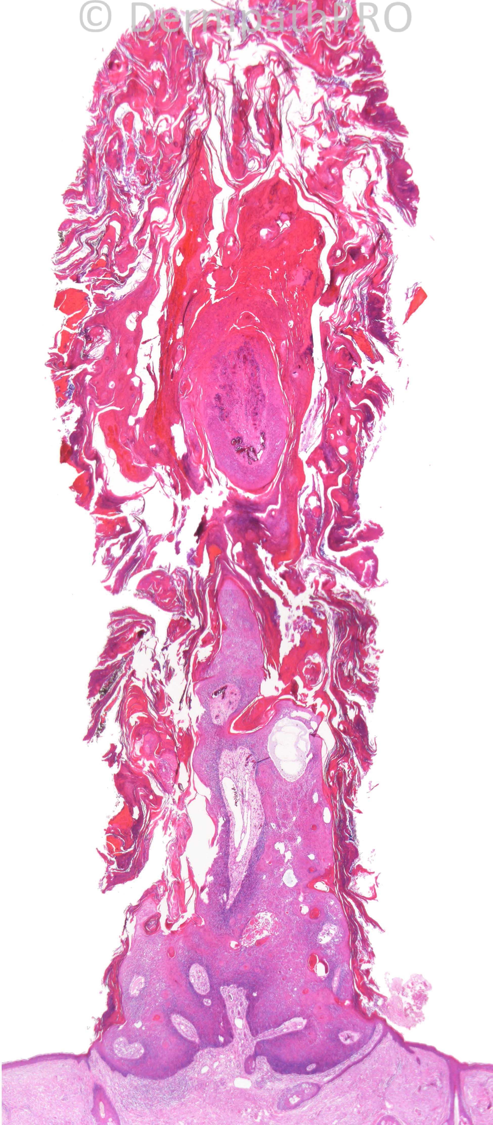
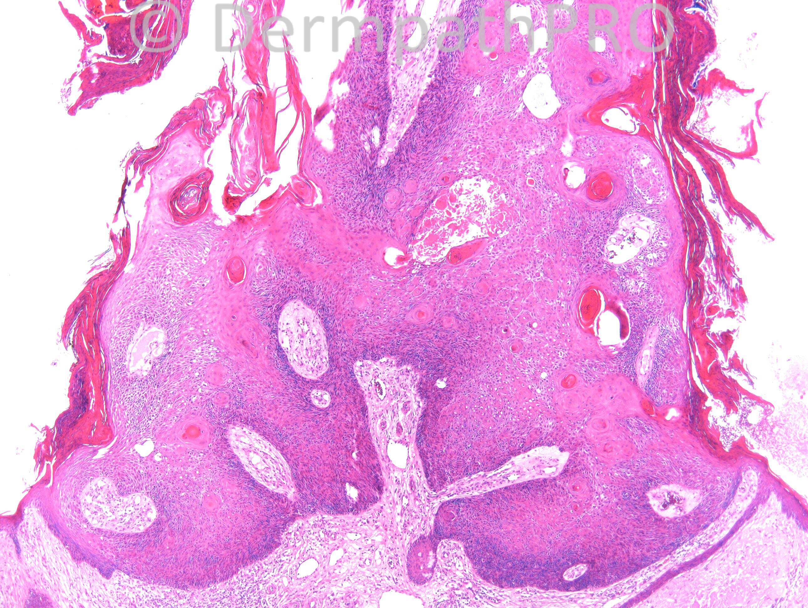
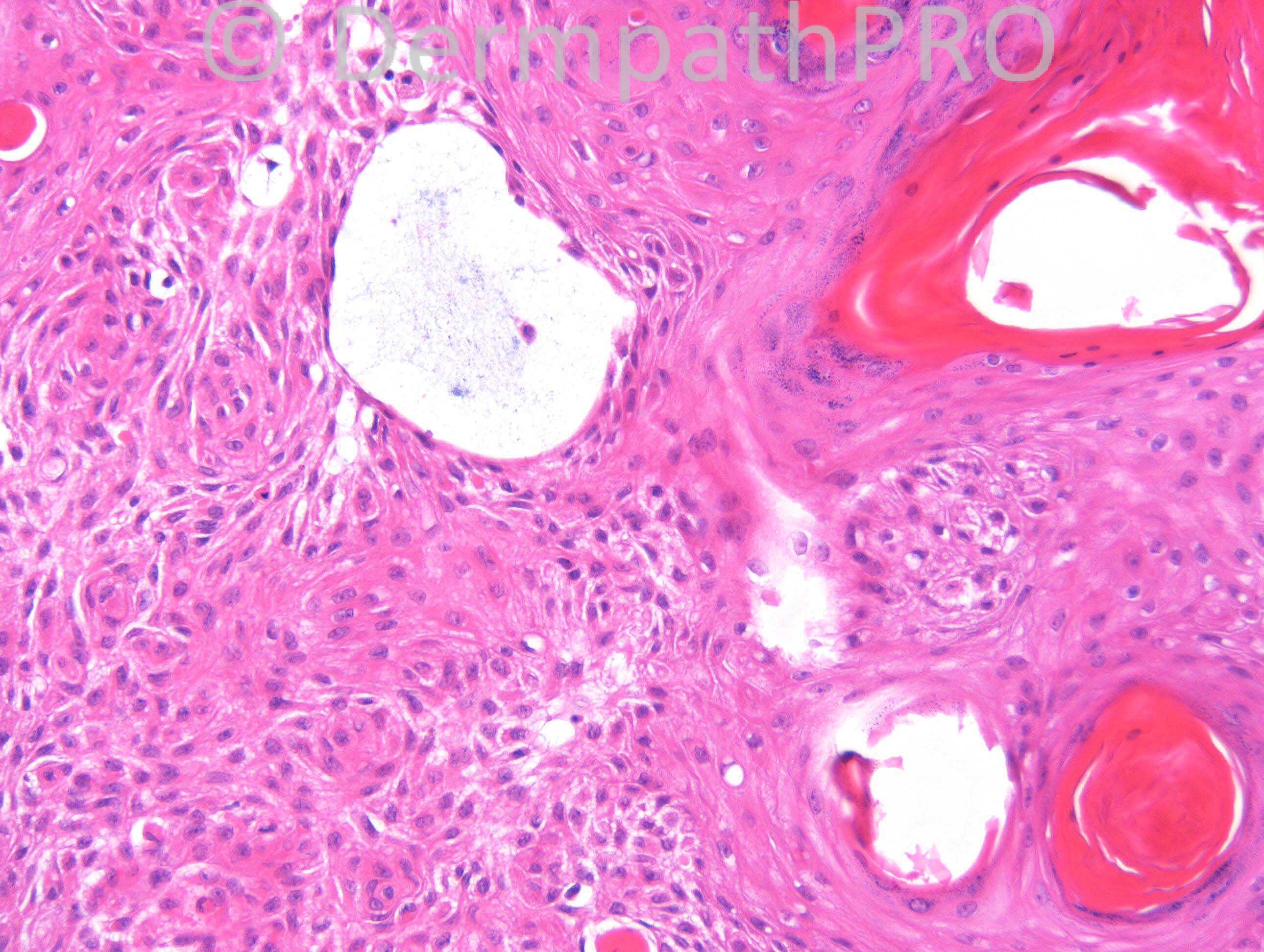
F89. Keratin horn on eyebrow.

Case posted by Dr. Richard Carr.

  Report Record

User Feedback


Guest Dr.Yüksel Okumuş

Posted

Cutaneous horn overlies an irritated seborrheic keratosis

Share this comment


Link to comment
share_externally

Guest Marwa Fawzy

Posted

[color=#1C2837][font=arial, verdana, tahoma, sans-serif][size=4]Cutaneous horn on top of an irritated seborrheic keratosis[/size][/font][/color]

Share this comment


Link to comment
share_externally

Robledo F. Rocha

Posted

Irritated seborrheic keratosis topped by filiform keratotic horn. It also can be an irritated filiform wart.

Share this comment


Link to comment
share_externally

Guest Juan Carlos Garcés, Ecuador

Posted

Agree with irritated seborrheic keratosis

Share this comment


Link to comment
share_externally

Sasi Attili

Posted

Benign Cutaneous horn arising from an irritated Seborrhoeic keratosis

Share this comment


Link to comment
share_externally

Guest Romualdo

Posted

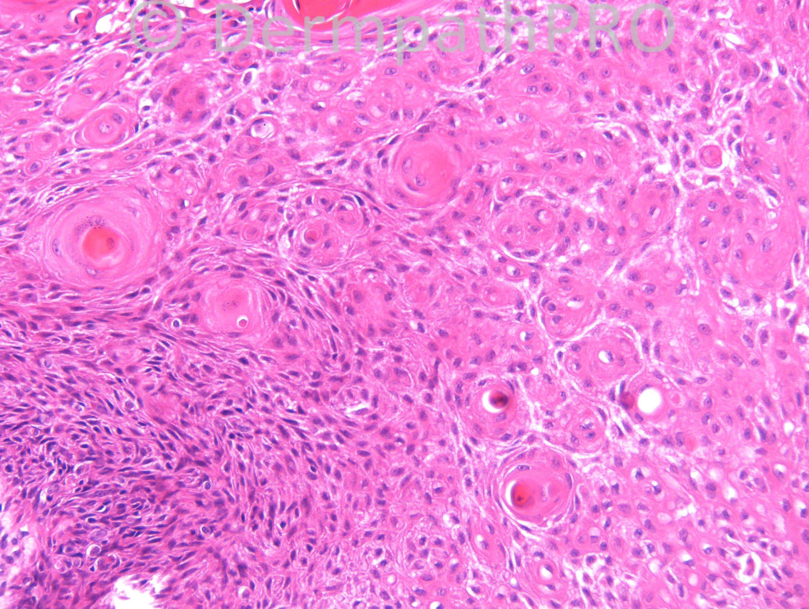
I prefer verruca vulgaris with squamous eddies over irritated seborrheic keratosis (inverted follicular keratosis). I think there are two clues to this diagnosis: rete ridges converging to the center and fusing in the base of the lesion and hemorrhages within the horn.

Share this comment


Link to comment
share_externally

Dr. Mona Abdel-Halim

Posted

Cutaneous horn overlying an old wart with squamous eddies. Controversy exists about inverted follicular keratosis and its relation to warts...

Share this comment


Link to comment
share_externally

Guest Jim Davie MD

Posted

Agree with the above (ISK vs. filiform irritated SK).

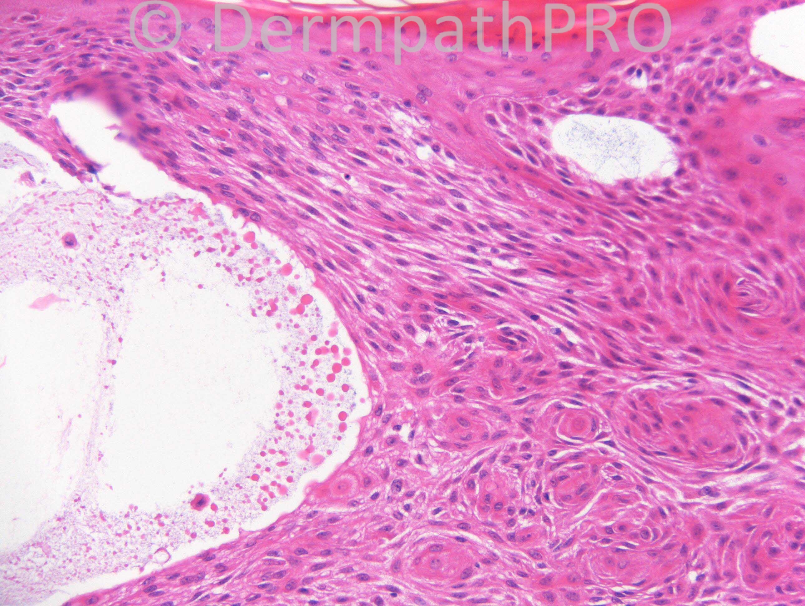
For sake of an expanded differential, I would also consider a markedly irritated trichilemmoma. There is focal clear cell changes (low power images) and a thickened eosinophilic basal layer. CD34 may be useful to evaluate.

Share this comment


Link to comment
share_externally

Guest Dr. Taskin Erknuresin

Posted

cutaneous horn overlying an old verruca vulgaris

Share this comment


Link to comment
share_externally

Guest Engin Sezer

Posted

IFK based on conspicious squamous eddies (which is already a seb ker).

Share this comment


Link to comment
share_externally

Guest Mazen charaf iddin

Posted

I've looked in lever dermatopathology book, Isee it come in the contexts of hyperkeratotic type of SK, I couldn't see the clear signs of Warts

Share this comment


Link to comment
share_externally

Dr. Richard Carr

Posted

This is an inverted follicular keratosis with nice follicular mucin. CD34 was negative which is also my experience with these lesions although they clearly are very close to tricholemmoma. It is quite a case as most keratin horns of this degree are usually related to underlying actinic keratosis. Clearly the name (inverted) needs to be taken with a pinch of salt.

Share this comment


Link to comment
share_externally


Join the conversation

You can post now and register later. If you have an account, sign in now to post with your account.

Guest
Add a comment...

×   Pasted as rich text.   Paste as plain text instead

  Only 75 emoji are allowed.

×   Your link has been automatically embedded.   Display as a link instead

×   Your previous content has been restored.   Clear editor

×   You cannot paste images directly. Upload or insert images from URL.

Loading...

×
×
  • Create New...