Jump to content
In this section we have spot diagnoses posted on a daily basis since June 2010, now over 4000! You can review the archived cases and read the suggested diagnoses by users and the final comment by the contributors.
Case are uploaded each week day by 10 am UK time with the correct diagnosis will generally be posted at 8 pm UK time. Why not view the most recent spot diagnosis and proffer a diagnosis?

Case Number : Case 896 - 25th November Posted By: Guest

Please read the clinical history and view the images by clicking on them before you proffer your diagnosis.
Submitted Date :
   (0 reviews)

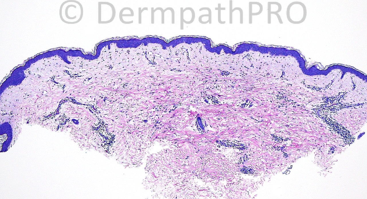
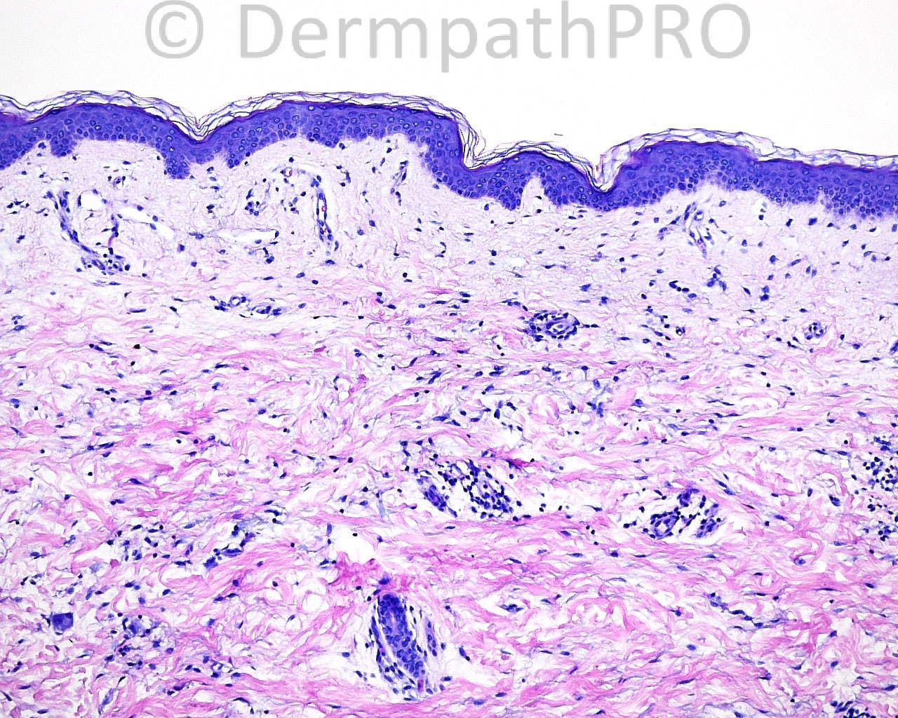
The patient is a 59 year old woman with punch biopsies taken from the right groin fold.

Case posted by Dr. Mark Hurt


  Report Record

User Feedback


Guest Marwa Fawzy

Posted

[font=tahoma,geneva,sans-serif][b]Mucin deposition ?? may consider granuloma annular ;interstitial type..[/b][/font]
[font=tahoma,geneva,sans-serif][b]A bit difficult without clinical description for the lesions.[/b][/font]

Share this comment


Link to comment
share_externally

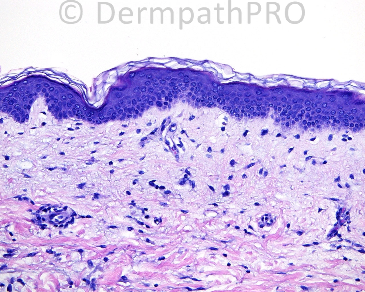
Mucin deposition associated with an increased number of fibroblasts, some of them multinucleated.
I think this is Papular mucinosis.

Share this comment


Link to comment
share_externally

Guest Dr. Taskin Erkinuresin

Posted

dermal mucinosis. primarily papular mucinosis

Share this comment


Link to comment
share_externally

Guest Juan Carlos Garcés, Ecuador

Posted

[b]Agree with papular mucinosis[/b]

Share this comment


Link to comment
share_externally

Robledo F. Rocha

Posted

Localized lichen myxedematosus (papular mucinosis). There is fibroblast proliferation and mucin deposition in the upper dermis, particularly pronounced in the subepidermal zone. Fibrosis is insufficient to scleromyxedema.

Share this comment


Link to comment
share_externally

Guest Jim Davie MD

Posted

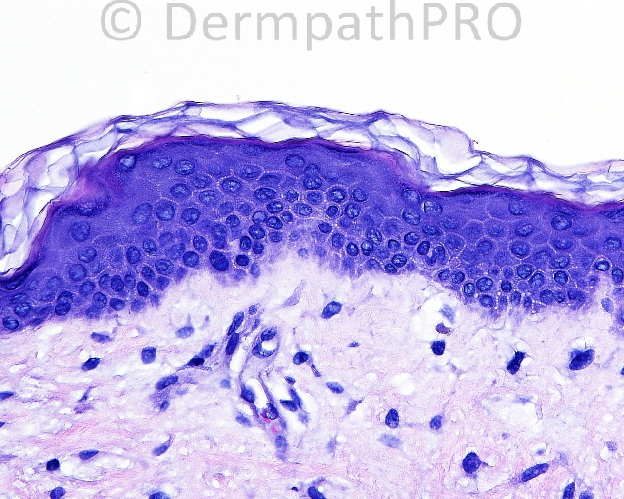
I will add macular mastocytosis (TMEP) to the differential. There is an increased number of subtly granulated mononuclear cells and a few rare stellate multinucleated cells, although the resolution limits make it difficult to differentiate the latter's lineage (mast cell, plasma cell, fibroblast).

Share this comment


Link to comment
share_externally

Robledo F. Rocha

Posted

[size=4]Ooops, this is vitiligo. No melanocytes nor melanin pigment were highlighted.
But what about the bluish material splitting collagen fibers in the upper dermis? Maybe it is l[/size][font=arial, verdana, tahoma, sans-serif][size=4]ocalized lichen myxedematosus involving an area of previous known vitiligo..[/size][/font][color=#1C2837][font=arial, verdana, tahoma, sans-serif][size=4].[/size][/font][/color]

Share this comment


Link to comment
share_externally

Sasi Attili

Posted

Indeed- looks like vitiligo. However I am a bit puzzled like everyone else with the dermal changes. Are these incidental??

Share this comment


Link to comment
share_externally

Sasi Attili

Posted

It is indeed quite amusing to see the entire dermpath community on the website being fooled by this case. Having said that Vitiligo is certainly not a histological diagnosis as CPC is always required and essentially it is a diagnosis of exclusion: [url="http://www.ncbi.nlm.nih.gov/pubmed/18613870"]http://www.ncbi.nlm.nih.gov/pubmed/18613870[/url].

Do remember that in the groin/ mucosal area, histology is often required to rule out Vitiligoid Lichen Sclerosus: [url="http://www.ncbi.nlm.nih.gov/pubmed/18388368"]http://www.ncbi.nlm.nih.gov/pubmed/18388368[/url]

Share this comment


Link to comment
share_externally

Sasi Attili

Posted

Indeed Dr. Hurt, that is what I meant. Certainly did not mean that 'you' had tried to fool us. I was referring to the 'case' having fooled us. Apologies if I came across differently. Vitiligo is considered in the differential of 'invisible dermatoses' (though in the early stages- the features are inflammatory, lichenoid). However in this case, something else was visible which made us all go off on a tangent. We do often see associated features in various dermatoses and neoplasms. However, when we know the history and feel that the associated changes are of no clinical relevance, we sub-consciously or consciously ignore these changes, for the sake of not confusing the clinician. In this case, without the knowledge of the clinical request- we all concentrated on the 'associated' change!

Blind reporting is certainly the way I was taught and is the best practice. It makes us think; In this case- I wondered, if there is indeed some mucin in the dermis (which you might not have tested for), does the patient have associated myxedema (related to another autoimmune, thyroid disease)?

Of course it is easy to also imagine things when we are looking blind and I have often made a complete fool of myself (even in not so invisible dermatoses!). But, I am sure we would all be interested to know the status of the dermal mucin in this case.

Share this comment


Link to comment
share_externally


Join the conversation

You can post now and register later. If you have an account, sign in now to post with your account.

Guest
Add a comment...

×   Pasted as rich text.   Paste as plain text instead

  Only 75 emoji are allowed.

×   Your link has been automatically embedded.   Display as a link instead

×   Your previous content has been restored.   Clear editor

×   You cannot paste images directly. Upload or insert images from URL.

Loading...

×
×
  • Create New...