Jump to content
In this section we have spot diagnoses posted on a daily basis since June 2010, now over 4000! You can review the archived cases and read the suggested diagnoses by users and the final comment by the contributors.
Case are uploaded each week day by 10 am UK time with the correct diagnosis will generally be posted at 8 pm UK time. Why not view the most recent spot diagnosis and proffer a diagnosis?

Case Number : Case 858 - 1st October Posted By: Guest

Please read the clinical history and view the images by clicking on them before you proffer your diagnosis.
Submitted Date :
   (0 reviews)

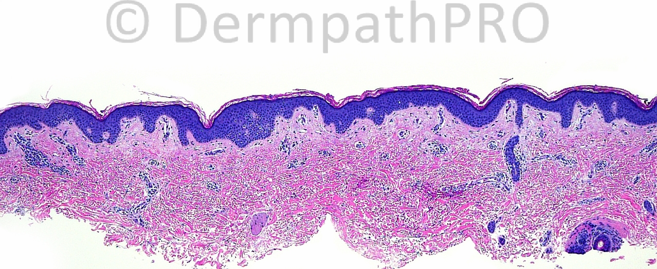
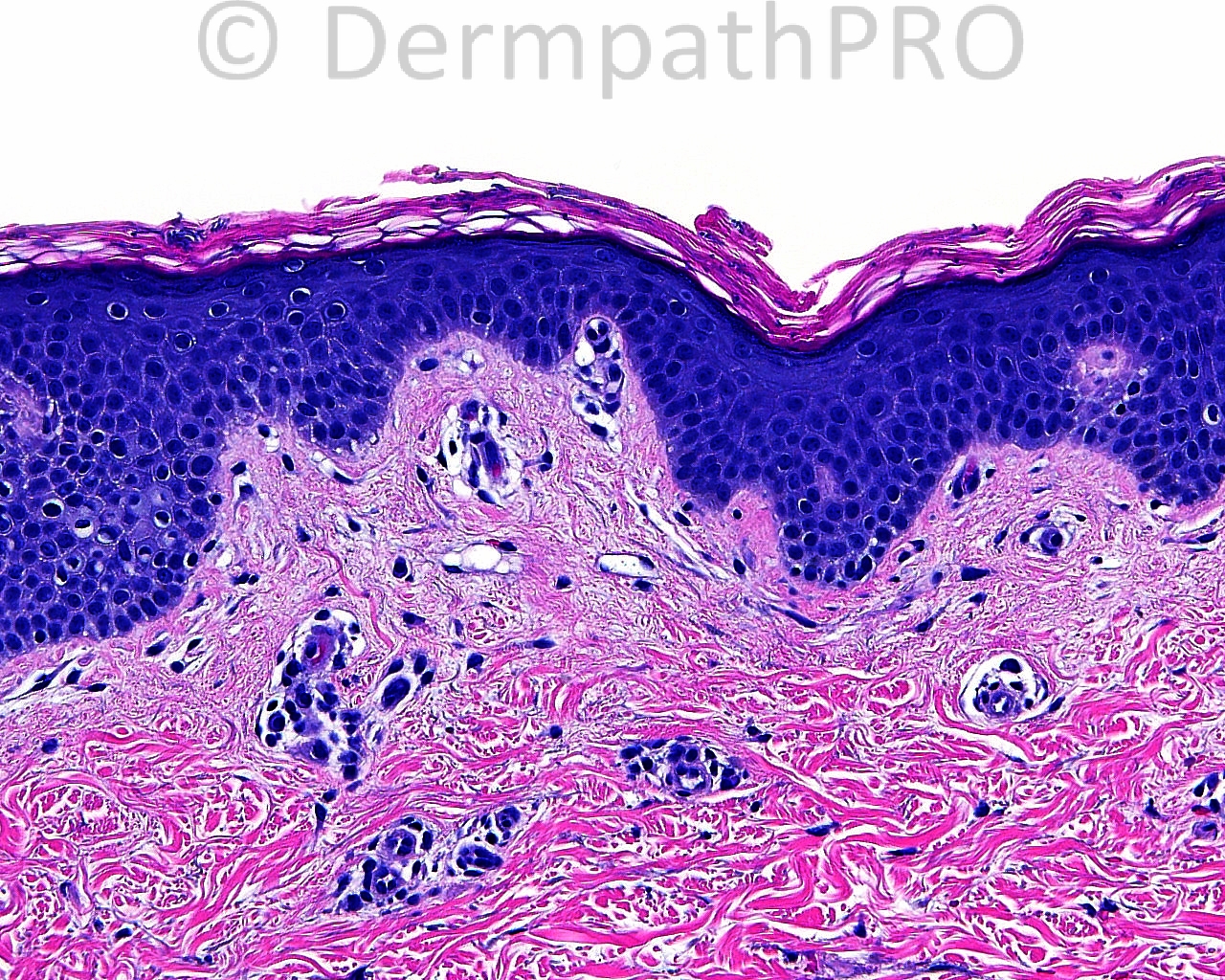
The patient is a 35 year old woman with a shave biopsy taken from the abdomen.

Case posted by Dr. Mark Hurt.


  Report Record

User Feedback


Guest Marwa Fawzy

Posted

Is the clue in the SC.. Could i say fungal infection ,need PAS to exclude and clinical data

Share this comment


Link to comment
share_externally

Guest Maria George

Posted

Tinea with [color=#000000][font=arial, helvetica, clean, sans-serif][size=3]intralymphatic histiocytosis or the so-called reactive intravascular angioendotheliomatosis?[/size][/font][/color]

Share this comment


Link to comment
share_externally

Guest Engin Sezer

Posted

How about tinea nigra? Is skin scraping performed?

Share this comment


Link to comment
share_externally

Sasi Attili

Posted

I find this tricky. My initial impression on low power was that we are seeing the edge of a naevus with just a few dermal nests scattered here and there. However looking at the higher power, the cells appear to be intra/perivascular. They do look lymphocytic. I was looking for mast cells but couldn't convince myself. Some of these lymphocytes do seem to be going up into the epidermis and surrounded by halo's. Not sure if I am starting to see things :) Are we dealing with something in the CTCL spectrum? I am however stumped by the history and the lack of a dense infiltrate. I might be completely on the wrong track, but will be requesting immunos before reporting this, if this was my case!

P.S: Given the clinical- I am presuming the yeasts seen on the surface are incidental?

Share this comment


Link to comment
share_externally

Guest Jim Davie MD

Posted

Agree with Engin: Tinea nigra.

Septate hyphae in stratum corneum without inflammatory reaction. I don't see anything of pigmentary interest below the stratum corneum. Potassium hydroxide scraping should show septate pigmented yeast and hopefully bicellular oval yeast.
Clinical hx would be good for tinea nigra given that it is often a large 1-2cm dark lesion frequently mistaken for melanoma, lentigo, or junctional nevi. The site is almost always palms and soles...very rarely other sites.

Share this comment


Link to comment
share_externally

Robledo F. Rocha

Posted

At scanning magnification, an illusion of normal skin, except for a subtle compact cornified layer, where pigmented hyphae and spores can be found at higher magnification.
I go with tinea nigra, a rare dematiaceous mycosis in a rare location. In its typical sites, to wit, the volar skin of palms and soles, clinical diagnosis invariably go with acral junctional melanocytic nevus or acral in-situ melanoma.

Share this comment


Link to comment
share_externally

Guest Romualdo

Posted

The clinical impression of melanoma/ nevus and the presence of fungi in the stratum corneum goes well with tinea nigra as the correct diagnosis.

Share this comment


Link to comment
share_externally

Dr. Mona Abdel-Halim

Posted

Tinea nigra,, lovely case :-)

Share this comment


Link to comment
share_externally

Guest Saleem Taibjee

Posted

Agree, fungal i.e. tinea nigra. Would be interested to know if the organism was cultured.

Share this comment


Link to comment
share_externally

Guest Jim Davie MD

Posted

[size=4]It is true that one can't see unequivocal brown pigment in the images (including the immunostain and PAS), yet I am sure some of us were impressed by: (1) hyphae were dark and very easy to see on H&E, without PAS/GMS, and (2) minimal inflammatory reaction; both would narrow down the likely differential to tinea versicolor or tinea nigra. The clinical history of melanoma versus nevus (implying significant pigment increase), absence of tinea versicolor-type hyphae/spore clusters, and the presence of characteristic blocky, short, and occasionally fractured hyphae (best seen on the PAS) all seem to fit with tinea nigra. ( I think we are handicapped in part by the image saturation and resolution limits of the digital images, in evaluating the fungal and epidermal pigmentation levels. Is there any pigmentation of the hyphae on H&E ? ).[/size]

Share this comment


Link to comment
share_externally


Join the conversation

You can post now and register later. If you have an account, sign in now to post with your account.

Guest
Add a comment...

×   Pasted as rich text.   Paste as plain text instead

  Only 75 emoji are allowed.

×   Your link has been automatically embedded.   Display as a link instead

×   Your previous content has been restored.   Clear editor

×   You cannot paste images directly. Upload or insert images from URL.

Loading...

×
×
  • Create New...