Jump to content
In this section we have spot diagnoses posted on a daily basis since June 2010, now over 4000! You can review the archived cases and read the suggested diagnoses by users and the final comment by the contributors.
Case are uploaded each week day by 10 am UK time with the correct diagnosis will generally be posted at 8 pm UK time. Why not view the most recent spot diagnosis and proffer a diagnosis?

Case Number : Case 861 - 4th October Posted By: Guest

Please read the clinical history and view the images by clicking on them before you proffer your diagnosis.
Submitted Date :
   (0 reviews)

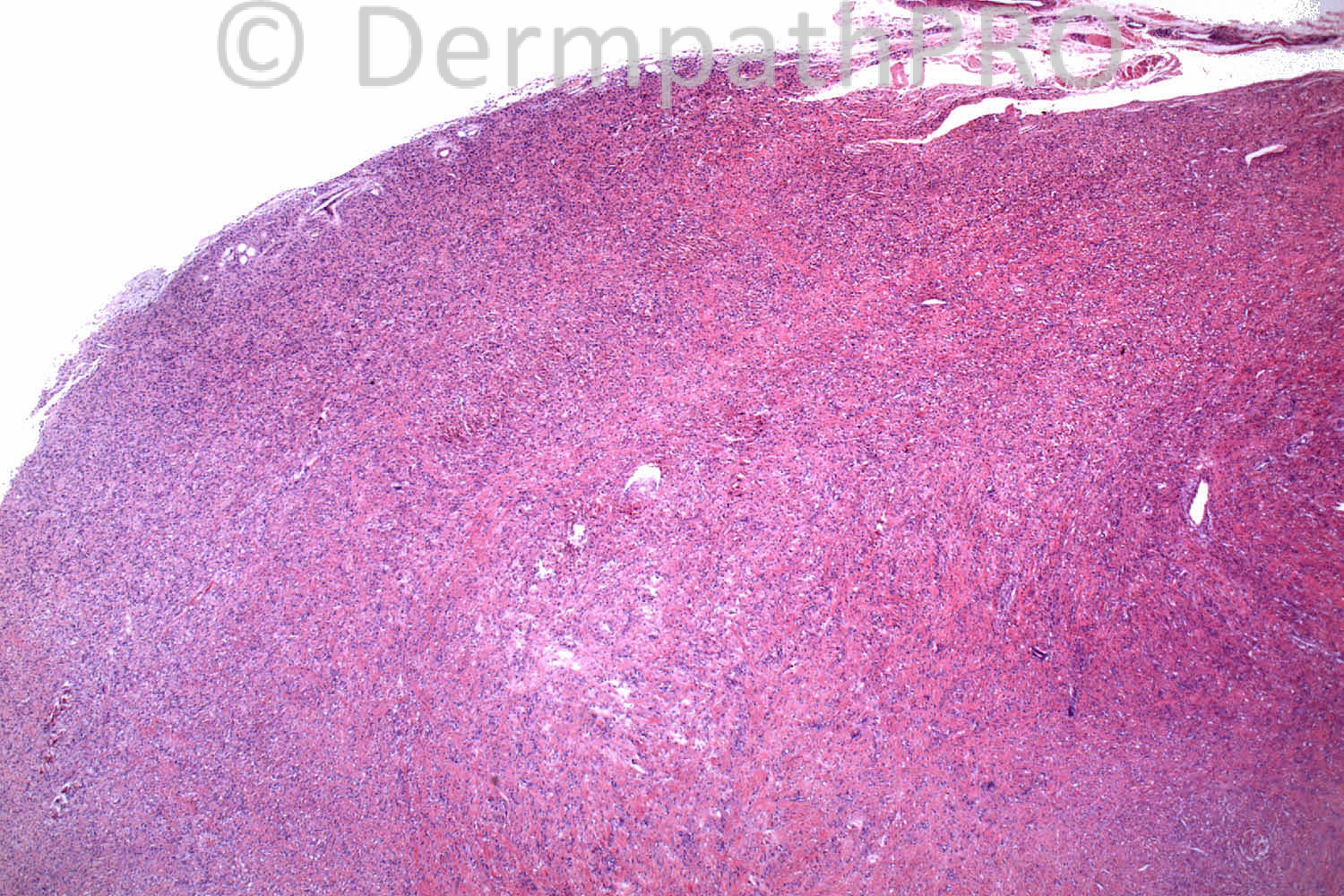
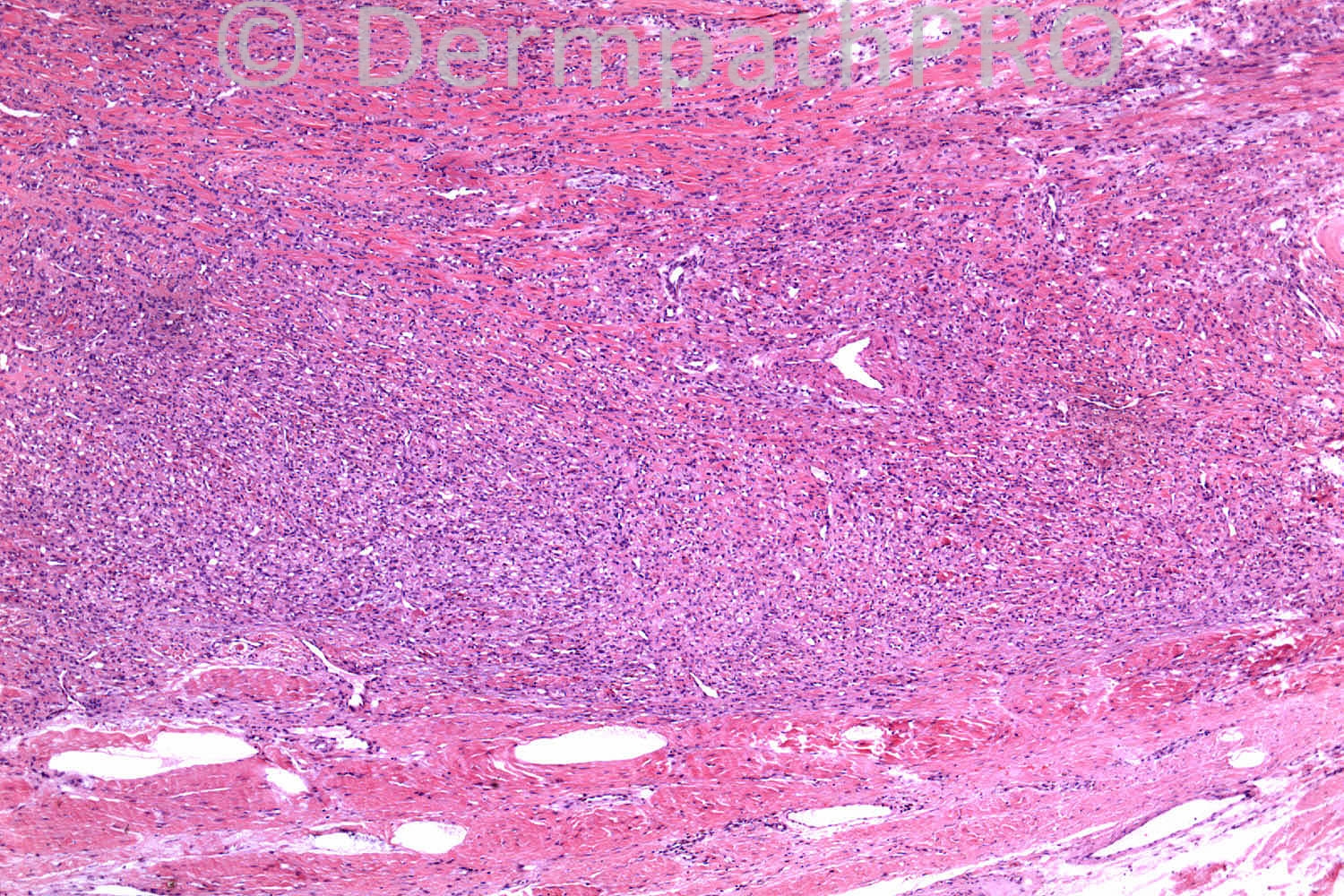
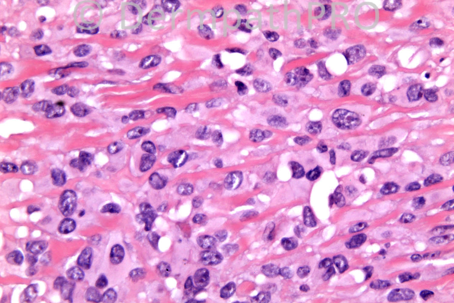
The patient is female 89 years old. Left index finger. ?digital cyst but macro 2.8cm solid mass with lobulated outline and uniform white cut surface.

Case posted by Dr. Richard Carr.


  • Like 1
  Report Record

User Feedback


Guest Maria George

Posted

[color=#444444][font=arial, sans-serif][size=1][b][right]Epithelioid sarcoma.[/right][/b][/size][/font][/color]

Share this comment


Link to comment
share_externally

Arti Bakshi

Posted

A well circumscribed lesion composed of a mixture of oval to epithelioid cells with variable cellularity.
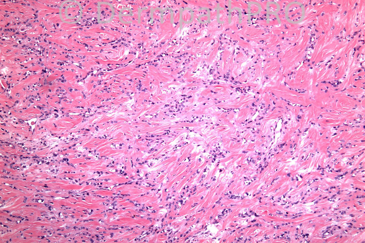
Image 3 shows a more collagenous stroma with a vague storiform pattern. No atypia or mitoses.
Epithelioid perineurioma is a distinct possibility. d/d- epithelioid fibrous histiocytoma and maybe cellular neurothekoma.
Immunos needed, would include EMA and NKIC3 in the panel.

Many thanks Richard for allowing me to look at your fantastic slide collection and your excellent teaching! 4 days were far too less and will be back again.

Share this comment


Link to comment
share_externally

Robledo F. Rocha

Posted

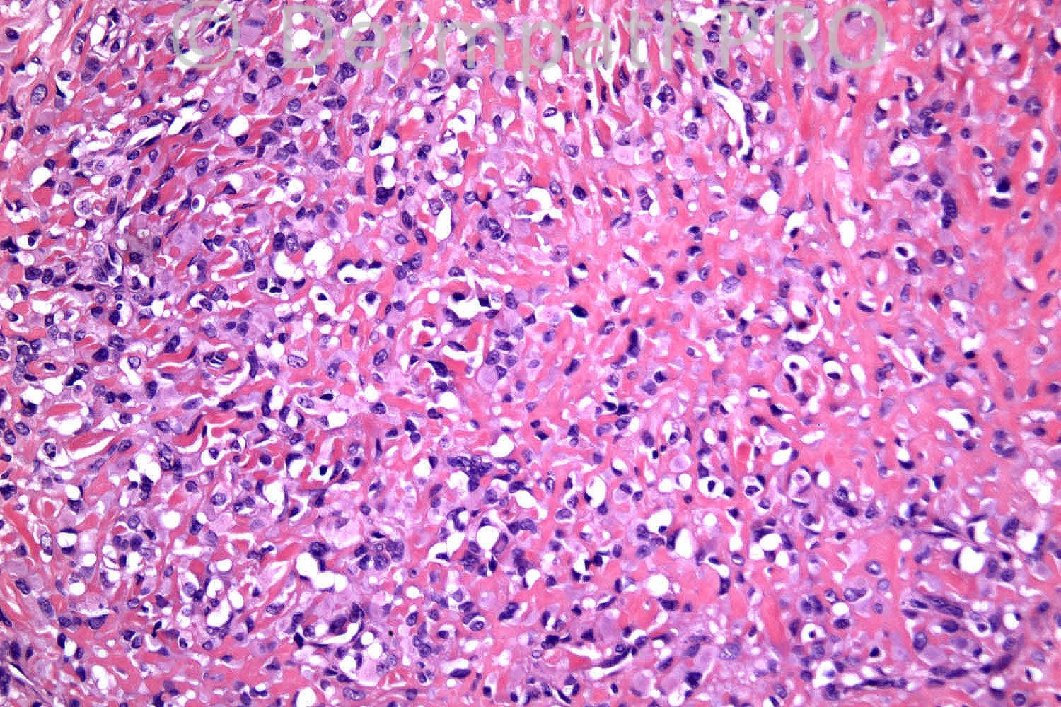
Epithelioid sarcoma is uncommon in old patients. What about epithelioid sarcoma-like hemangioendothelioma? I think intracellular lumens, an evidence of vascular differentiation, can be seen in Picture #6.

Share this comment


Link to comment
share_externally

Dr. Mona Abdel-Halim

Posted

I thought of epithelioid hemangioendothelioma.

Share this comment


Link to comment
share_externally

Dr. Richard Carr

Posted

Dear Arti, thank you so much for the kind comments, it was a pleasure and looking forward to seeing you again soon. This case is difficult!

Share this comment


Link to comment
share_externally

Sasi Attili

Posted

I agree with all the differentials. The digital location, wiry stromal collagen and the vascularity of the tumour (though it isn't quite hemangiopericytomatous) made me wonder about minute synovial sarcoma. However there aren't any mast cells that I can see and the tumour is not quite like the pictures in the books. Looking forward to the answer......

Share this comment


Link to comment
share_externally

Dr. Mona Abdel-Halim

Posted

Re-examining the images especially the third one, reconsidering my diagnosis: epithelioid perineurioma with areas of sclerosing perineurioma....

Share this comment


Link to comment
share_externally

Guest Romualdo

Posted

I agree with Robledo. The cells are epithelioid and somewhat atypical. Although I can't see red blood cells in them, the small round vacuoles apparently compress and distort the nuclei and suggest intracytoplasmic endothelial lumens. I go with epithelioid sarcoma-like hemangioendothelioma and epithelioid hemangioendothelioma as the second possibility.

Share this comment


Link to comment
share_externally

Guest Jim Davie MD

Posted

To add to the above excellent differentials (especially if this is a 'difficult' lesion):
1. Atypical example of Giant cell vs. fibrous tumor of tendon sheath. I would not expect this to be so short on hemosiderin and giant cells simultaneously (or have a white cross section grossly).
2. Cellular dermatofibroma.

Share this comment


Link to comment
share_externally

Dr. Richard Carr

Posted

Okay a little help is required. This is a soft tissue tumour. Usually negative for all markers except vimentin. The clues are the epithelioid cells and the distinctive stroma.

Share this comment


Link to comment
share_externally

Dr. Mona Abdel-Halim

Posted

Ok Dr Carr, from ur comment it means that we all above are wrong. Let me try using the given clues, soft tissue tumor, nests and cords of epithelioid cells, some show cytoplasmic vacuoles, sclerosing hyalinized stroma. Negative for all markers except Vimentin, this must be sclerosing epithelioid fibrosarcoma. Low grade type of fibrosarcoma, usually with little nuclear pleomorphism, Am I right?!!!! I hope so :-)

Share this comment


Link to comment
share_externally

Guest Maria George

Posted

[b]
low-grade fibromyxoid sarcoma (Evans tumor)( LGFS) and [color=#1C2837][font=arial, verdana, tahoma, sans-serif][size=4]sclerosing epithelioid fibrosarcoma (SEF) are thought to be identical or at least very similar.[/size][/font][/color][/b]

Share this comment


Link to comment
share_externally

Dr. Richard Carr

Posted

Yes well done Mona. The diagnosis is sclerosing epithelioid fibrosarcoma.[font=Times New Roman][size=3][color=#000000] [/color][/size][/font]
This is an old case that I sent to Chas Mangham, soft tissue pathologist who made the diagnosis. [font=Times New Roman][size=3][color=#000000] [/color][/size][/font]
Maria is right there does appear to be lesions of LGFMS that have features that are overlapping with SEF
References:
Meis-Kindblom Am J Surg Pathol 1995;19:979-993 and Eydon et al Histopathology 1998; 33: 354-360

Doyle LA, Möller E, Dal Cin P, Fletcher CD, Mertens F, Hornick JL.
MUC4 is a highly sensitive and specific marker for low-grade fibromyxoid sarcoma. Am J Surg Pathol. 2011 May;35(5):733-41.

Doyle LA, Wang WL, Dal Cin P, Lopez-Terrada D, Mertens F, Lazar AJ, Fletcher CD, Hornick JL. MUC4 is a sensitive and extremely useful marker for sclerosing epithelioid fibrosarcoma: association with FUS gene rearrangement. Am J Surg Pathol. 2012 Oct;36(10):1444-51.

Share this comment


Link to comment
share_externally


Join the conversation

You can post now and register later. If you have an account, sign in now to post with your account.

Guest
Add a comment...

×   Pasted as rich text.   Paste as plain text instead

  Only 75 emoji are allowed.

×   Your link has been automatically embedded.   Display as a link instead

×   Your previous content has been restored.   Clear editor

×   You cannot paste images directly. Upload or insert images from URL.

Loading...

×
×
  • Create New...