Jump to content
In this section we have spot diagnoses posted on a daily basis since June 2010, now over 4000! You can review the archived cases and read the suggested diagnoses by users and the final comment by the contributors.
Case are uploaded each week day by 10 am UK time with the correct diagnosis will generally be posted at 8 pm UK time. Why not view the most recent spot diagnosis and proffer a diagnosis?

Case Number : Case 863 - 8th October Posted By: Guest

Please read the clinical history and view the images by clicking on them before you proffer your diagnosis.
Submitted Date :
   (0 reviews)

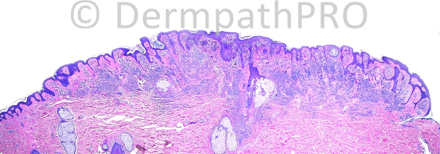
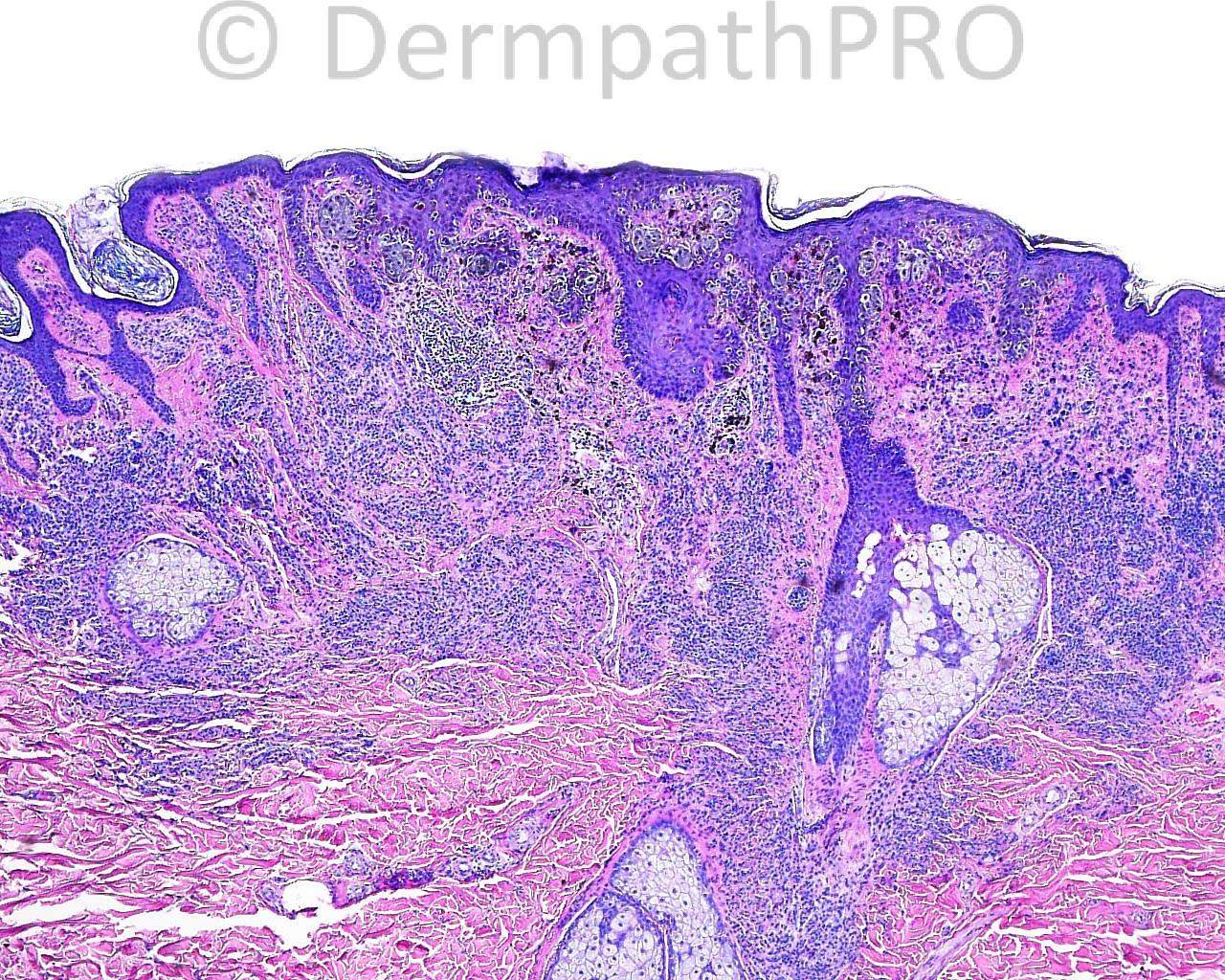
The patient is a 53 year old man with an excision with margin exam of an irregular brown papule taken from the right trapezial neck.

Case posted by Dr. Mark Hurt.


  Report Record

User Feedback


Guest Giovanni Falconieri

Posted

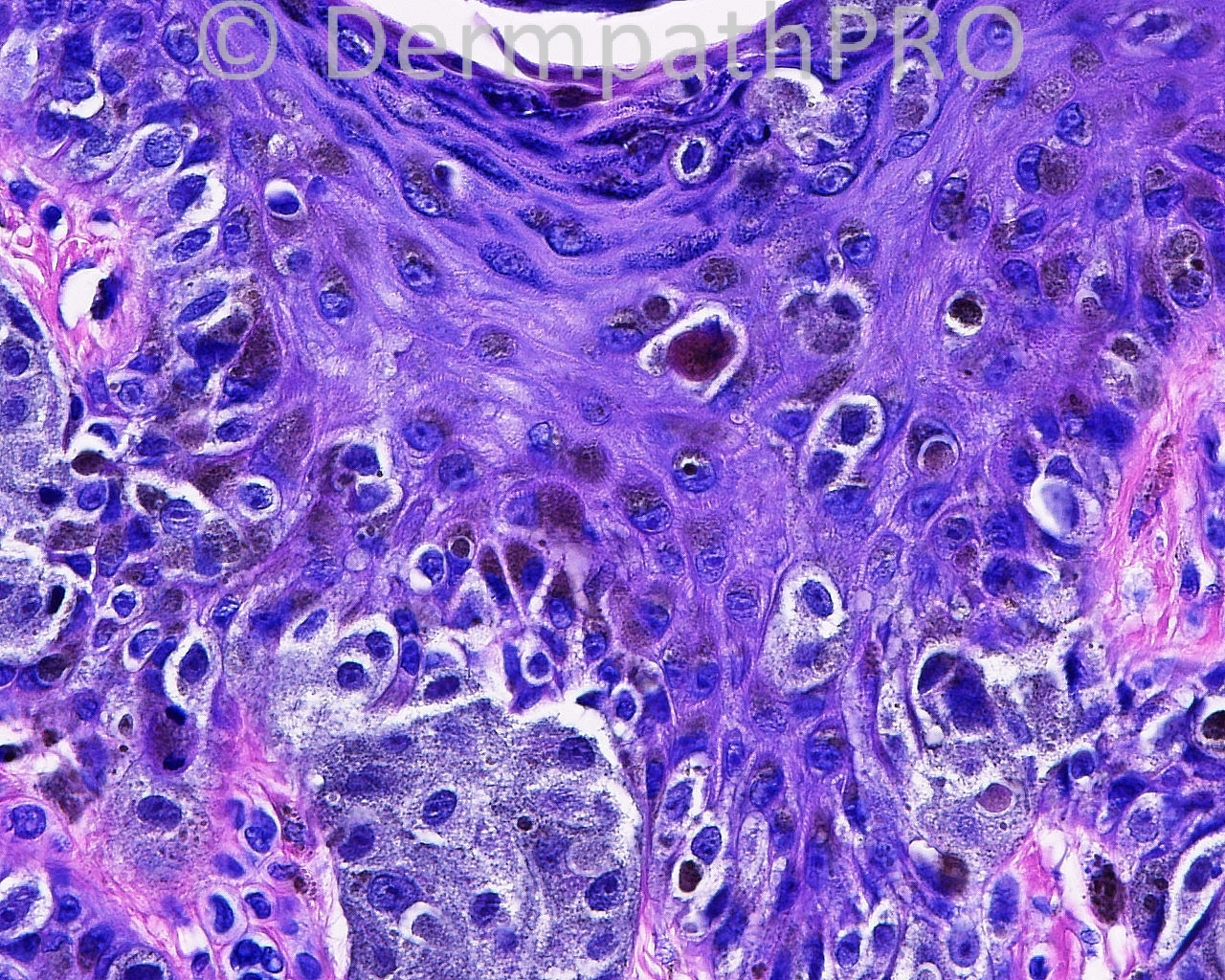
This looks basically a congenital type of nevus harboring a central worrisome focus made up of junctional spitzoid cells. Mostly nested, with dust-like pigment, and limited pagetoid spread. Patchy inflammation and fibrosis within the subjacent dermis. I am inclined to call this benign. My threshold for MM is usually low, especially in back skin lesions from adults, yet in this particular case I would not move too much away from a combined nevus with likely superficial reactive/repair changes

Share this comment


Link to comment
share_externally

Guest Maria George

Posted

I assume that this is a dysplastic congenital compound nevus (nevus on special site)
Niether archetictural no cytological features make me worry about melanoma.

Share this comment


Link to comment
share_externally

Guest Romualdo

Posted

Melanocytic lesions continue to be a great problem to pathologists, certainly to me. In my opinion this is melanoma in situ (pagetoid) over intradermal nevus. I think this grade of pagetoid invasion of the epidermis is not allowed in benign melanocytic proliferations.

Share this comment


Link to comment
share_externally

Hanan Vaknine MD

Posted

I also favor the diagnosis of MMIS arisning in the background of compund nevus with congenital features as did Romualdo (especially if there is a history of "mole with changing charecter")

Share this comment


Link to comment
share_externally

Arti Bakshi

Posted

Agree with Romualdo.
The central area of atypical lentiginous proliferation with pagetoid spread is concerning for melanoma in situ, particularly in the absence of any other obvious cause for this (ie no underlying scar or features to suggest trauma).

Share this comment


Link to comment
share_externally

A simple congenital nevus or a displastic nevus should not have this lentiginous and pagetoid proliferation, so I believe this is a Melanoma "in situ" associated with a nevus.

Share this comment


Link to comment
share_externally

Robledo F. Rocha

Posted

Melanocytic lesion on the back of a man is always a source of concern, mainly if it is an asymmetric one.
In this case, I found a superficial and deep congenital nevus with pagetoid spread restricted to the center of the lesion and appendageal involvment by lentiginous melanocytic proliferation. I think those and others atypical findings are too focal and slight to be construed as evidence of melanoma. They would be better interpreted as signs of trauma or abnormalities due to flexural location.
I would call this Zitelli's nevus.

Share this comment


Link to comment
share_externally

Sasi Attili

Posted

People hedging for this being a melanoma can never go wrong as the lesion irrespective of its nature (benign or malignant or uncertain!) has a low risk of metastasis and one cannot know what the true nature of this tumor is, unless it metastasizes. Therefore even if it does not metastasize, you cannot go wrong if you call it melanoma! Folk saying that this is benign have a small chance of being proved wrong if it does metastasize (terrible luck!).

On that statistical basis it would be wise to say that this is a melanoma based on the worrying central pagetoid spread. [u]You cannot go wrong![/u]

For what it is worth (nothing, in the bigger scheme of things!) in my opinion (based on my teachers and the slides I have seen. [url="https://dermpathpro.com/blog/4/entry-114-the-milk-of-human-kindness-and-the-diagnosis-of-melanocytic-lesions/"]Read Dr. Diwan's excellent blog if you haven't already![/url]), the central pagetoid spread of single cells, in the context of a benign looking symmetrical compound nevus, is not a feature that predicts malignancy.

Share this comment


Link to comment
share_externally

Dr. Mona Abdel-Halim

Posted

I am not expert with melanoma but I am not pleased with the variable sized nests with some atypia, also the follicular lentiginous component and the pagetoid spread and the scattered lentiginous activity that I can perceive some of them as large cells. I will better call it melanoma in situ evolving on top of a nevus.

Share this comment


Link to comment
share_externally

Dr. Richard Carr

Posted

Assuming this is representative I doubt very much I'd sign it out as melanoma in practice - even though as Sasi says one can make a clear-cut case! I would certainly admit to some uncertainty i.e. I do not know the biological significance of the changes but given small size and superficial nature of the lesion (and presuming no evidence of a vertical growth phase dermal component in additional levels) would certainly expect benign outcome (fingers crossed!!). Excision looks complete and no further management is required.

Share this comment


Link to comment
share_externally

Guest Bostjan

Posted

Melanoma in situ in the background of a compound naevus

Share this comment


Link to comment
share_externally

Guest nitinkhirwadkar

Posted

Looks like an in-situ melanoma on a back ground of a nevus. The latter shows some features of a congenital nevus. The melanocytes within the epidermis are non-atrophic.

Share this comment


Link to comment
share_externally

Guest Jim Davie MD

Posted

I agree with philosophy of cannot-rule-out MMIS (or superficially invasive MM if those are similar atypical large Spitzoid cells in the dermis off to the right) with Spitzoid features, arising in a background of congenital pattern nevus. P16 stains may be useful to help support malignant, but only if negative.

A few features that suggest malignancy, taken as a whole rather than just individually:
1. Age of patient. 50+ years is most probably a malignant rather than benign new Spitzoid proliferation.
2. Heterogeneity of cell populations and pigment, and abundant dusty grey cytoplasm.
3. Prominent and chronic pagetoid scatter. I don't think this is trauma related, as pagetoid cells go straight up into the stratum corneum, suggesting a constant, unrelenting pagetoid scatter process.
4. There is a mitotic figure possibly representing a melanocyte (photo resolution limits a proper ID) above the basal layer in the bottom right photo (immediately under the copyright © symbol).
5. Poorly circumscribed and messy lentiginous component.
6. Targeted host chronic inflammatory reaction.

Share this comment


Link to comment
share_externally

Guest Saleem Taibjee

Posted

Clinical history might be helpful i.e. recent change in longstanding lesion. But even so, I favour the beginnings of melanoma-in-situ arising in naevus rather than a combined naevocellular and Spitz based on the limited number of images seen. In any case, the prognosis is likely to be excellent.

Share this comment


Link to comment
share_externally

Robledo F. Rocha

Posted

Dr. Hurt, what did you take into account to diagnose this as melanoma and not as benign congenital nevus with atypical features?

Share this comment


Link to comment
share_externally

Sasi Attili

Posted

I guessed that this would be the answer Dr. Hurt. However, I hope you agree that there is not enough scientific data out there to be objective enough regarding the nature of such melanocytic lesions with conflicting 'opinions'.

Share this comment


Link to comment
share_externally

Guest Saleem Taibjee

Posted

I am coming around to Mark's view that dysplastic naevus is often a problematic term (although I am still guilty of using it in many reports!). I think there are many lesions, often in the elderly, which in reality are low grade melanomas, as confirmed by the clinical behaviour of slow and steady change/increase in size, which to my mind is not really compatible with naevus. Lentiginous melanoma is one example. I think many of these lesions are being signed out as dysplastic naevi by the majority(?) given the lack of significant cytological atypia or clear-cut histological criteria for malignancy, but where clinicopathological correlation including dermoscopic monitoring is helpful. The prognosis is usually excellent, but the reality is still that these are still likely to be melanomas, a small proportion of which become aggressive.

Share this comment


Link to comment
share_externally

Sasi Attili

Posted

Both of you might very well be right. However, the point I am making is that the reason melanocytic lesions are so controversial is because there not enough scientific papers out there to prove or disprove either theory. Thus we are having to rely on the opinion of 'experts' and it is well known that there is huge disparity even between 'experts', when it comes to borderline lesions.

As a scientifically trained doctor it does make me uncomfortable not to be able to be objective about an important diagnosis like a melanoma. In evidence based medicine [url="http://www.eortc.org/sites/default/files/POL019%20v%201.01%20NS.pdf"]'expert opinion' is a level 4/ Grade C (least preferred)[/url]. However, given the complexity of parameters used to judge melanocytic lesions, I am not sure if we are ever going to be able to be more objective about the diagnosis!. It might be safer to call all melanocytic lesions which are not benign- malignant and grade the risk and treat/ follow-up accordingly.

Share this comment


Link to comment
share_externally


Join the conversation

You can post now and register later. If you have an account, sign in now to post with your account.

Guest
Add a comment...

×   Pasted as rich text.   Paste as plain text instead

  Only 75 emoji are allowed.

×   Your link has been automatically embedded.   Display as a link instead

×   Your previous content has been restored.   Clear editor

×   You cannot paste images directly. Upload or insert images from URL.

Loading...

×
×
  • Create New...