Jump to content
In this section we have spot diagnoses posted on a daily basis since June 2010, now over 4000! You can review the archived cases and read the suggested diagnoses by users and the final comment by the contributors.
Case are uploaded each week day by 10 am UK time with the correct diagnosis will generally be posted at 8 pm UK time. Why not view the most recent spot diagnosis and proffer a diagnosis?

Case Number : Case 864 - 9th October Posted By: Guest

Please read the clinical history and view the images by clicking on them before you proffer your diagnosis.
Submitted Date :
   (0 reviews)

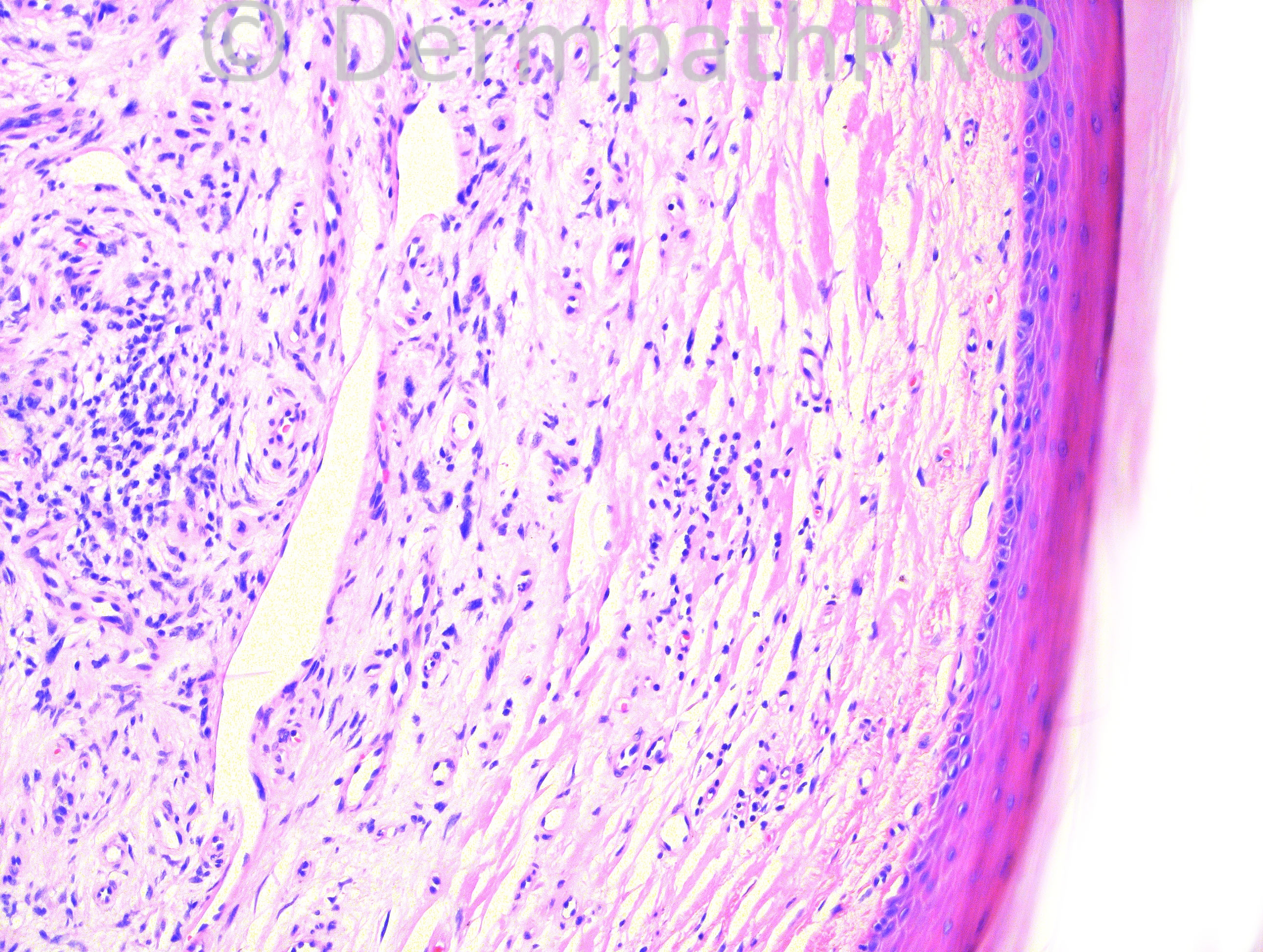
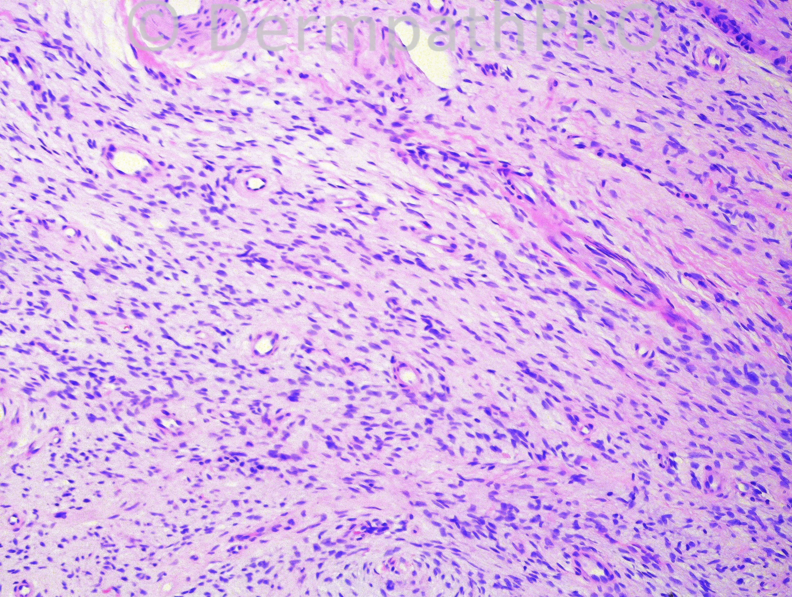
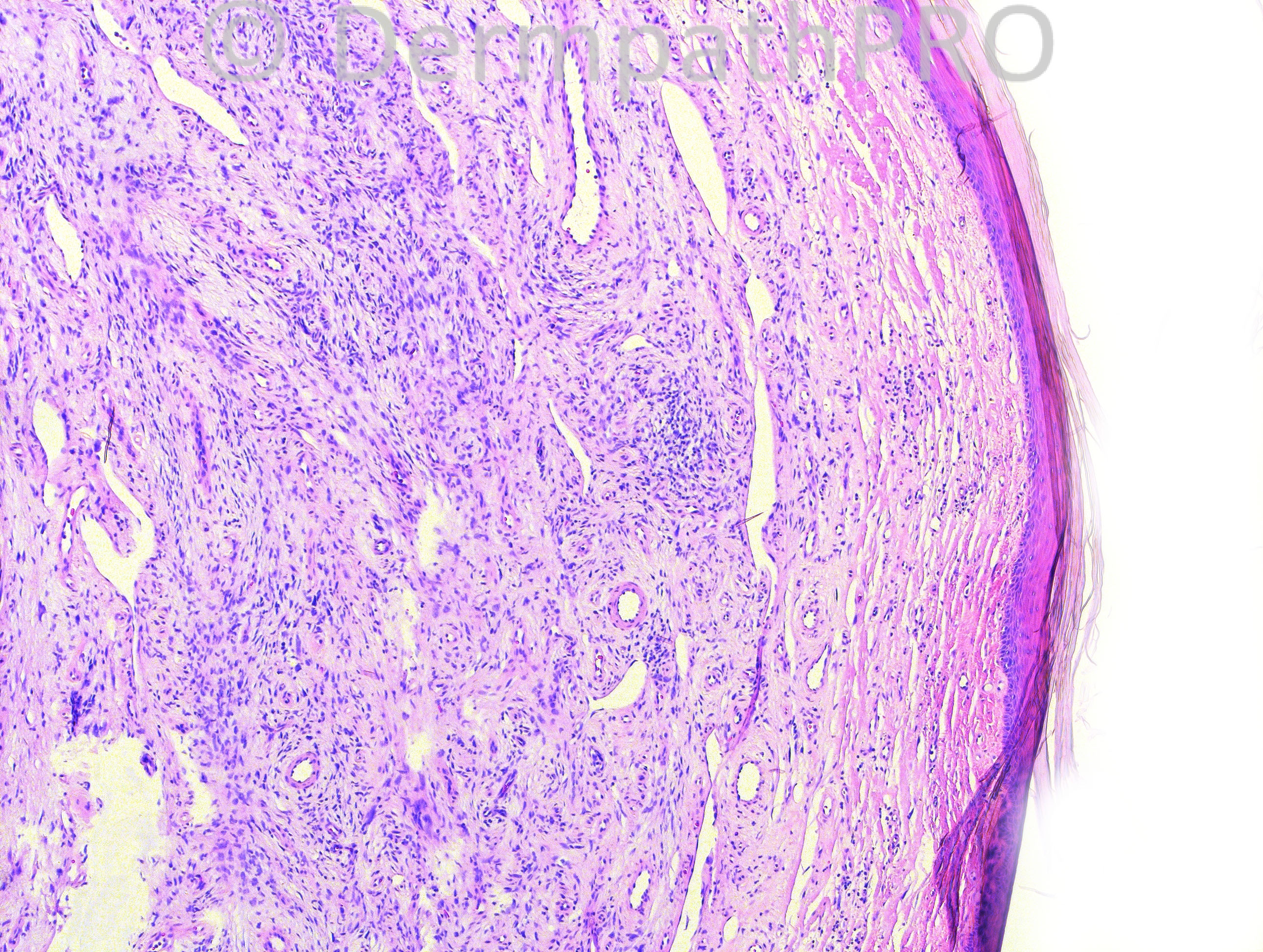
61 year-old male with lesion on right 2nd toe.

Case posted by Dr. Hafeez Diwan


  Report Record

User Feedback


Guest Romualdo

Posted

I think this spindle cell proliferation excessively cellular. I go with cellular digital fibroma.

Share this comment


Link to comment
share_externally

Guest Bostjan

Posted

Agree with superficial acral fibromyxoma (digital fibromyxoma), if lesional cells are CD34 positive.Just in case, not to miss minute synovial sarcoma (usually arising in the web space area) I would also do cytokeratin staining.

Share this comment


Link to comment
share_externally

Guest Josko Bezic

Posted

Digital Fibromyxoma (Superfitial Acral Fibromyxoma). Detailed characterization of 124 cases is recently published in Am J Surg Path (Hollmann TJ, and al. Am J Surg Path 2012; 36:789-798).

Share this comment


Link to comment
share_externally

This diagnosis is fancy nowadays! I agree. Some authors consider Superficial acral fibromyxoma and Cellular digital fibroma the same entity. CD34 must be positive.

Share this comment


Link to comment
share_externally

Guest Marwa Fawzy

Posted

[color=#1C2837][font=arial, verdana, tahoma, sans-serif][size=4]Superfitial Acral Fibromyxoma[/size][/font][/color]

Share this comment


Link to comment
share_externally

Robledo F. Rocha

Posted

Cellular digital fibroma and superficial acral fibromyxoma are overlapping entities.
http://www.ncbi.nlm.nih.gov/m/pubmed/17083699/
A similar case has already been discussed here:
https://dermpathpro.com/spot-diagnosis-1/_/2011-spot-diagnoses/august-2011/case-300-r118

Share this comment


Link to comment
share_externally

Dr. Mona Abdel-Halim

Posted

Agree with Romualdo that the lesion is very cellular, so favor calling it cellular digital fibroma

Share this comment


Link to comment
share_externally

Guest Jim Davie MD

Posted

[font=arial,helvetica,sans-serif]Agree with the above. [color=#1C2837][size=4]Cellular digital fibroma vs. superficial acral fibromyxoma. There is focal multinucleation and vascular proliferation with what appears to be mixed fibrous and mucinous stromal elements. Some hyperchromasia without mitotic activity or inflammation.[/size][/color][/font]

Share this comment


Link to comment
share_externally

Dr. Hafeez Diwan

Posted

I called this cellular digital fibroma, and as pointed out above that this overlaps with superficial acral fibromyxoma.

Share this comment


Link to comment
share_externally

Guest Mazen charaf iddin

Posted

may be it's ture fibromyxoma, if CD 34 + , but this bundling with some faint focal aggregation like in neural tumor suggest diffuse neurofibroma, and myxoid change can be seen in all soft tissue lesion

Share this comment


Link to comment
share_externally


Join the conversation

You can post now and register later. If you have an account, sign in now to post with your account.

Guest
Add a comment...

×   Pasted as rich text.   Paste as plain text instead

  Only 75 emoji are allowed.

×   Your link has been automatically embedded.   Display as a link instead

×   Your previous content has been restored.   Clear editor

×   You cannot paste images directly. Upload or insert images from URL.

Loading...

×
×
  • Create New...