Jump to content
In this section we have spot diagnoses posted on a daily basis since June 2010, now over 4000! You can review the archived cases and read the suggested diagnoses by users and the final comment by the contributors.
Case are uploaded each week day by 10 am UK time with the correct diagnosis will generally be posted at 8 pm UK time. Why not view the most recent spot diagnosis and proffer a diagnosis?

Case Number : Case 866 - 14th October Posted By: Guest

Please read the clinical history and view the images by clicking on them before you proffer your diagnosis.
Submitted Date :
   (0 reviews)

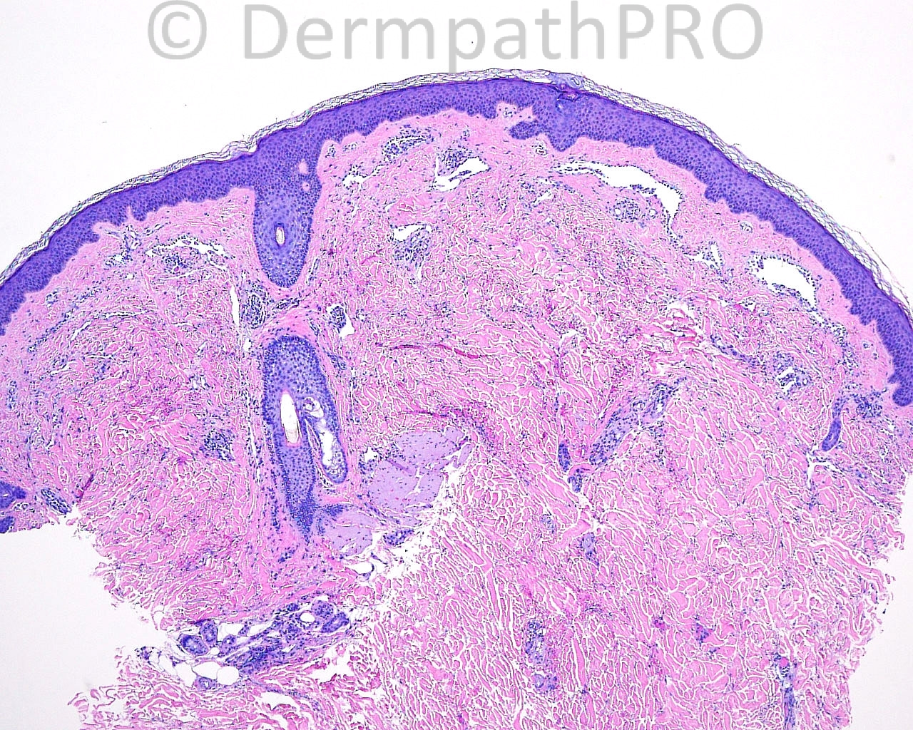
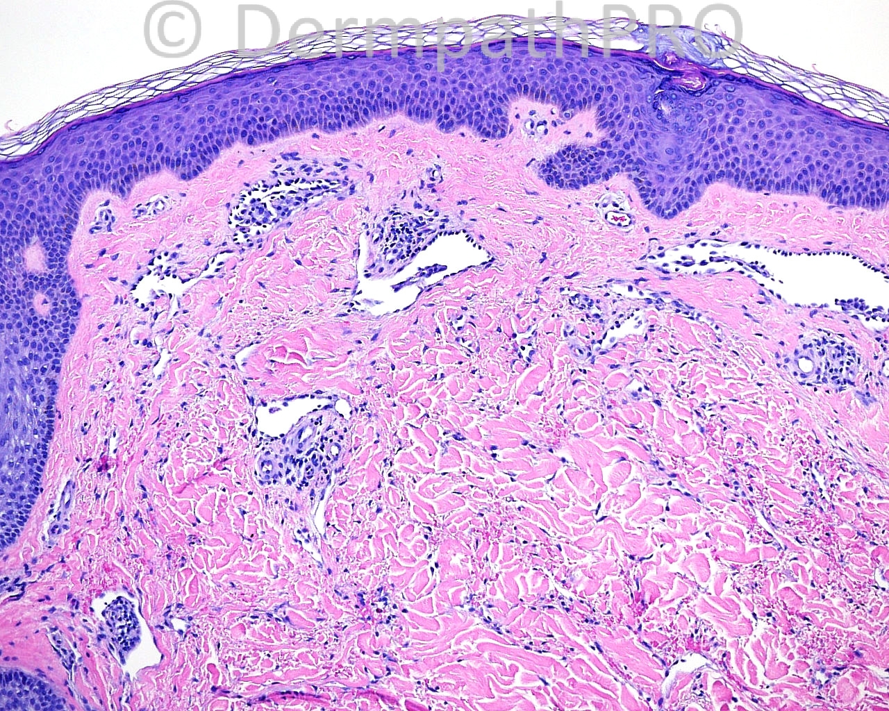
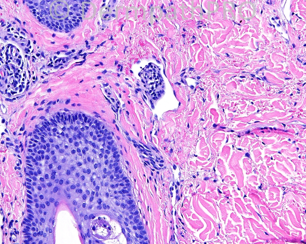
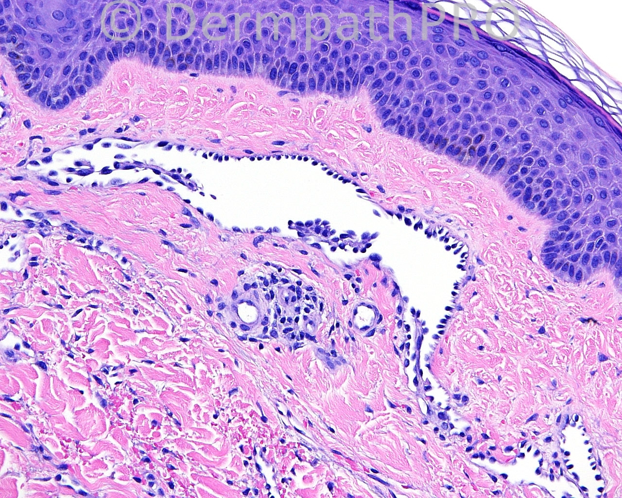
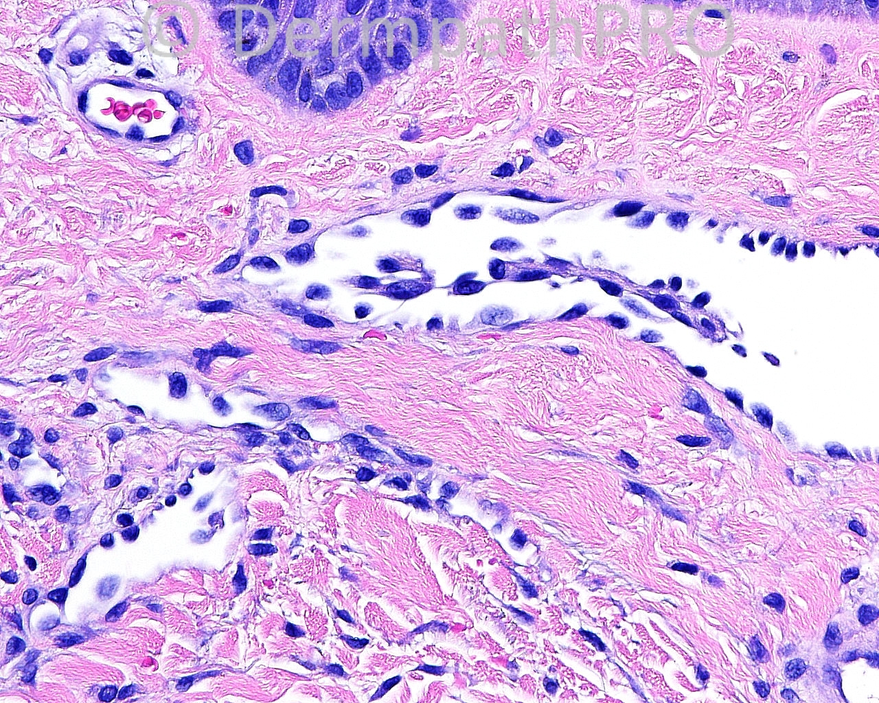
The patient is a 36 year old white woman with a punch biopsy taken from the lateral aspect of the right lower leg.

Case posted by Dr. Mark Hurt.


  Report Record

User Feedback


Dr. Mona Abdel-Halim

Posted

Papillary intralymphatic angioendothelioma (PILA).

Share this comment


Link to comment
share_externally

Hobnail haemangioma Targetoid hemangiotic haemangioma

Share this comment


Link to comment
share_externally

Guest Romualdo

Posted

Hobnail (Dabska-retiform) hemangioendothelioma.

Share this comment


Link to comment
share_externally

Sasi Attili

Posted

[color=#1C2837][font=arial, verdana, tahoma, sans-serif][size=4]Hobnail haemangioma[/size][/font][/color]

Share this comment


Link to comment
share_externally

Robledo F. Rocha

Posted

Hobnail hemangioma / targetoid hemosiderotic hemangioma. Age and site are typical. Also there's no rete testis-like appearance, a feature that would drive to the diagnosis of retiform hemangioendothelioma, and the presence of plump endothelial cells lining intraluminal papillary projections rules out patch Kaposi's sarcoma.

Share this comment


Link to comment
share_externally

Dr. Mona Abdel-Halim

Posted

What about the lymphocytic collections adjoining the dilated spaces?

Share this comment


Link to comment
share_externally

Guest Romualdo

Posted

I think this is a hobnail hemangioendothelioma, not a hobnail hemangioma (targetoid hemosiderotic hemangioma). I am not able to see hemosiderin. By the other hand there are elongated vascular spaces lined by hobnailed cells with large hyperchromatic nuclei. There are no red blood cells within these vascular spaces. Papillary projections and lymphocytes are also seen. So, I agree with Mona: I think this is a hobnail hemangioendothelioma, a term that encompasses retiform hemangioendothelioma and papillary intralymphatic angioendothelioma (PILA).

Share this comment


Link to comment
share_externally

Guest Jim Davie MD

Posted

[font=arial,helvetica,sans-serif][size=4]Retiform hemangioendothelioma, with differential only slightly less favouring hobnail hemangioma (targetoid hemosiderotic hemangioma).

Histology and clinical history is equivocal for these two, in my opinion.
Proliferation of collapsed spaces with papillary lumenal architecture. Proliferation is predominantly superficial > mid reticular dermis (assuming that there is no extension into the subcutis; not shown). Extravasated red cells, sparse patchy lymphocytic inflammatory response, no epidermal pseudocarcinomatous changes. No significant hemosiderin (common).[/size][/font]

Share this comment


Link to comment
share_externally


Join the conversation

You can post now and register later. If you have an account, sign in now to post with your account.

Guest
Add a comment...

×   Pasted as rich text.   Paste as plain text instead

  Only 75 emoji are allowed.

×   Your link has been automatically embedded.   Display as a link instead

×   Your previous content has been restored.   Clear editor

×   You cannot paste images directly. Upload or insert images from URL.

Loading...

×
×
  • Create New...