Jump to content
In this section we have spot diagnoses posted on a daily basis since June 2010, now over 4000! You can review the archived cases and read the suggested diagnoses by users and the final comment by the contributors.
Case are uploaded each week day by 10 am UK time with the correct diagnosis will generally be posted at 8 pm UK time. Why not view the most recent spot diagnosis and proffer a diagnosis?

Case Number : Case 867 - 15th October Posted By: Guest

Please read the clinical history and view the images by clicking on them before you proffer your diagnosis.
Submitted Date :
   (0 reviews)

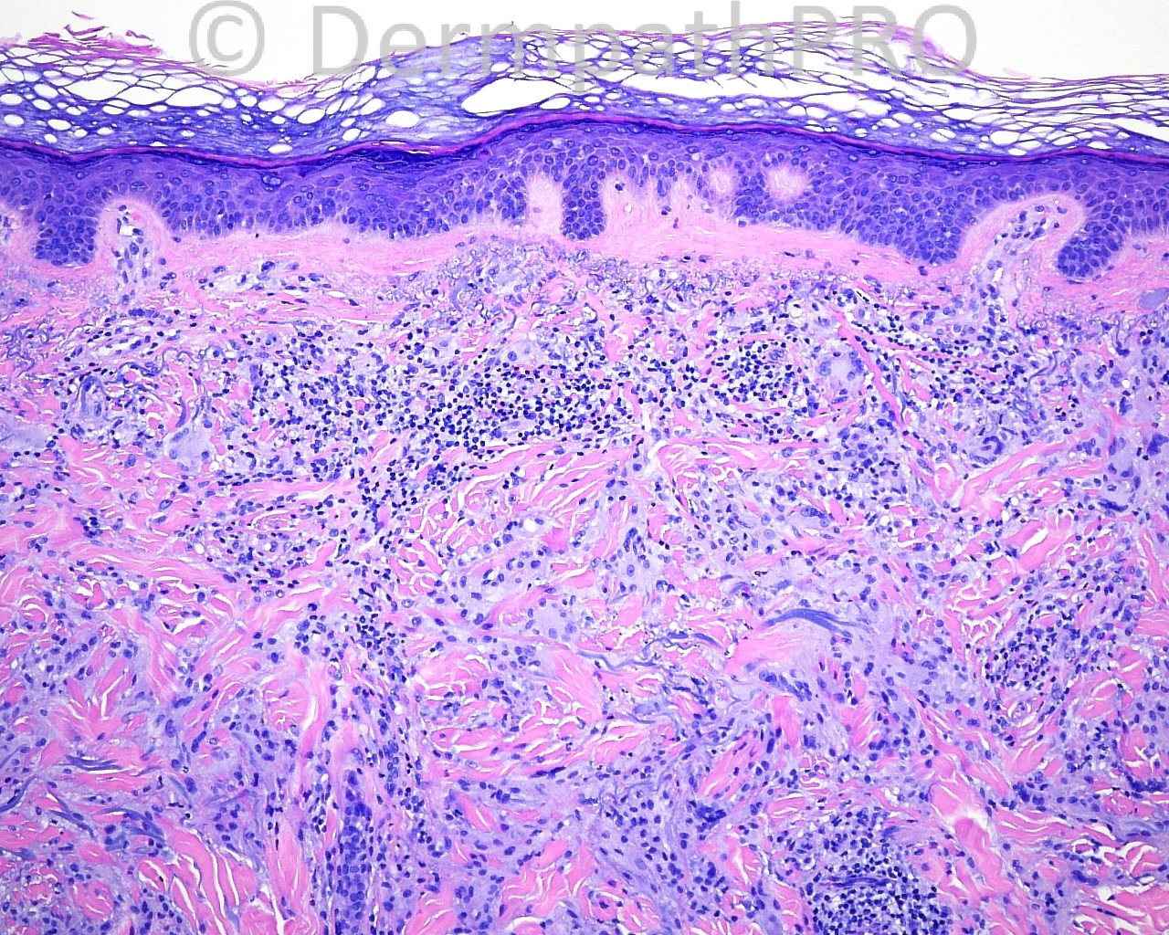
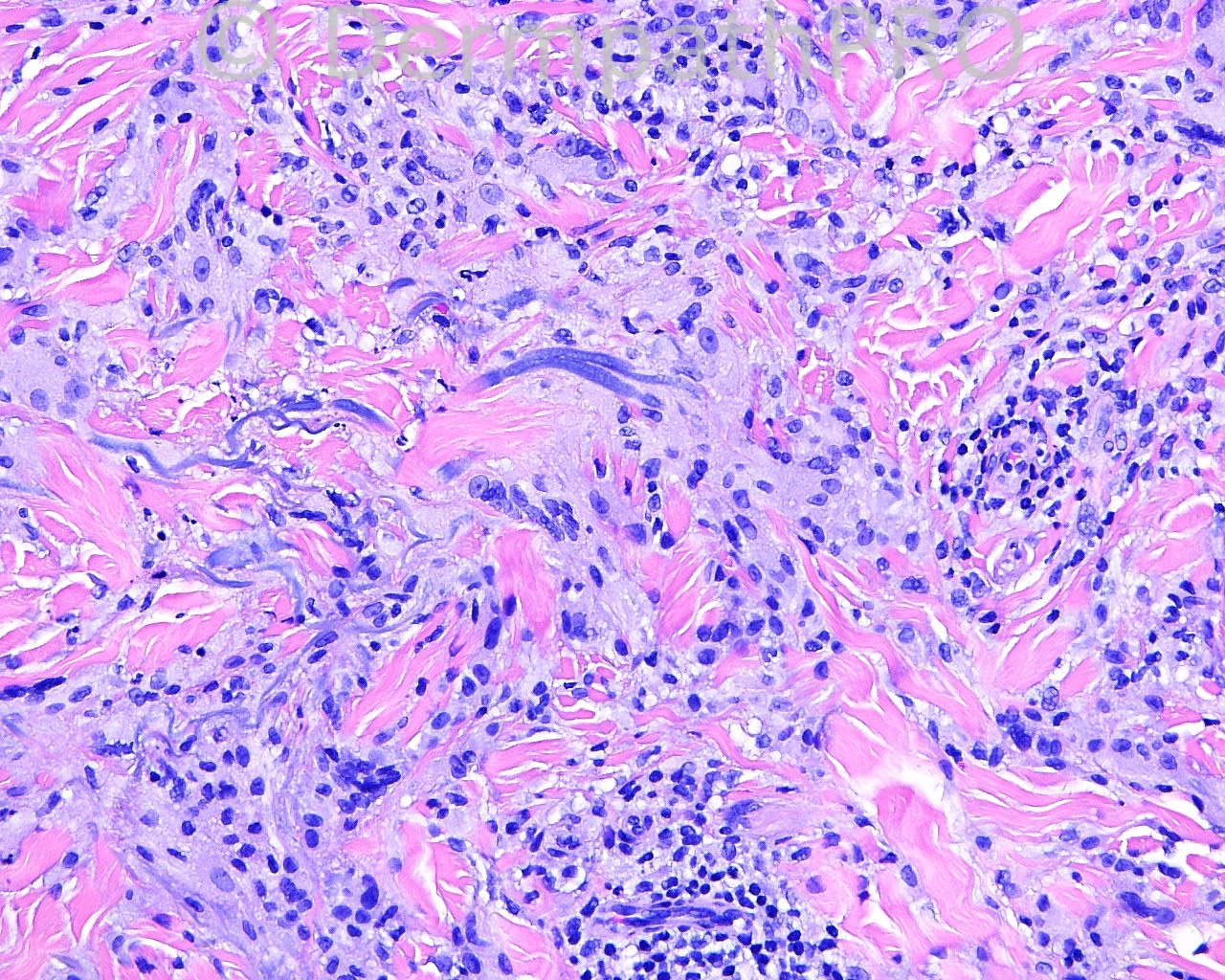
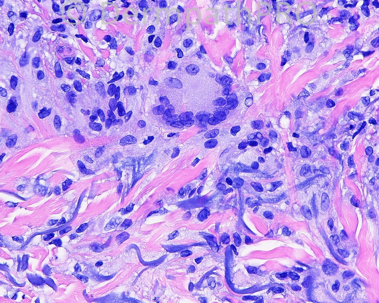
The patient is a 54 year old woman who has a few papules that are skin colored and depressed in the center on the dorsum of her hands. A shave biopsy is taken from the dorsum of the left hand.

Case posted by Dr. Mark Hurt.


  Report Record

User Feedback


Sasi Attili

Posted

[color=#003366][font=arial, sans-serif][size=4][b][i]Annular elastolytic giant cell granuloma[/i][/b][/size][/font][/color]

Share this comment


Link to comment
share_externally

[b]Actinic granuloma / [b]Annular elastolytic giant cell granuloma.[/b][/b]

Share this comment


Link to comment
share_externally

Guest Romualdo

Posted

Granuloma annulare at severely sun damaged skin.

Share this comment


Link to comment
share_externally

Dr. Mona Abdel-Halim

Posted

Agree with actinic granuloma/annular elastolytic giant cell granuloma

Share this comment


Link to comment
share_externally

Robledo F. Rocha

Posted

Agree with Romualdo. Actinic granuloma and its synonyms merely represent granuloma annulare in sun-damaged skin.

Share this comment


Link to comment
share_externally

Guest Jim Davie MD

Posted

Annular elastolytic giant-cell granuloma (actinic granuloma annulare).

Although usually on head/neck, clinical history of multifocal, bilateral involvement of both dorsal hand would be more consistent with diagnosis of AGA, and help exclude a mycobacterial process. Histologically, there is a giant cell component, and distinct absence of mucin or necrobiotic/altered collagen that would suggest a primary granuloma annulare with incidental (bystander) elastolytic component. I would suspect that an elastin stain would show reduced stainable elastin within the central areas.

Share this comment


Link to comment
share_externally


Join the conversation

You can post now and register later. If you have an account, sign in now to post with your account.

Guest
Add a comment...

×   Pasted as rich text.   Paste as plain text instead

  Only 75 emoji are allowed.

×   Your link has been automatically embedded.   Display as a link instead

×   Your previous content has been restored.   Clear editor

×   You cannot paste images directly. Upload or insert images from URL.

Loading...

×
×
  • Create New...