Jump to content
In this section we have spot diagnoses posted on a daily basis since June 2010, now over 4000! You can review the archived cases and read the suggested diagnoses by users and the final comment by the contributors.
Case are uploaded each week day by 10 am UK time with the correct diagnosis will generally be posted at 8 pm UK time. Why not view the most recent spot diagnosis and proffer a diagnosis?

Case Number : Case 870 - 18th October Posted By: Guest

Please read the clinical history and view the images by clicking on them before you proffer your diagnosis.
Submitted Date :
   (0 reviews)

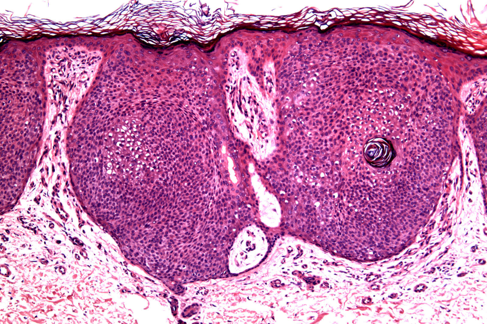
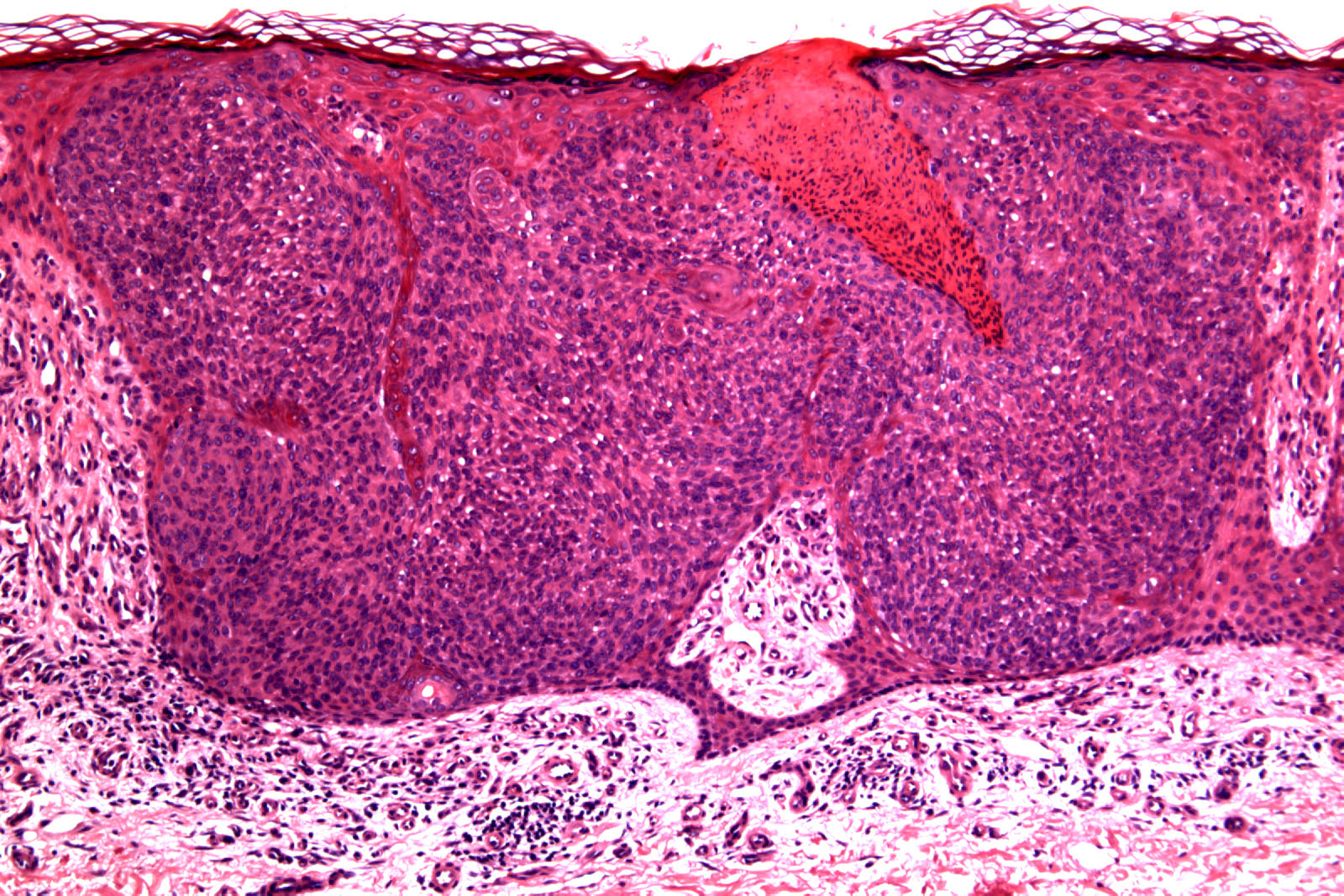
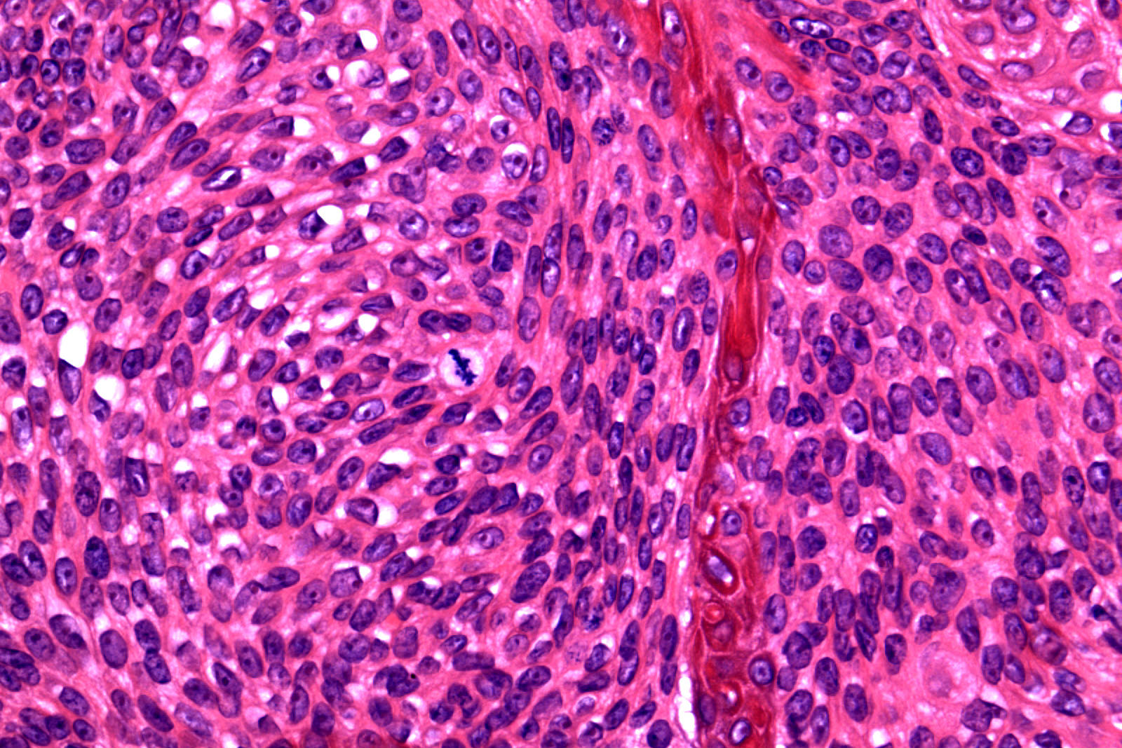
78 year-old female. Rigth shin, lesion 5 years duration, 19 x 15mm.


Case posted by Dr. Richard Carr.

  Report Record

User Feedback


Guest Saleem Taibjee

Posted

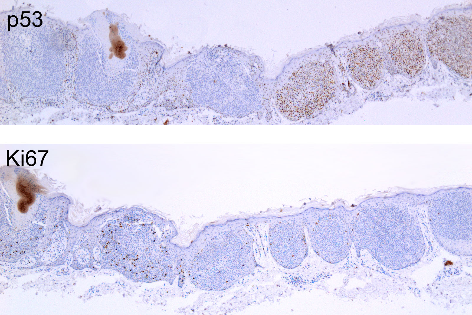
Dysplasia (bowenoid), as supported by the p53, arising within a clonal lesion. The distinct areas would suggest there is a pre-existing clonal seborrhoeic keratosis.

Share this comment


Link to comment
share_externally

Guest Maria George

Posted

There are things to be considered here but first 2 for me are hidraacanthoma simplex and seborheric keratosis.

Share this comment


Link to comment
share_externally

Robledo F. Rocha

Posted

In-situ porocarcinoma arising within a preexisting hidroacanthoma simplex.
I found very instructive the arrangement of the pictures because they show comparatively two populations of cells with distinct morphological appearance, distinct mitotic index and distinct expression of p53.

Share this comment


Link to comment
share_externally

The neoplastic cells looks Poroid and It seems to have a ductular formation on the lower third of the second picture. Also, as far as I know, p53 is not a good marker to differentiate between an Ecrine poroma and a Porocarcinoma.
Ki-67 index and the histologic findings are compatible with an Hidroacanthoma simplex.

Share this comment


Link to comment
share_externally

Don't think this lesion is dysplastic. Agree with differential diagnoses above of clonal seb k or intraepidermal eccrine poroma.

Share this comment


Link to comment
share_externally

Guest Romualdo

Posted

I agree with Dr. Saleem: clonal Bowen's disease arising in the context of clonal seborrheic keratosis. The slightly higher cellularity and the parakeratotic/ necrotic focus seen in Picture # 2 together with p53 positivity seen in the majority of the cells in the four nests at the right of Picture # 5 compared with the almost complete negativity in the cell nests at the left are in concert with this diagnosis. An apparent paradox: the p53 positive cells are less Ki67 positive than expected.

Share this comment


Link to comment
share_externally

Guest Jim Davie MD

Posted

Clonal seborrheic keratosis. Clinical is helpful....stable 5 year old lesion.

Agree with the above excellent observations. I agree with Romualdo and Mark that the P53 strong-positive population (right side of IPEX photos) is also paradoxically Ki67 negative, whereas the P53-negative population (left side of IPEX photos) shows a higher (but still unimpressive) Ki67 staining percentage. These two populations show identical low- and high-power features, lacking high grade atypia or (significant) mitotic or apoptotic activity. The cells show simple clear-cell change (clustered in the center of one nest, in the low power first photo)...without definitive poroid differentiation, in my opinion.

This case seems to be a good example of an assertion that P53 and Ki67 are of limited value in differentiating poroma vs. porocarcinoma vs. clonal SK.

[size=3][color=#008080] link: [/color][b][url="http://www.ncbi.nlm.nih.gov/pubmed/21587033"][color=#008080]http://www.ncbi.nlm....pubmed/21587033[/color][/url][/b]
[color=#008080] The "intraepidermal epithelioma" revisited: immunohistochemical study of the Borst-Jadassohn phenomenon.
Lora V, Chouvet B, Kanitakis J. Am J Dermatopathol. 2011 Jul;33(5):492-7. doi: 10.1097/DAD.0b013e3181fe6f9[/color][/size]

[size=3][color=#008080][i] ..."Cell nests in cSK, cBD, hidroacanthoma simplex (HS), and porocarcinoma (PC) showed strong expression of epidermal growth factor-receptors (EGF-R), Ki-67, p63, and p53."[/i][/color][/size]

Share this comment


Link to comment
share_externally

Dr. Richard Carr

Posted

Shame the nice low powers I also submitted were not included that showed the two lesions incombination. The top two left images are clonal seborrhoeic keratosis (no glands or ducts other than trapped acrosyringia on H&Es and IHC). The top two right images are subtle clonal bowen's disease. What you all missed though was the null p53 on the left supports the diagnosis of Bowen's. The p53 staining on the right is wild type (+30%/++20%, unfortunately the photography greatly over estimates the staining) in a seborrhoeic keratosis. The Ki67 is a little more in the bowen's but not particularly helpful diagnostically other than showing the lesions are different. While diffuse strong p53 can be supportive of Bowen's it is only about 40 to 50% sensitive in my experience. A completely negative p53 (null phenotype) is uncommon but also supports a diagnosis of Bowen's/malignancy. p16 can also be helpful (frequently diffusely strongly positive in Bowen's, negative in seborrhoeic keratosis) but not in this particular case.

Share this comment


Link to comment
share_externally


Join the conversation

You can post now and register later. If you have an account, sign in now to post with your account.

Guest
Add a comment...

×   Pasted as rich text.   Paste as plain text instead

  Only 75 emoji are allowed.

×   Your link has been automatically embedded.   Display as a link instead

×   Your previous content has been restored.   Clear editor

×   You cannot paste images directly. Upload or insert images from URL.

Loading...

×
×
  • Create New...