Jump to content
In this section we have spot diagnoses posted on a daily basis since June 2010, now over 4000! You can review the archived cases and read the suggested diagnoses by users and the final comment by the contributors.
Case are uploaded each week day by 10 am UK time with the correct diagnosis will generally be posted at 8 pm UK time. Why not view the most recent spot diagnosis and proffer a diagnosis?

Case Number : Case 872 - 22nd October Posted By: Guest

Please read the clinical history and view the images by clicking on them before you proffer your diagnosis.
Submitted Date :
   (0 reviews)

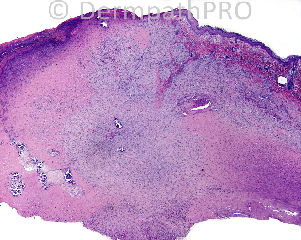
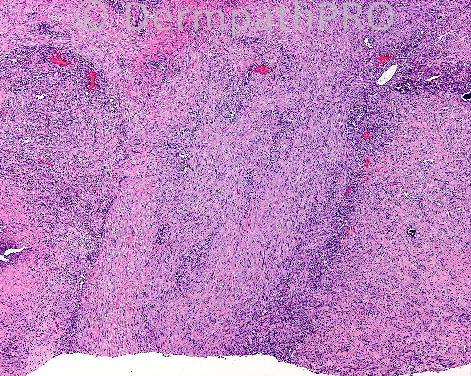
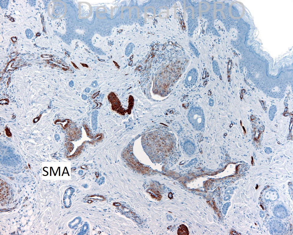
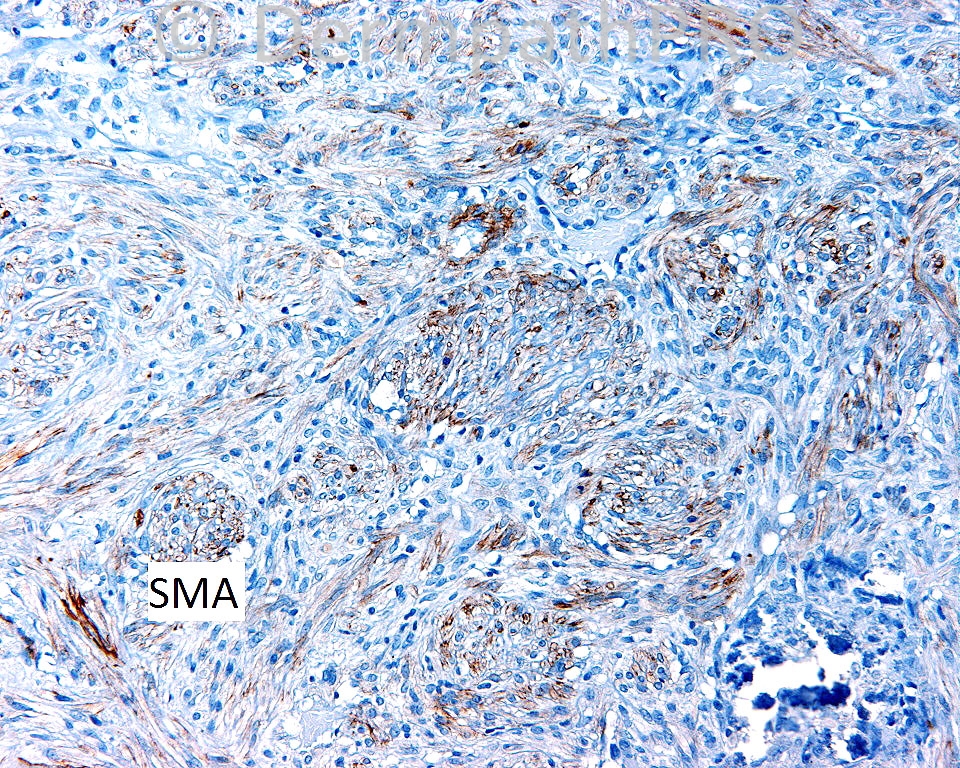
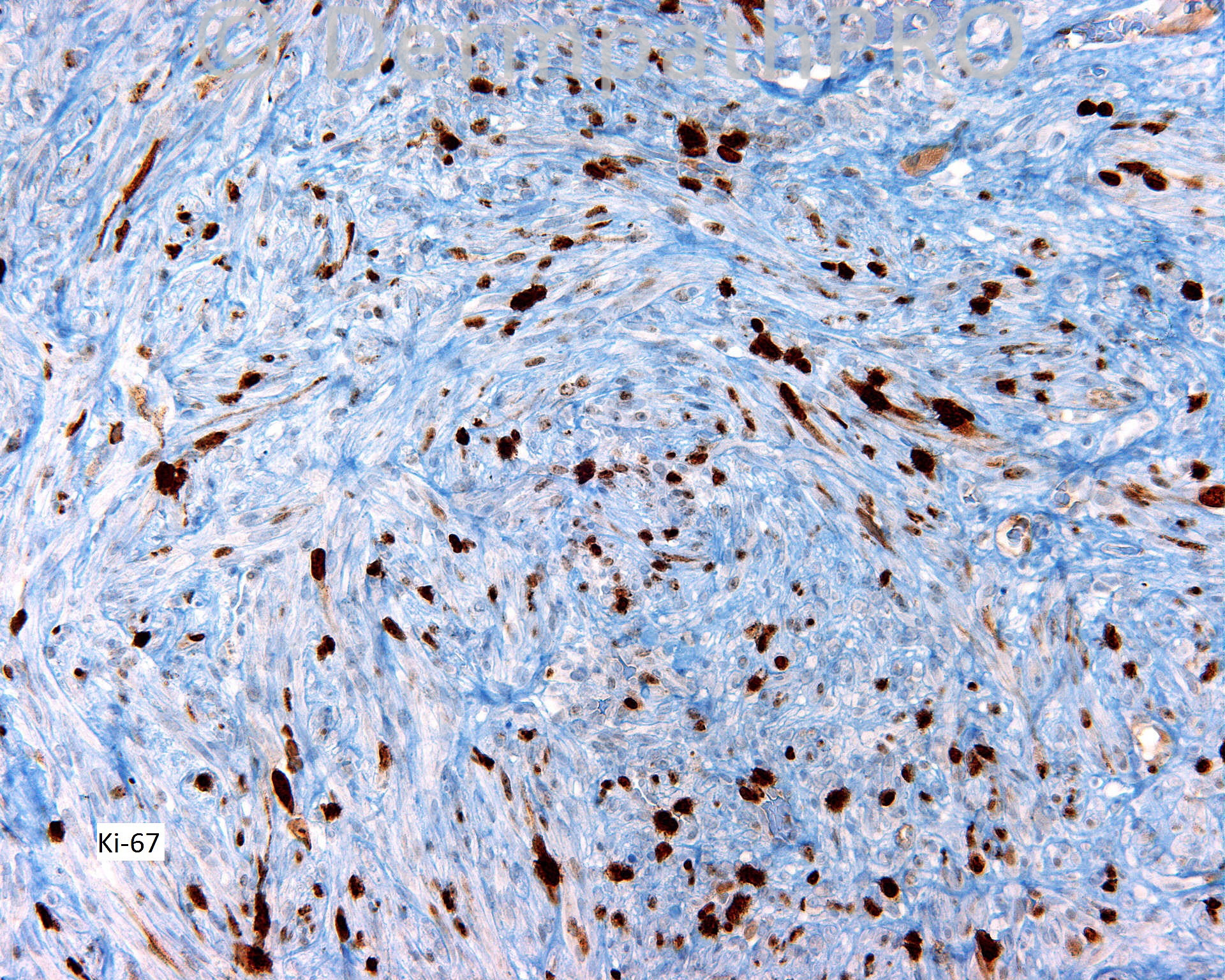
The patient is a five day old baby with a shave biopsy from the left thigh.

Case posted by Dr. Mark Hurt.


  Report Record

User Feedback


Guest Saleem Taibjee

Posted

myofibroma - biphasic pattern is supportive. The infant would need a work-up to assess for multiple lesions including imaging.

Share this comment


Link to comment
share_externally

Sasi Attili

Posted

[color=#1C2837][font=arial, verdana, tahoma, sans-serif][size=4]infantile myofibromatosis/ myofibroma. Nice case....[/size][/font][/color]

Share this comment


Link to comment
share_externally

Robledo F. Rocha

Posted

[size=5][color=#000000][font=Palatino Linotype, serif]Solitary myofibroma displaying beautiful intravascular myofibroblastic clusters and foci of calcification. Increased expression of Ki-67 is expect in a few-days-aged infant.[/font][/color]
[font=Palatino Linotype, serif][color=#000000]As pointed out by Dr. Taibjee, it would be prudent if an in-depth investigation were carried out in order to look for [/color][color=#000000]multiple lesions [/color][color=#000000]involv[/color][color=#000000]ing[/color][color=#000000] superficial soft tissues, [/color][color=#000000]bones,[/color] [color=#000000]and[/color] [color=#000000]visceral organs, once solitary lesions are not common away from head and neck.[/color][/font][/size]

Share this comment


Link to comment
share_externally

Mark A. Hurt MD

Posted

Yes, this was a myofibroma, and the only one in this patient, to my knowledge. I really learned a lot from this patient, as I misdiagnosed this initially as leiomyosarcoma. I was overly concerned with the massive necrosis in the lesion, as it is rarely shown or discussed. After reading Stout's early descriptions of the condition, and with the aid of some soft tissue experts, I was set on the right course.

Share this comment


Link to comment
share_externally

Robledo F. Rocha

Posted

[quote name='Mark A. Hurt MD' timestamp='1382468747']
I was overly concerned with the massive necrosis in the lesion, as it is rarely shown or discussed.
[/quote]
Very interesting, Dr. Hurt. Although there's no supportive data from the literature, I got a sense that the more frequent the intravascular growth, the more extensive the necrosis. Perhaps this observation may spark interest among researchers.
Thanks for share this nice case.

Share this comment


Link to comment
share_externally

Mark A. Hurt MD

Posted

[quote name='Robledo F. Rocha' timestamp='1382494388']

Very interesting, Dr. Hurt. Although there's no supportive data from the literature, I got a sense that the more frequent the intravascular growth, the more extensive the necrosis.
Thanks for share this nice case.
[/quote]

Robledo,

You may well be right, although it is speculative. Perhaps it is an age-related phenomenon, as I get the distinct impression that necrosis may simply be a finding in very young children, as I don't often see necrosis in myofibromas presenting in adults. Yet, they are uncommon enough that my experience simply may not be rich enough to understand the natural history or the spectrum of these lesions. Anyone who has more experience is welcome to chime in!

Mark

Share this comment


Link to comment
share_externally


Join the conversation

You can post now and register later. If you have an account, sign in now to post with your account.

Guest
Add a comment...

×   Pasted as rich text.   Paste as plain text instead

  Only 75 emoji are allowed.

×   Your link has been automatically embedded.   Display as a link instead

×   Your previous content has been restored.   Clear editor

×   You cannot paste images directly. Upload or insert images from URL.

Loading...

×
×
  • Create New...