Jump to content
In this section we have spot diagnoses posted on a daily basis since June 2010, now over 4000! You can review the archived cases and read the suggested diagnoses by users and the final comment by the contributors.
Case are uploaded each week day by 10 am UK time with the correct diagnosis will generally be posted at 8 pm UK time. Why not view the most recent spot diagnosis and proffer a diagnosis?

Case Number : Case 874 - 24th October Posted By: Guest

Please read the clinical history and view the images by clicking on them before you proffer your diagnosis.
Submitted Date :
   (0 reviews)

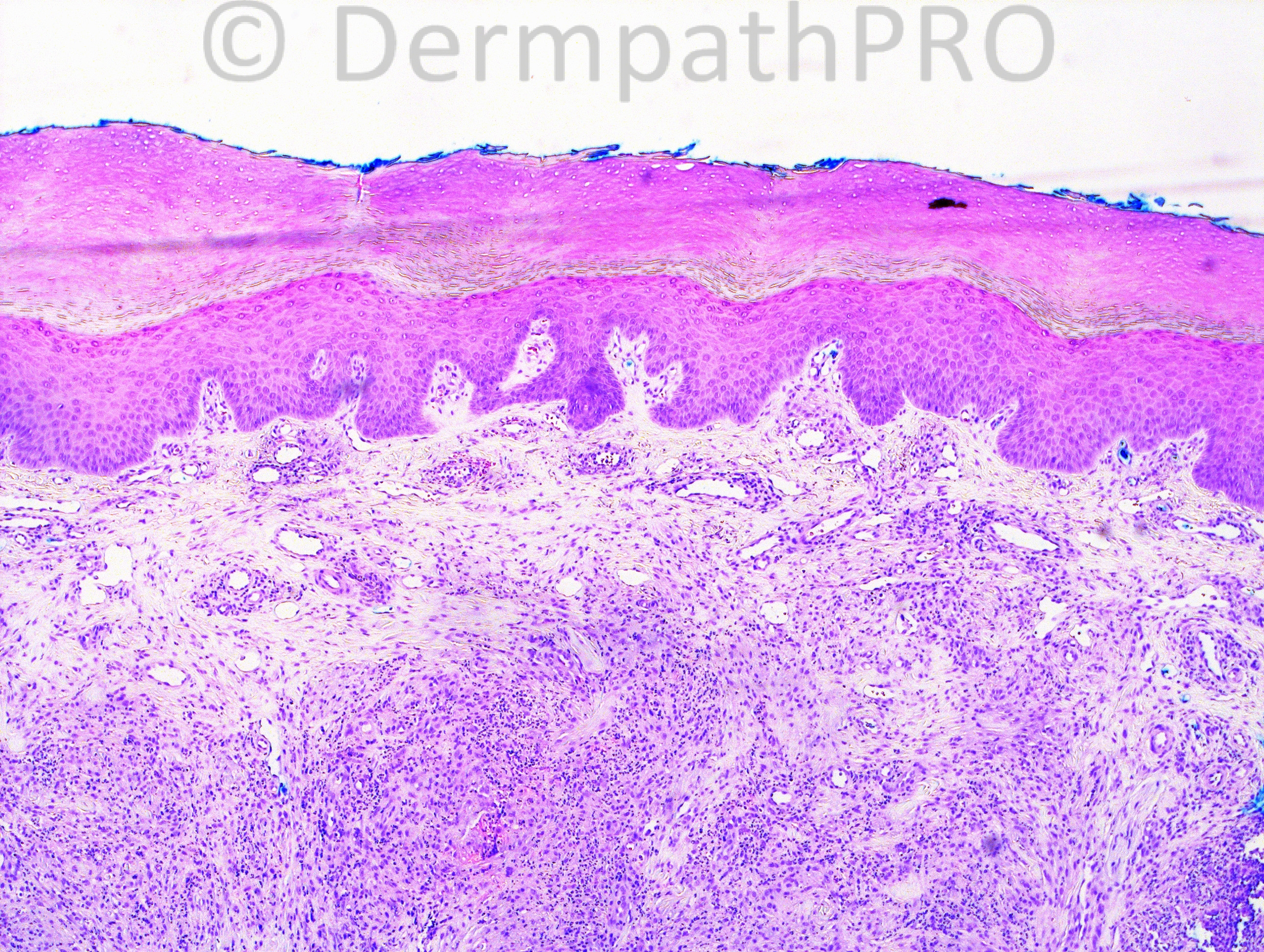
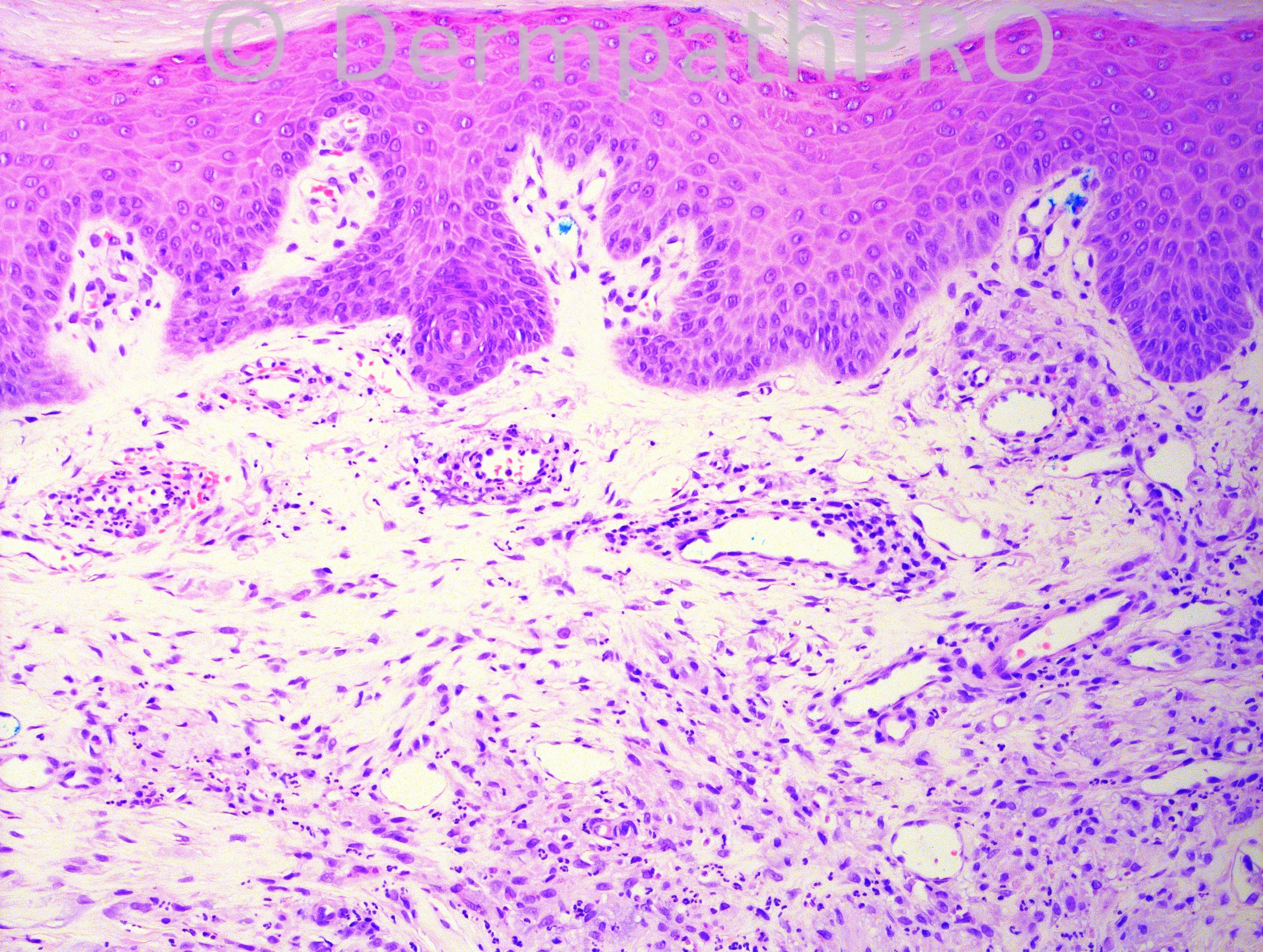
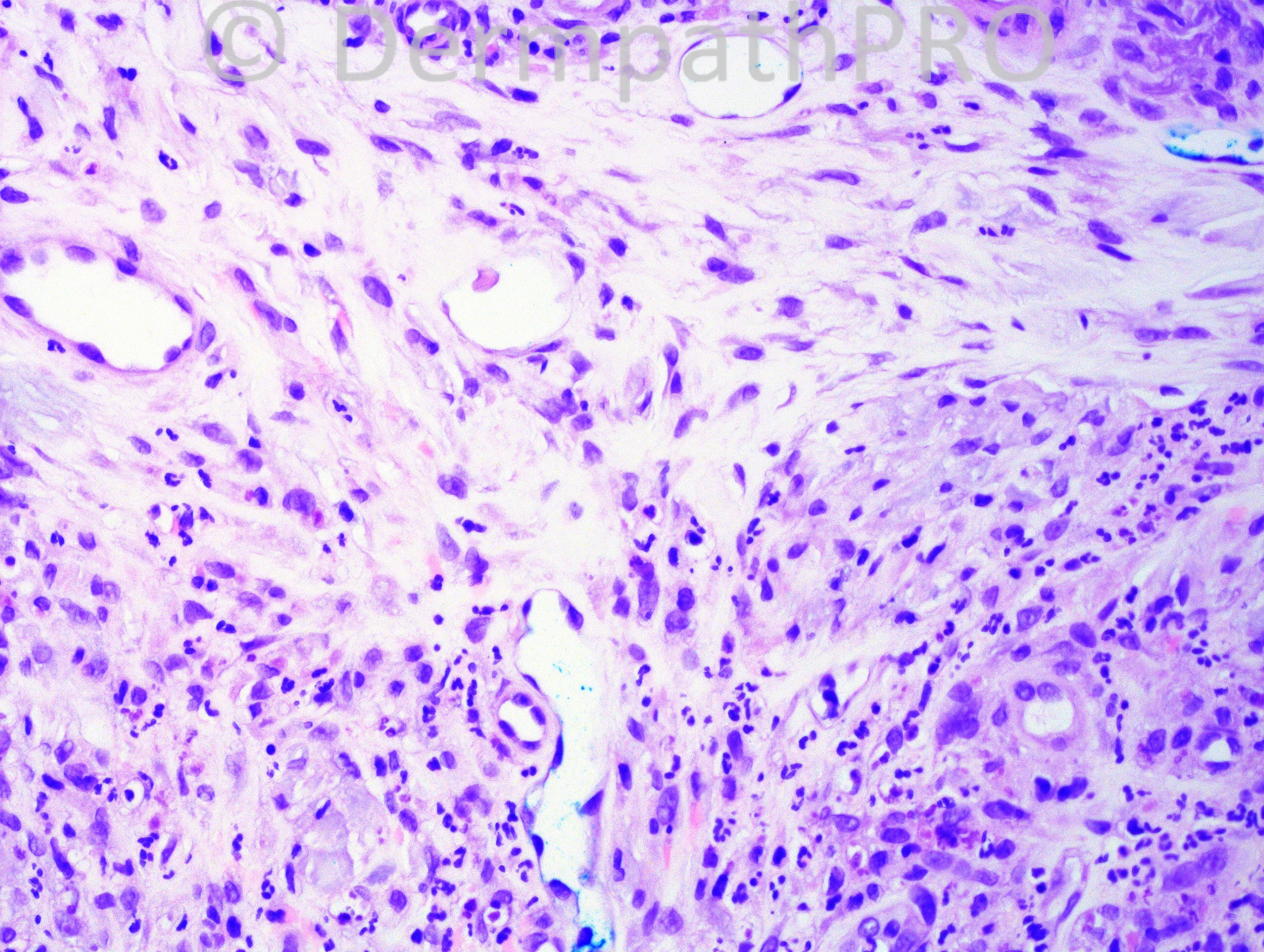
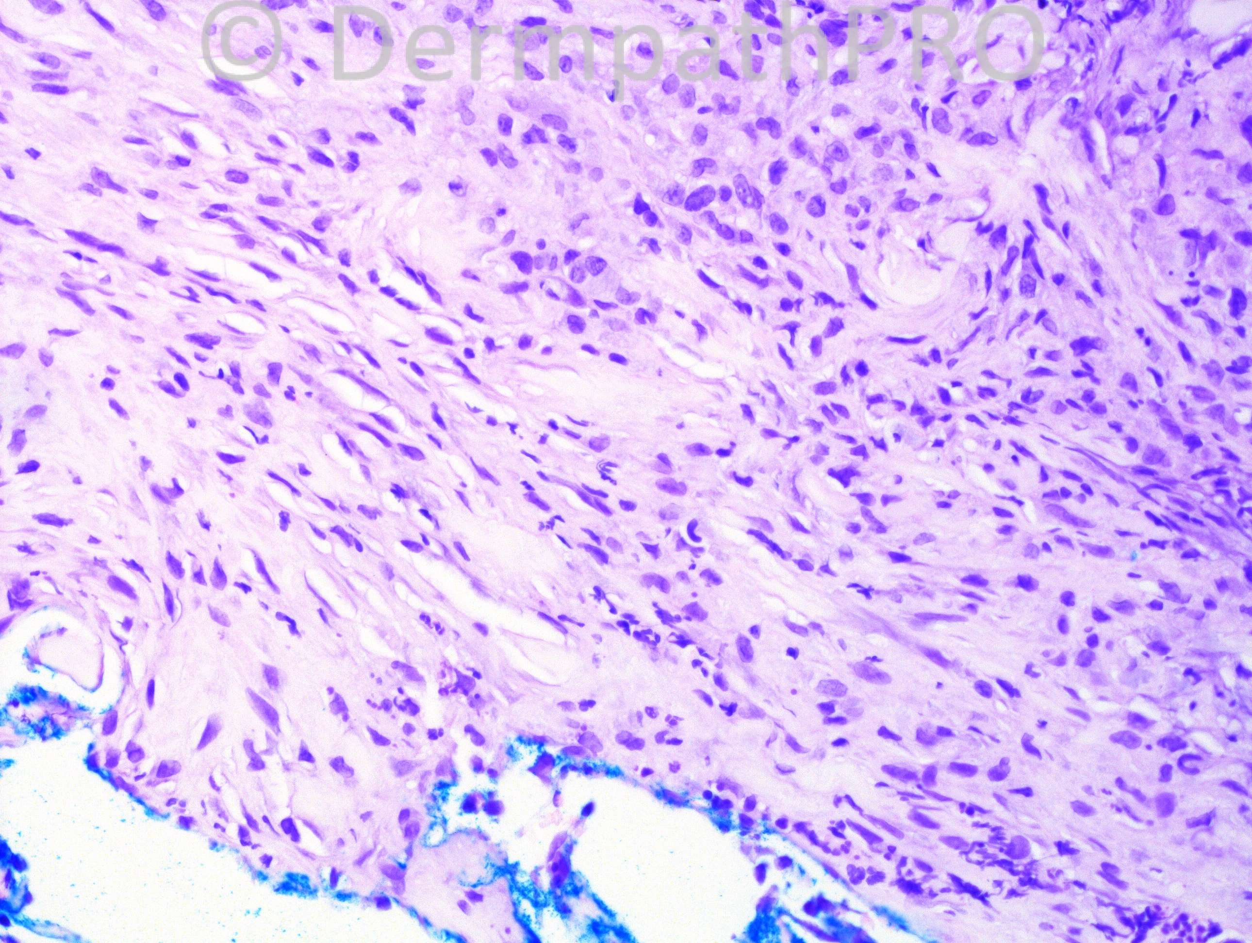
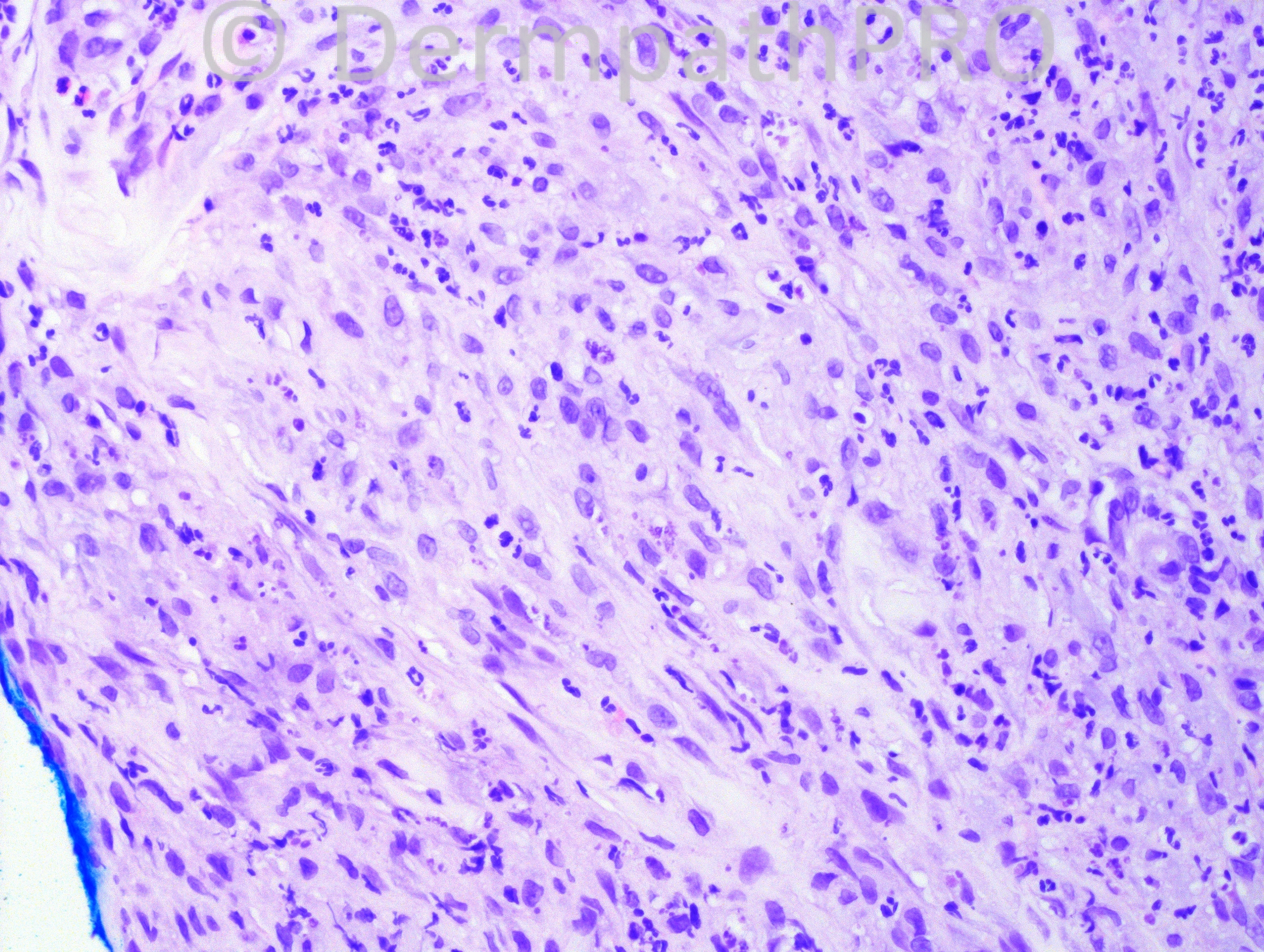
Thumb biopsy from a 62-year-old male. The clinical is “rule out sarcoid vs Rosai-Dorfman.”

Case posted by Dr. Hafeez Diwan


  Report Record

User Feedback


Guest Romualdo

Posted

I think this is neutrophilic dermatosis of the hands. It is necessary to exclude infectious causes.

Share this comment


Link to comment
share_externally

Guest Mazen charaf iddin

Posted

Histiocytes .. Xantomatous reaction, or mast cells .... mastocytosis??

Share this comment


Link to comment
share_externally

Sasi Attili

Posted

The neutrophilic infiltrate in my opinion is not dense enough for Neutrophilic dermatosis of the hands, BUt CPC needed. How about EED?

Share this comment


Link to comment
share_externally

I think the correct diagnosis is Erythema elevatum diutinum. I have seen cases of EED very similar and at the same location (extensor surfaces at acral location). As EED can show red to violaceous nodular configuration, It can be confused with RD and Sarcoidosis.

Share this comment


Link to comment
share_externally

Guest Romualdo

Posted

I did not remind EED, suggested by Dr. Sasi. It is an excellent possibility. The fibroblastic proliferation better shown in the last two images fits well with this diagnosis.

Share this comment


Link to comment
share_externally

Guest Juan Carlos Garcés, Ecuador

Posted

Agree with localized chronic fibrosing vasculitis

Share this comment


Link to comment
share_externally

Robledo F. Rocha

Posted

[font="Arial, sans-serif"][size="4"]Erythema elevatum diutinum characterized by granulation tissue containing neutrophilic-rich inflammatory infiltrate and vascular damage.[/size][/font]

Share this comment


Link to comment
share_externally

Guest Jim Davie MD

Posted

Agree with EED.
- Sarcoidosis and RD would be a common clinical consideration in EED, as Igor indicated.
- Mid-dermal inflammatory fibrosis, leukocytoclastic debris, neutrophil-predominant infiltrate without significant eosinophils, extravasated red cells, and hint of vasculitis in the first low-power image.

Share this comment


Link to comment
share_externally

Guest Saleem Taibjee

Posted

Agree EED, relatively acute stage. Associated paraproteinaemia should be excluded.

Share this comment


Link to comment
share_externally


Join the conversation

You can post now and register later. If you have an account, sign in now to post with your account.

Guest
Add a comment...

×   Pasted as rich text.   Paste as plain text instead

  Only 75 emoji are allowed.

×   Your link has been automatically embedded.   Display as a link instead

×   Your previous content has been restored.   Clear editor

×   You cannot paste images directly. Upload or insert images from URL.

Loading...

×
×
  • Create New...