Jump to content
In this section we have spot diagnoses posted on a daily basis since June 2010, now over 4000! You can review the archived cases and read the suggested diagnoses by users and the final comment by the contributors.
Case are uploaded each week day by 10 am UK time with the correct diagnosis will generally be posted at 8 pm UK time. Why not view the most recent spot diagnosis and proffer a diagnosis?

Case Number : Case 875 - 25th October Posted By: Guest

Please read the clinical history and view the images by clicking on them before you proffer your diagnosis.
Submitted Date :
   (0 reviews)

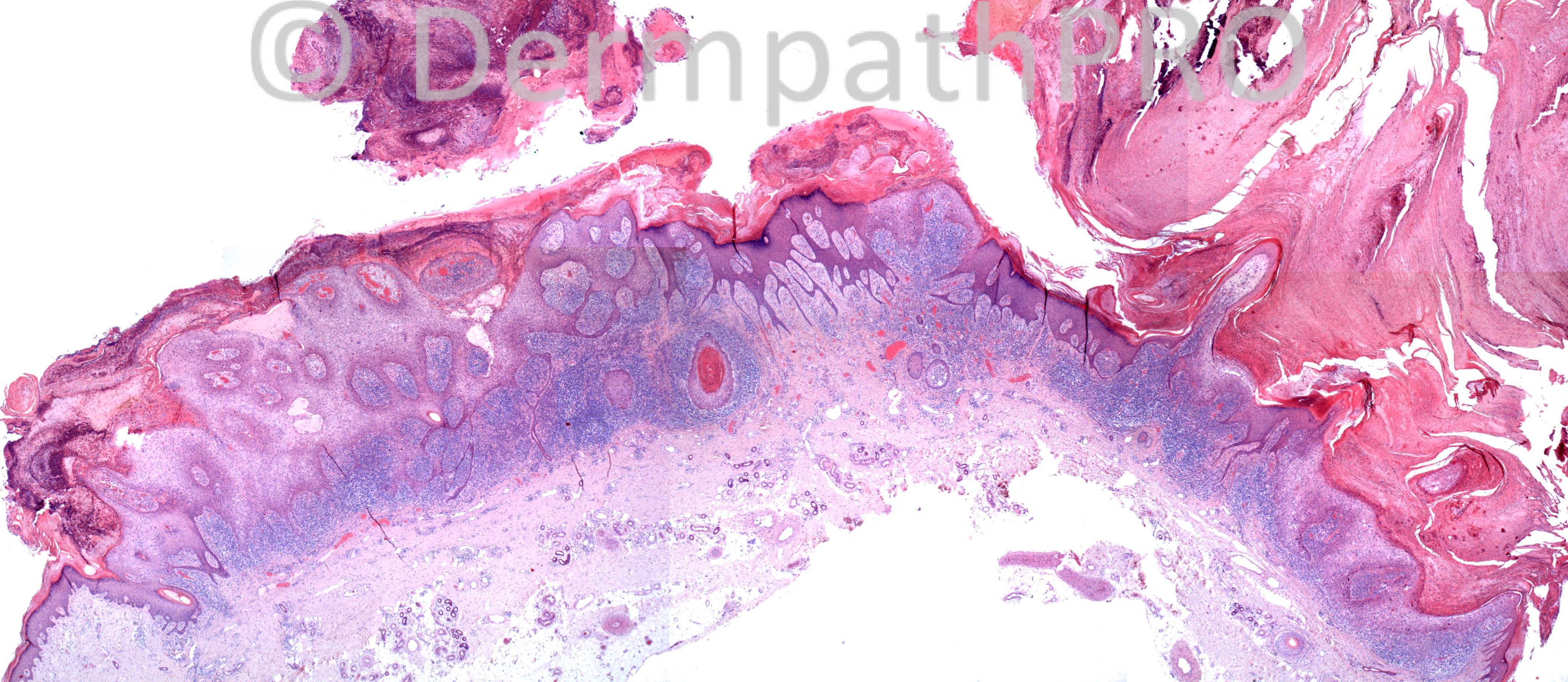
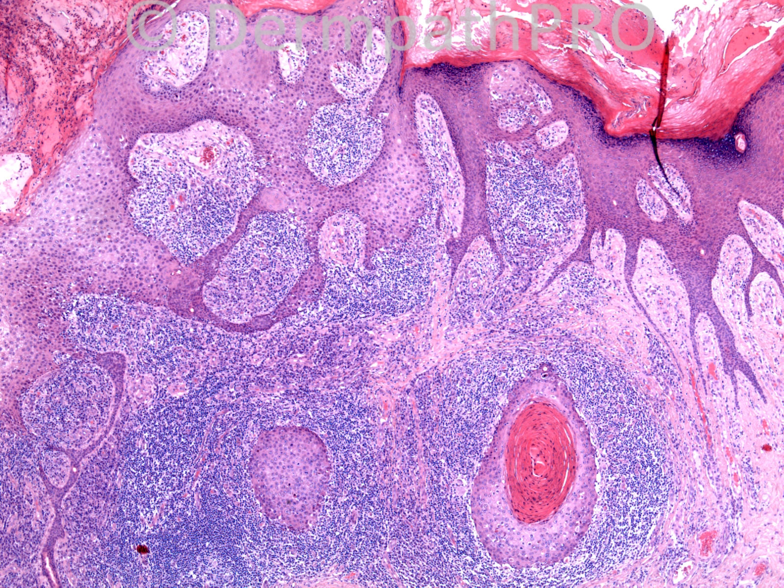
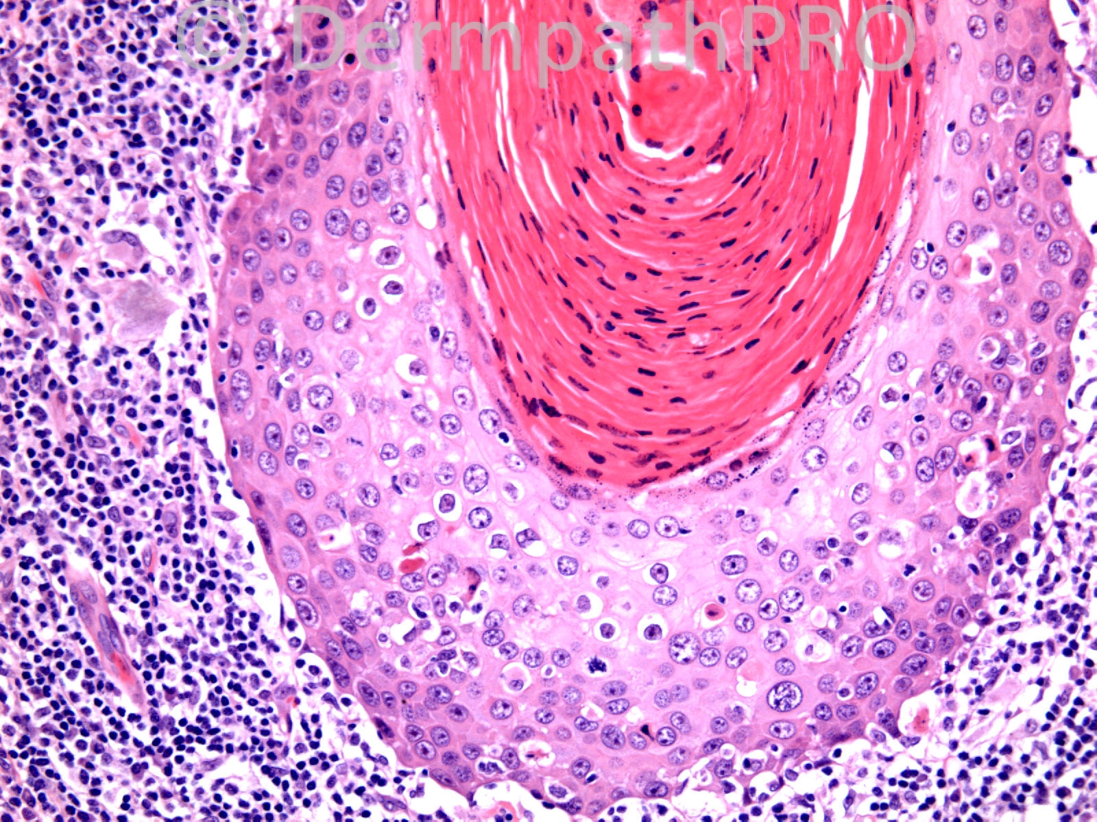
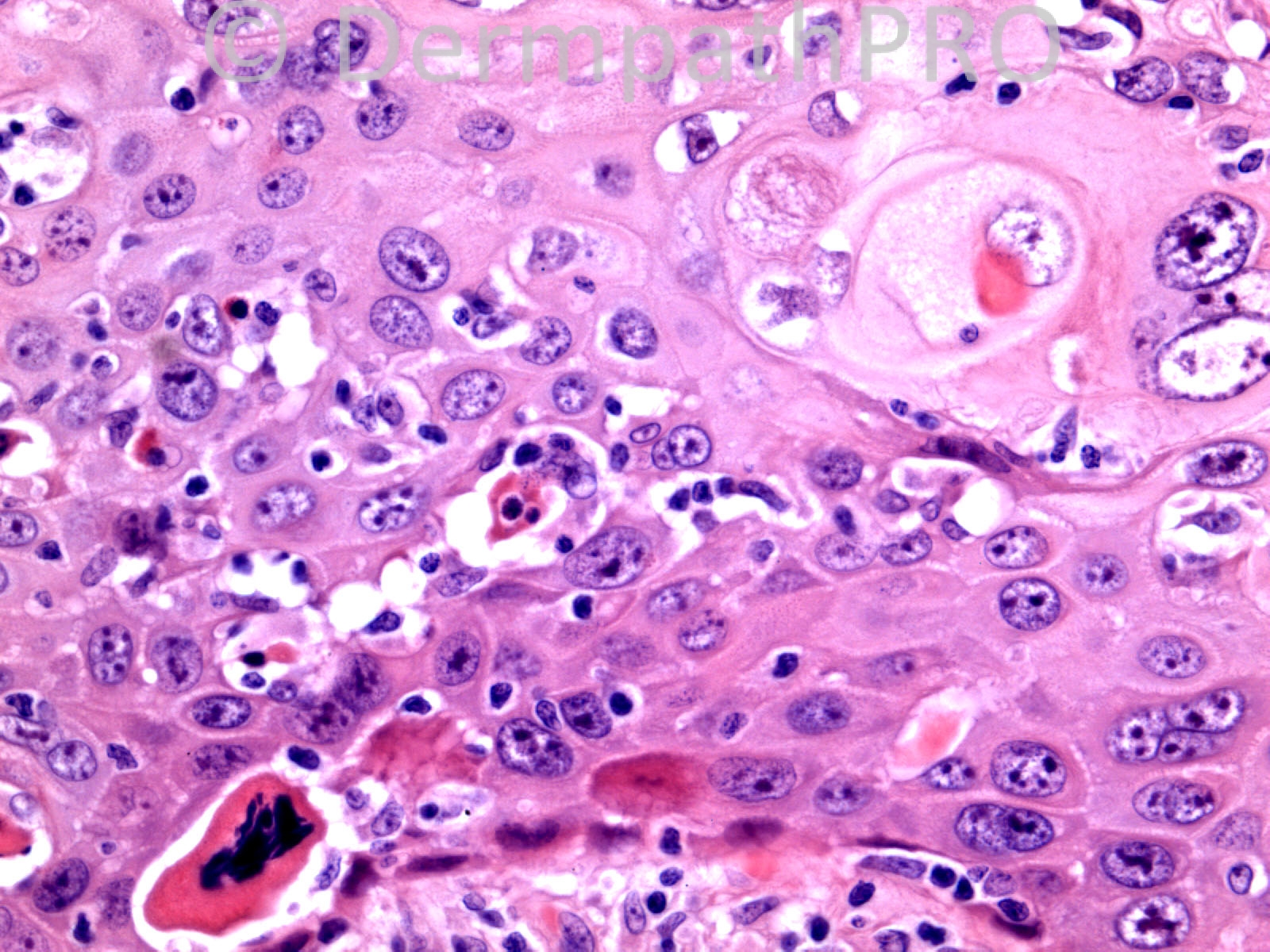
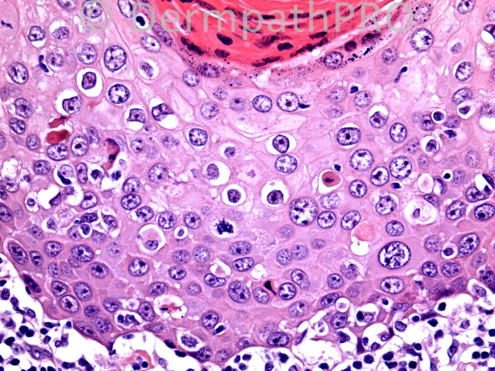
74 years old male. 3cm keratotic nodule right hand.

Case posted by Dr. Richard Carr.

  Report Record

User Feedback


Dr. Mona Abdel-Halim

Posted

Squamous cell carcinoma in situ (Bowen's Disease), with involvement of adnexal (follicular) epithelium.

Share this comment


Link to comment
share_externally

Sasi Attili

Posted

Bowenoid in-situ SCC suspicious of early invasion.(island to the left of the hair follicle where the tumour is extending down..in picture on top right). However need to see a close up of it to be sure.

I would like to take this opportunity to express my thoughts (following on from a discussion I had with Dr. Carr recently) regarding invasive Bowens disease which is supposed to be a high risk SCC.

I was looking for scientific evidence of this notion (that this is a high risk SCC) and Richard pointed out the paper by Cassarino DS, Derienzo DP, Barr RJ. Cutaneous squamous cell carcinoma: a comprehensive clinicopathologic classification. Part one. J Cutan Pathol. 2006 Mar;33(3):191-206.

In the paper, the authors themselves say:

'However, up to 5% of Bowen’s disease may become invasive, and up to 13–20% of those patients develop metastases, although these numbers are based on small studies and may reflect referral bias.7,116,117,120.'

The statement at the end of the para 'Although large outcome studies have not been performed, given the relatively high rates of metastasis reported with invasive Bowen’s disease, it should be considered a potentially high-risk variant of SCC.', is a fail-safe rather than evidence based approach.

The refs are all ancient and are small studies indeed. So I don't think the notion that 'Bowen's disease (BD) with early invasion, is a high risk SCC' is a proven fact. However, if there is adjacent/ contiguous BD in a patient with a barn door SCC, that might be a different case, and carry a worse prognosis as it may suggest field change.

In fact most clinicians I know (including me) would consider BD with early papillary dermal invasion (not just the pushing margins, but proper single cells or small nests invading into the papillary dermis) to be of good prognosis once excised/ treated. [i]Maybe Breslow has a role to play here[/i]. But I don't think BD itself is a proven independent RF for SCC prognosis in these cases. I think there are a number of similar other unsubstantiated notions prevailing in SCC (incl KA) that need addressing....

Would certainly like to know what others think.......

Share this comment


Link to comment
share_externally

I would call this an early well differentiated squamous cell carcinoma with extensive in situ component (BD) in routine practice but am interested to know what others think.

Share this comment


Link to comment
share_externally

Guest Jim Davie MD

Posted

Agree with SCC in-situ. I agree with Mark that tangential sections of in situ carcinoma may account for the "superficially invasive" component. The multifocal/clonal nature the proliferation, and high-grade atypia help exclude the other low-power differential diagnosis considerations of hypertrophic lichenoid actinic keratosis or hypertrophic lichenoid keratosis.

Share this comment


Link to comment
share_externally

Sasi Attili

Posted

Thanks Dr. Hurt. I have just read Ackerman's papers you have referenced. This does not still answer my question as to why we should/ shouldn't accept the common notion that invasive BD is a high risk SCC. I suppose you are hinting at the fact that this point is moot because both are SCC (a view the Ackerman purported). If this is the case, I am afraid I have to disagree, as the nomenclature/ diagnosis has therapeutic and prognostic implications (as per existing literature anyway). If this is called BD, this is a low-risk tumour with excellent prognosis once treated. If we call this invasion, then this is classed as a 'high- risk' tumour as per existing notion/ literature and management would be different.

I don't want this discussion to overshadow the interesting spot today but since you have mentioned his papers, in the absence of an alternative forum I shall reply to these interesting points raised.

I do not quite agree that prognostication is the duty of epidemiologists (As Ackerman also hints in his papers) and pathologists have no responsibility. As a clinician I can say the same thing and say that it is my duty as a clinician to just treat the patient and I do not need to prognosticate as i am not an epidemiologist! That would be silly for me to then refer the patient to an epidemiologist, or worse, leave the patient with an open ended diagnosis of cancer!

Certain tumour parameters determine prognosis. Prognosis determines the treatment modality. Its all interlinked. For ex. Low-risk in-situ SCC's and some superficial BCC's ([url="https://dermpathpro.com/blog/12/entry-119-superficial-bcc-why-it-is-important-to-measure-depth/"]my recent blog post on this[/url]) can be treated by non-surgical means; SCC's with neurovascular invasion/ high risk, might need adjuvant radiotherapy etc, and hence risk-stratification is important and this is the role of both the clinician and pathologist (hence we have MDT's in the UK- hopefully its the same in the US).

Regarding the concept of the phalanx , Ackerman says (I hope it is not one particular phalanx he is talking about!) :):

'That is not the case, however, for so-called squamous cell carcinoma
in situ in which, in vivo, neoplastic keratocytes
do not move as discrete aggregations from the
epidermis into the dermis, but when they do extend
ever so slowly deeper and deeper into the dermis,
they do that in the form of a phalanx, thereby
making it impossible for a histopathologist, no
matter how skilled, to determine where the presumed
in situ’ malignancy ends and the supposed
invasive’ malignancy begins.'

But, the same can be said about melanocytes, and often it is difficult to differentiate in situ from 'invasive' disease. So I am not sure I necessarily agree with him on this, unless I can't really understand what he is trying to explain. From what I understand, he is trying to draw a distinction between invasion of cells into the dermis and cells pushing into the dermis (not pushing borders), when in fact both are the same pathological processes wherein a tumour cells lose their usual homeostatic relation with their native surrounding cells and develop the ability to survive in tissue's that are not native, thus migrating up or down! This is the same whether the tumour cell is a melanocyte or keratinocyte. I cannot see the distinction. Both have the same (albeit difference in grade) prognostic implications. I have utmost respect for Dr. Ackerman, but on this occasion, I am afraid he has lost me!

His arguments re in-situ SCC still being an SCC are well known and understood. However prognostically and clinically there are huge implications for the patient and the distinction has to be made. Perhaps topical/ non-surgical modalities weren't popular in his time. However, times have changed and so should we :)

P.S: I don't mind the new terminology for Breslow being named after me :D

Share this comment


Link to comment
share_externally

Dr. Richard Carr

Posted

I called this adnexotropic bowenoid actinic keratosis (although "warty" carcinomas can also occur at this site). I did not mention the mild syringofibroadenomatous hyperplasia (I like not to confuse the clinicians any more than absolutely necessary) but thanks Mark for noticing it. There were four other blocks of this large lesion and I saw no convincing dermal invasion. I don't use the term carcinoma in situ as it promotes a knife mentality and as Sasi says there are other therapeutic options. I do mention the depth of adnexotropic downgrowths from the granular layer as I think this might prompt a surgical approach to some cases where this feature is extreme (i.e. thicker than 1mm). Happy for this to be known in future as "Carr's pragmatic mearsurement". I agree with Sasi that we need a better system of stratifying invasive Bowenoid SCC for prognostication. The current WHO approach obviously has many flaws and the RCPath dataset does not mention follicular SCC subtype yet (it appears that this is the dominant SCC subtype on the head and neck in my practice).

Share this comment


Link to comment
share_externally

Dr. Mona Abdel-Halim

Posted

But isn't it by definition that in actinic keratosis dysplastic cells do not involve the adnexal structures but only descend on the outer surface of acrosyringial structures and hair follicles. I will consider the extensive involvement of the follicular epithelium as a point in favour of Bowen's (SCC in situ), would love to know Dr Carr's opinion in this point...

Share this comment


Link to comment
share_externally

Sasi Attili

Posted

Dr. Mona. Bowenoid Actinic Keratosis is used synonymously with BD. It is used for full thickness dysplasia (Bowen's disease) in the context of a sun-exposed site (in contrast to non-sun exposed legs where classical BD is supposed to occur). So essentially this is Bowen's disease, but in a sun-exposed setting. Richard will correct me if I am wrong.....

Share this comment


Link to comment
share_externally

Sasi Attili

Posted

Thanks dr. Hurt. Re- Pathologists not having any clinical responsibility reminded me of this cartoon I saw recently :)
[img]https://lh3.googleusercontent.com/-TxQgD4kG85c/Um0e7kyBRpI/AAAAAAAAhPw/ADKC0lr4rQM/w855-h553-no/1235424_533230903431893_1564887619_n.jpg[/img]


I still do not understand Dr. Ackerman's theory regarding the Phalanx though! Why are epithelial cells not changing compartments, when moving from the epidermis to the dermis. Does he mean that they are taking the epidermis with them and the entire epidermis is moving into the dermis? Am I missing something very obvious? O.o

Share this comment


Link to comment
share_externally

Dr. Richard Carr

Posted

Dear Mona,

Sasi is right. I use the term bowenoid epidermal dysplasia for full thickness dysplasia on non-sun exposed sites (or on exposed skin without prominent solar elastosis e.g. lower leg often) and Bowenoid actinic keratosis for cases like the one here on sun-exposed sites with accompanying moderate to severe solar elastosis. I don't remember seeing adnexotropic actinic keratosis so I think the adnexotropism must just reflect the grade of dysplasia (in bowenoid actinic keratosis or bowenoid epidermal dysplasia).

Share this comment


Link to comment
share_externally


Join the conversation

You can post now and register later. If you have an account, sign in now to post with your account.

Guest
Add a comment...

×   Pasted as rich text.   Paste as plain text instead

  Only 75 emoji are allowed.

×   Your link has been automatically embedded.   Display as a link instead

×   Your previous content has been restored.   Clear editor

×   You cannot paste images directly. Upload or insert images from URL.

Loading...

×
×
  • Create New...