Jump to content
In this section we have spot diagnoses posted on a daily basis since June 2010, now over 4000! You can review the archived cases and read the suggested diagnoses by users and the final comment by the contributors.
Case are uploaded each week day by 10 am UK time with the correct diagnosis will generally be posted at 8 pm UK time. Why not view the most recent spot diagnosis and proffer a diagnosis?

Case Number : Case 838 - 3rd September Posted By: Guest

Please read the clinical history and view the images by clicking on them before you proffer your diagnosis.
Submitted Date :
   (0 reviews)

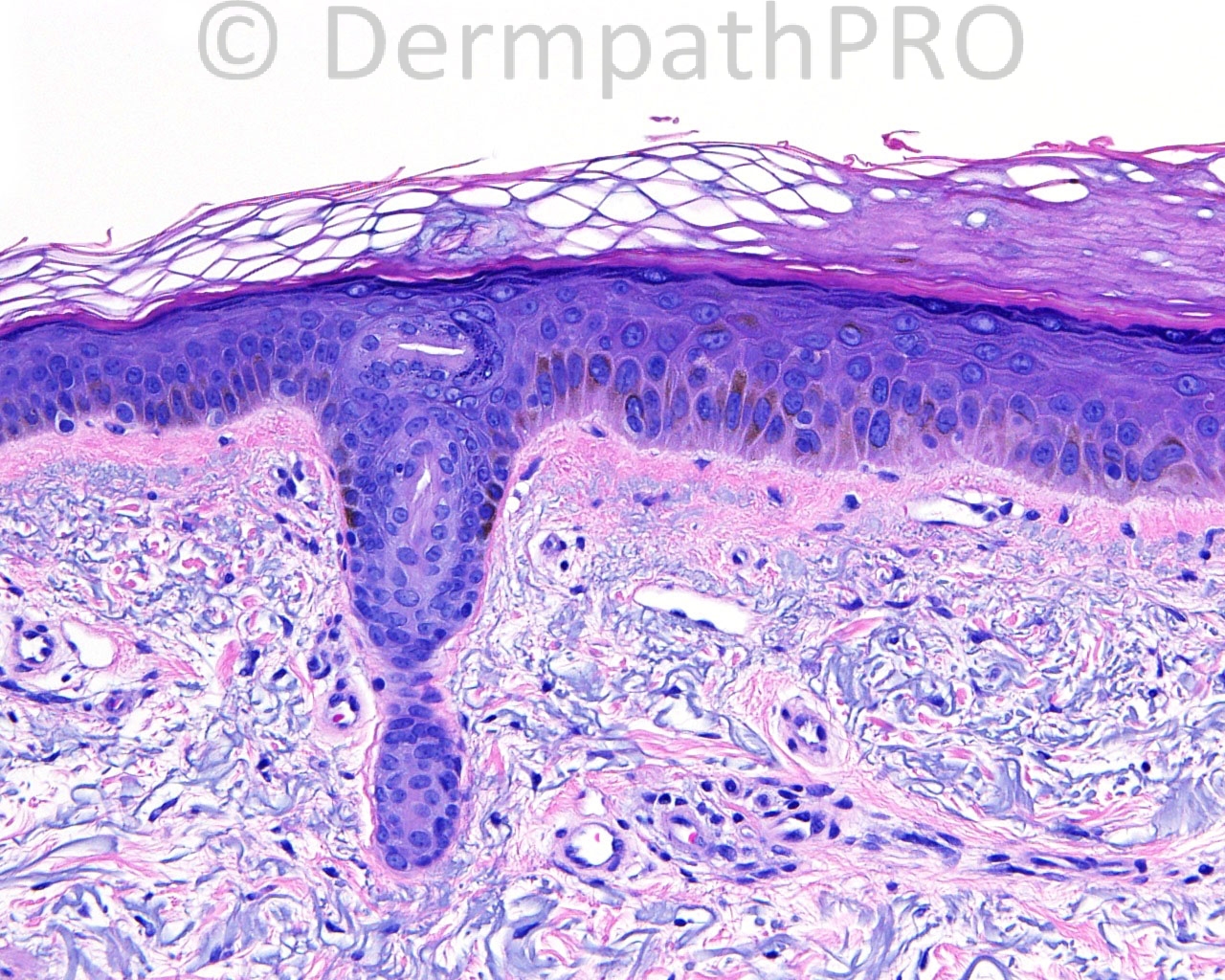
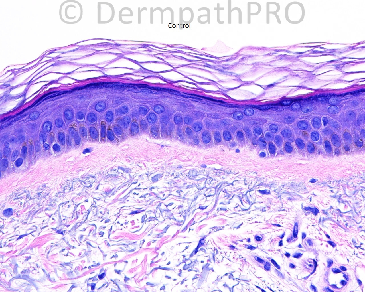
The patient is a 75 year old woman with an excision of a pigmented macule from the left arm.

Case posted by Dr. Mark Hurt.

  Report Record

User Feedback


Guest Maria George

Posted

I shall give her the benefit of doubt.Malignant melanoma insitu.

Share this comment


Link to comment
share_externally

Guest Maria George

Posted

I shall give her the benefit of doubt.Malignant melanoma insitu.

Share this comment


Link to comment
share_externally

Arti Bakshi

Posted

agree with large cell acanthoma.
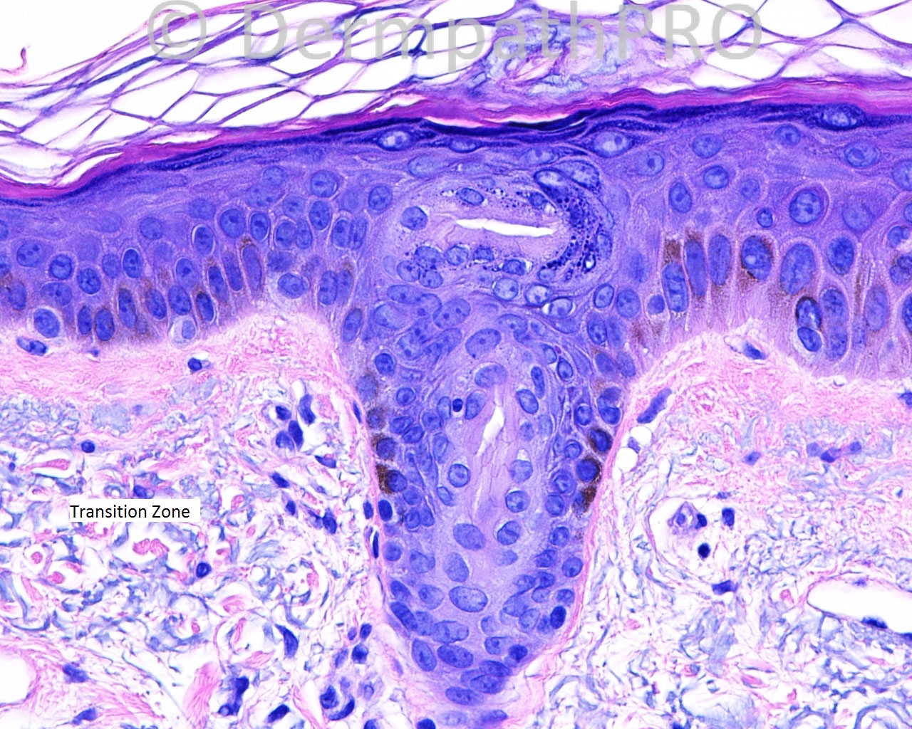
image labelled as 'transition zone' is instructive!

Share this comment


Link to comment
share_externally

Robledo F. Rocha

Posted

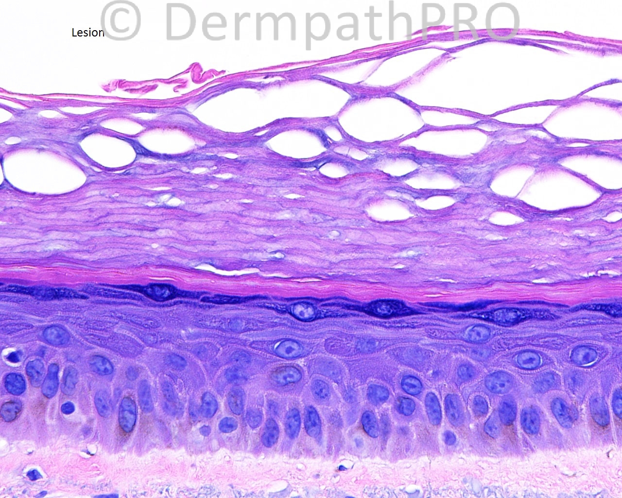
Large cell acanthoma. A circumscribed area of epidermis with enlarged keratinocytes topped by hyperorthokeratosis. Like Dr. Bakshi, I found the transition zone very instructive.

Share this comment


Link to comment
share_externally

Guest Jim Davie MD

Posted

- Large cell acanthoma (pigmented variant).

[size=4] Nuclear and cytoplasmic enlargement of keratinocytes (maintaining similar nuclear/cytoplasmic ratio as normal keratinocytes), hypergranulosis, and hyperorthokeratosis.
Seeing a clonal proliferation of enlarged keratinocytes--well-demarcated against normal, smaller keratinocytes, as seen in the 'transition zone' photo above--with involvement of the basal keratinocytes, is helpful in differentiating it from simple reactive keratinocytes, which may result from chronic inflammation and/or irritation. When there is actinic dysplasia, the differential would then include actinic keratosis.
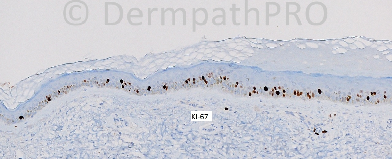
[MelanA and Ki67 stains show expected basal melanocytes, which are dwarfed by the surrounding enlarged keratinocytes, and absence of high-level scatter or high-level proliferative activity].[/size]

Share this comment


Link to comment
share_externally

Dr. Richard Carr

Posted

Agree with the "academic" diagnosis but the distinction seems to be rather academic from a relatively flat solar lentigo which, afterall, is probably just a thin seborrhoeic keratosis! For the clinicians (even most dermatologists are not over familiar with this diagnostic splitting) I like not to confuse them with some of our more "academic" diagnoses so I might label it "solar lentigo / thin seborrhoeic keratosis (so-called large cell acanthoma variant)".

Share this comment


Link to comment
share_externally


Join the conversation

You can post now and register later. If you have an account, sign in now to post with your account.

Guest
Add a comment...

×   Pasted as rich text.   Paste as plain text instead

  Only 75 emoji are allowed.

×   Your link has been automatically embedded.   Display as a link instead

×   Your previous content has been restored.   Clear editor

×   You cannot paste images directly. Upload or insert images from URL.

Loading...

×
×
  • Create New...