Jump to content
In this section we have spot diagnoses posted on a daily basis since June 2010, now over 4000! You can review the archived cases and read the suggested diagnoses by users and the final comment by the contributors.
Case are uploaded each week day by 10 am UK time with the correct diagnosis will generally be posted at 8 pm UK time. Why not view the most recent spot diagnosis and proffer a diagnosis?

Case Number : Case 840 - 5th September Posted By: Guest

Please read the clinical history and view the images by clicking on them before you proffer your diagnosis.
Submitted Date :
   (0 reviews)

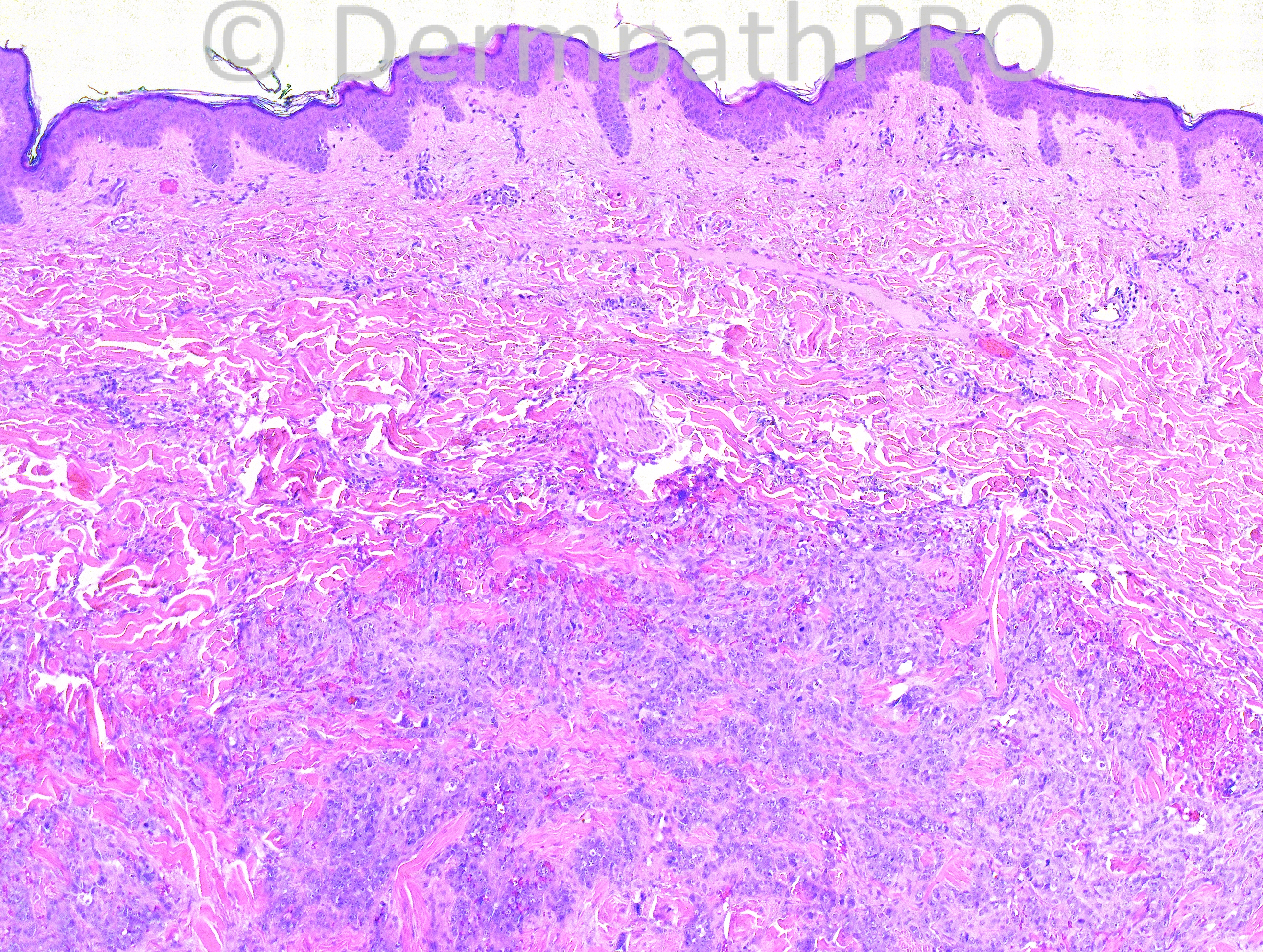
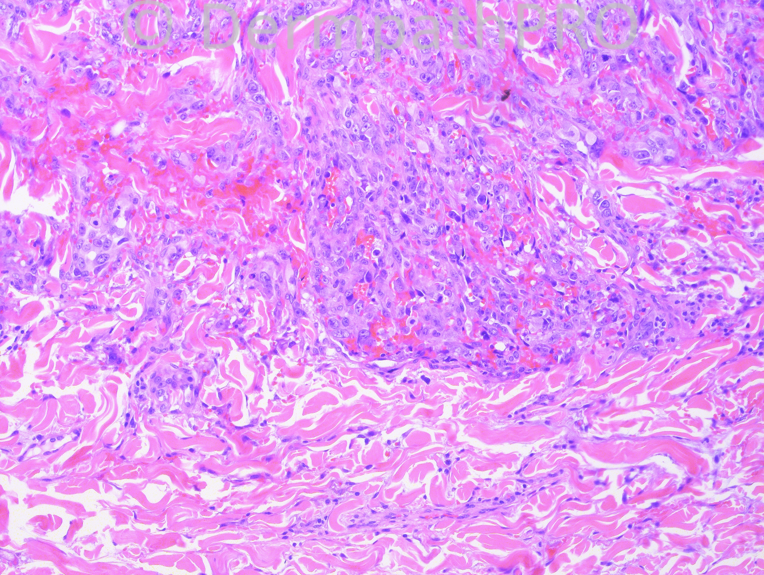
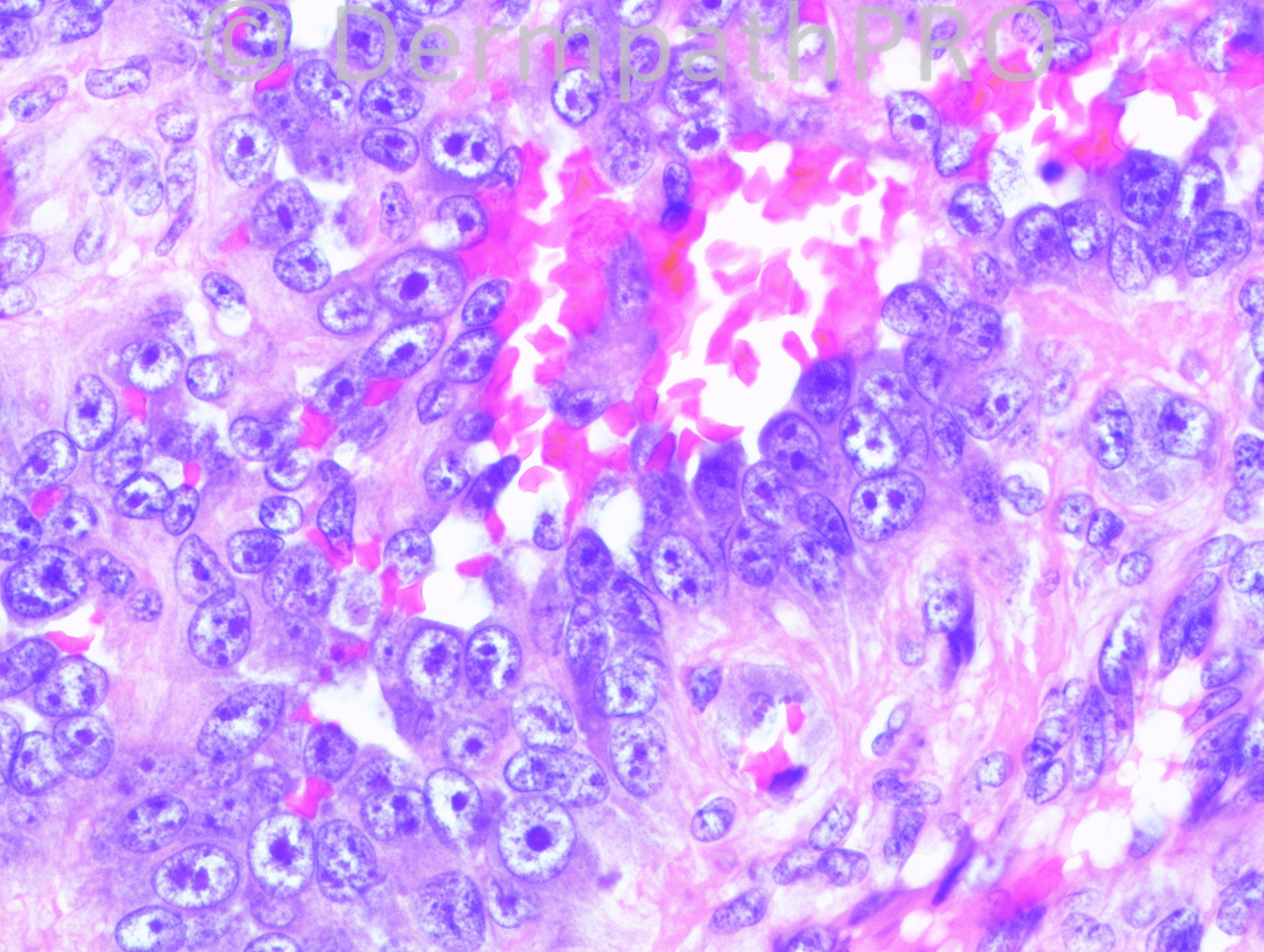
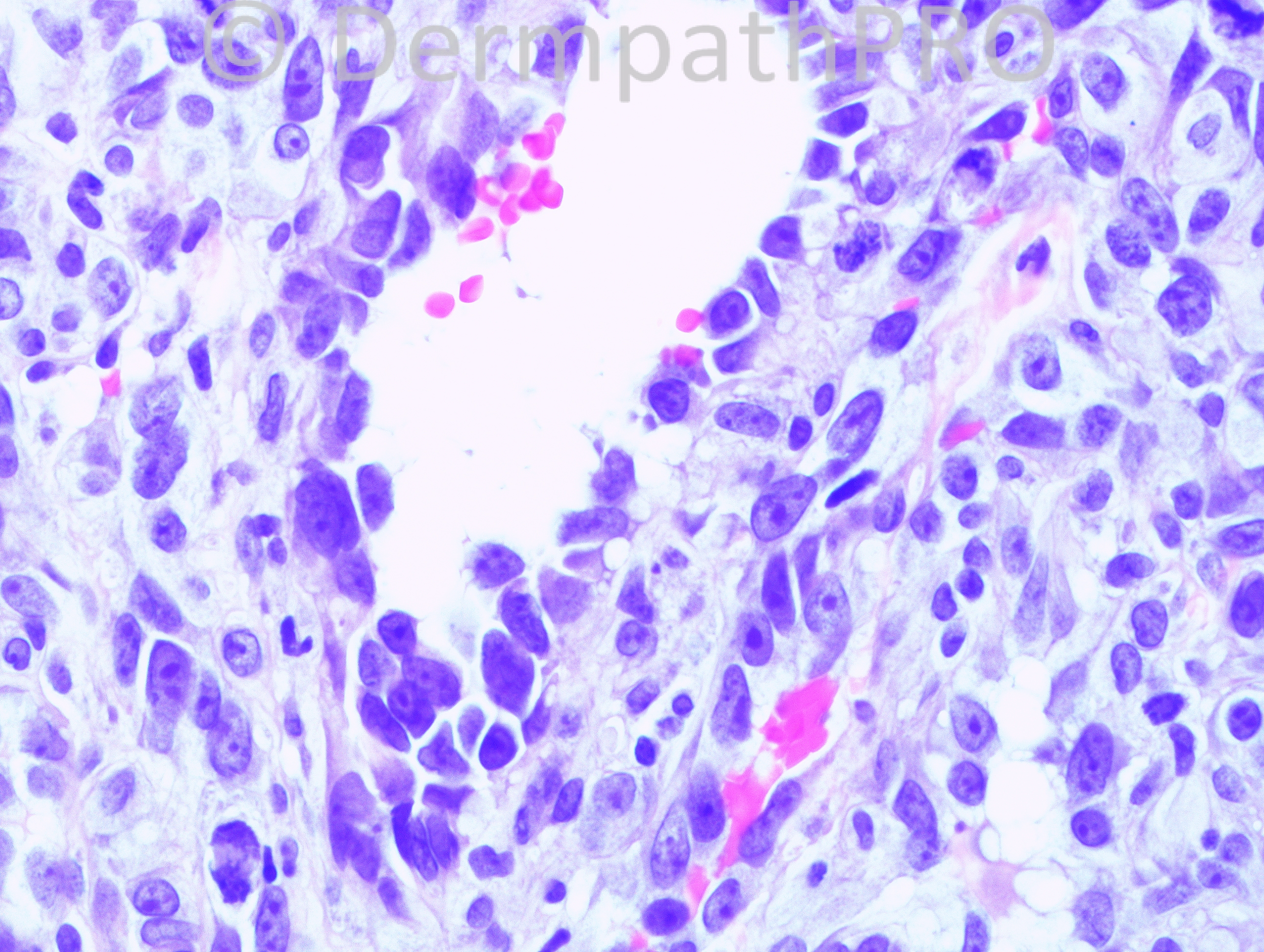
60 year old female with a skin lesion on the breast.

Case posted by Dr. Hafeez Diwan.


  Report Record

User Feedback


Guest Romualdo

Posted

Cutaneous angiosarcoma, possibly postradiation.

Share this comment


Link to comment
share_externally

Arti Bakshi

Posted

[quote name='Romualdo' timestamp='1378372323']
Cutaneous angiosarcoma, possibly postradiation.
[/quote]
Agree!

Share this comment


Link to comment
share_externally

Guest Giovanni Falconieri

Posted

Epithelioid angiosarcoma on top of differential, with nice pseudoglandular pattern. Perhaps radiation tx induced changes in the background.

Share this comment


Link to comment
share_externally

High grande angiosarcoma, postradiation (probably).

Share this comment


Link to comment
share_externally

Dr. Mona Abdel-Halim

Posted

Epithelioid angiosarcoma, mostly post radiation ..

Share this comment


Link to comment
share_externally

Guest Jim Davie MD

Posted

[size=3]Epithelioid angiosarcoma. (Looks like we are all in agreement! )

The predominantly polygonal and rounded cells show prominent nucleoli, pleomorphic size/shape, moderate to high nuclear/cytoplasmic ratios, and mitotic activity. Architecture shows infiltrative single cells and fascicles weaving through collagen bundles, with some single, spindled tumor cells closely applied to the surface of collagen bundles (promontory sign) at the peripheries of the infiltrate. Poorly formed large blood-filled vascular spaces are lined by one or more layers of polygonal tumor cells that show tombstone-like intracellular separation artefact. Intracellular spaces packed with red cells are present in some tumor cells.

I don't think it applies in this case given the classic histology, but the differential would usually include Kaposi's sarcoma (often positive for CD31), epithelioid hemangioendothelioma, primary breast carcinoma, or other metastatic carcinoma/melanoma simulating angiosarcoma. Immunostaining will help confirm the diagnosis.

Angiosarcoma is (like metastatic melanoma) notorious for occasionally mimicking other, less aggressive neoplasms such as breast carcinoma* or metastatic renal cell carcinoma, especially if there is common, anomalous positive staining for cytokeratins such as CK7, positive staining for renal cell carcinoma marker CD10 -- or anomalous negative staining for CD34-- to trap the unwary.** (CD31 is, in my experience, a reasonably reliable positive stain, but hanging the entire diagnosis on any one stain may be unwise for cases of equivocal AS).[/size]

[size=3]* [url="http://www.ncbi.nlm.nih.gov/pubmed/21124204"]Angiosarcoma with pseudoepidermotropism in a patient with breast cancer: a mimic of epidermotropic metastatic adenocarcinoma.[/url] A[i]m J Dermatopathol. 2011 Jun;33(4):400-2.[/i][/size]
[font=arial,helvetica,sans-serif][size=3]** [url="http://www.ncbi.nlm.nih.gov/pubmed/19813270"]Epithelioid angiosarcoma: a neoplasm with potential diagnostic challenges.[/url] [i]Diagn Cytopathol. 2010 Feb;38(2):154-8[/i][/size][/font]
[size=3][font=arial,helvetica,sans-serif]** [url="http://www.ncbi.nlm.nih.gov/pubmed/22943673"]The co-expression of cytokeratin and p63 in epithelioid angiosarcoma of the parotid gland: a diagnostic pitfall.[/url][i]Diagn Pathol. 2012 Sep 3;7:118.[/i][/font][/size]

Share this comment


Link to comment
share_externally

Guest Giovanni Falconieri

Posted

Just a couples of notes from the profane. According to soft tissue gurus (including Chris Fletcher) angiosarcomas at visceral location are far often more positive for keratins than those occurring in the skin. An expanded spectrum of cutaneous epithelioid angiosarcoma (and the differential thereof entailed) has been reported a few years ago by Carlos Bacchi et al in the AJSP 2010; 34(9):1334-43.

Share this comment


Link to comment
share_externally

Dr. Hafeez Diwan

Posted

Epithelioid angiosarcoma. Keratin was negative in this case.

Share this comment


Link to comment
share_externally


Join the conversation

You can post now and register later. If you have an account, sign in now to post with your account.

Guest
Add a comment...

×   Pasted as rich text.   Paste as plain text instead

  Only 75 emoji are allowed.

×   Your link has been automatically embedded.   Display as a link instead

×   Your previous content has been restored.   Clear editor

×   You cannot paste images directly. Upload or insert images from URL.

Loading...

×
×
  • Create New...