Jump to content
In this section we have spot diagnoses posted on a daily basis since June 2010, now over 4000! You can review the archived cases and read the suggested diagnoses by users and the final comment by the contributors.
Case are uploaded each week day by 10 am UK time with the correct diagnosis will generally be posted at 8 pm UK time. Why not view the most recent spot diagnosis and proffer a diagnosis?

Case Number : Case 841 - 6th September Posted By: Guest

Please read the clinical history and view the images by clicking on them before you proffer your diagnosis.
Submitted Date :
   (0 reviews)

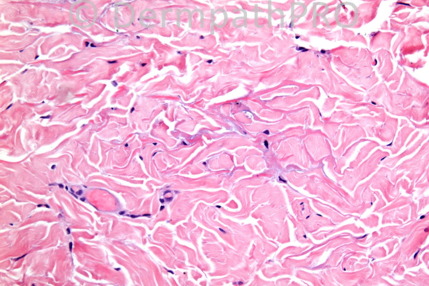
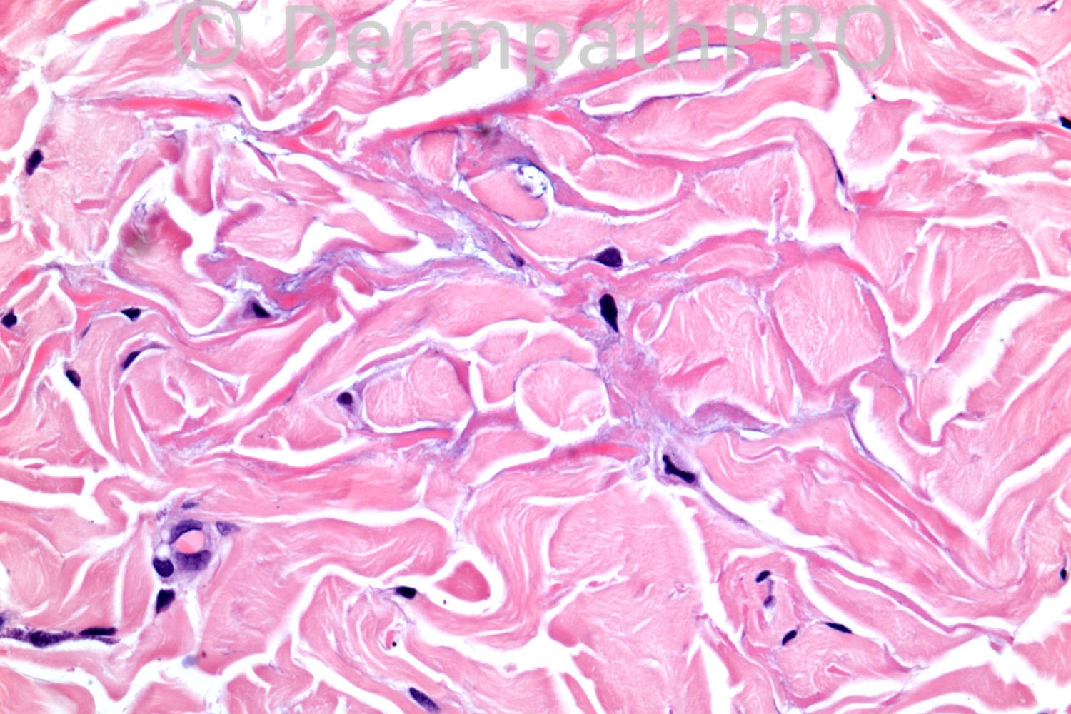
6 years old female. Barely visible pale plaques on torso.

Case posted by Dr. Richard Carr.


  Report Record

User Feedback


Wolter Mooi

Posted

I would be interested in an elastin stain. The pictures remind me of elastoma / Buschke-Ollendorf syndrome. But I have never diagnosed that lesion before :)

Share this comment


Link to comment
share_externally

guttate morphea

the fibroblasts that synthesize mucin ( can be seen in morphea) is visible between the thickened eosinophilic hylanized collagen bundles more prominant at the junction between the reticular dermis and fat tissue is typically seen.

There is mild superficial perivasualr lymphocystic infiltrate in the papillary dermis with normal epidermis
hair follicles and seb glands not seen in the center of the lesion, only the arrector pilorum is left alone

Share this comment


Link to comment
share_externally

Guest Engin Sezer

Posted

Sclerosis of deep dermis with mucin deposition between the collagen bundles suggestive of incipient morphoea

Share this comment


Link to comment
share_externally

Guest Maria George

Posted

While the fenestrations of the collagen fibers by mucin point toward a type of cutaneous mucinoses such as (sclerema, scleromyxedema , lupus, REM, ..etc), the combination of superficial perivascular infiltrate with mucin in between collagen make me think for sure that this is a case of Granuloma annulare.

Share this comment


Link to comment
share_externally

Guest Jim Davie MD

Posted

Connective tissue nevus (shagreen patch).

There is sometimes slight increase in fibroblasts and interstitial mucin in these lesions.
I don't see any elastin fibers in the high-power image (unless they are staining red), although an elastin stain would be the gold standard to help support the diagnosis.

The differential diagnosis would be tuberous sclerosis or (as Wolter said) Buschke-Ollendorff syndrome.
I favor tuberous sclerosis if the patches were discovered in childhood rather than postnatally, although the absence of other TS findings would favor BOS.

Share this comment


Link to comment
share_externally

Robledo F. Rocha

Posted

I opine for late morphea. Inflammatory infiltrate is less than scant and diminished plump fibroblasts can be found in the midst of thickened collagen bundles with increased mucin between them.

Share this comment


Link to comment
share_externally

Dr. Richard Carr

Posted

Well done Wolter!!! These are I believe elastomas of Buschke-Ollendorff. Thickened hyaline elastic fibres in zonal areas of chicken wire appearances around the collagen. Giving the elastic stain would make it far too easy for this crowd but I was a little surprised but the responses. Jim - sorry the elastic fibres are a bit red in these images. You also need to watch out for the haematoxylin in Warwick which exaggerates any hint of interstitial mucin especially the day after the stains are changed!!

I have to thank Dr Vivek Mudaliar for kindly sharing this case with me.
Enjoy your weekends

Share this comment


Link to comment
share_externally

As a clinician, I would like to point out that stretching the skin helps in visualising the elastomas of Buschke-Ollendorf syndrome.

Share this comment


Link to comment
share_externally


Join the conversation

You can post now and register later. If you have an account, sign in now to post with your account.

Guest
Add a comment...

×   Pasted as rich text.   Paste as plain text instead

  Only 75 emoji are allowed.

×   Your link has been automatically embedded.   Display as a link instead

×   Your previous content has been restored.   Clear editor

×   You cannot paste images directly. Upload or insert images from URL.

Loading...

×
×
  • Create New...