Jump to content
In this section we have spot diagnoses posted on a daily basis since June 2010, now over 4000! You can review the archived cases and read the suggested diagnoses by users and the final comment by the contributors.
Case are uploaded each week day by 10 am UK time with the correct diagnosis will generally be posted at 8 pm UK time. Why not view the most recent spot diagnosis and proffer a diagnosis?

Case Number : Case 842 - 9th September Posted By: Guest

Please read the clinical history and view the images by clicking on them before you proffer your diagnosis.
Submitted Date :
   (0 reviews)

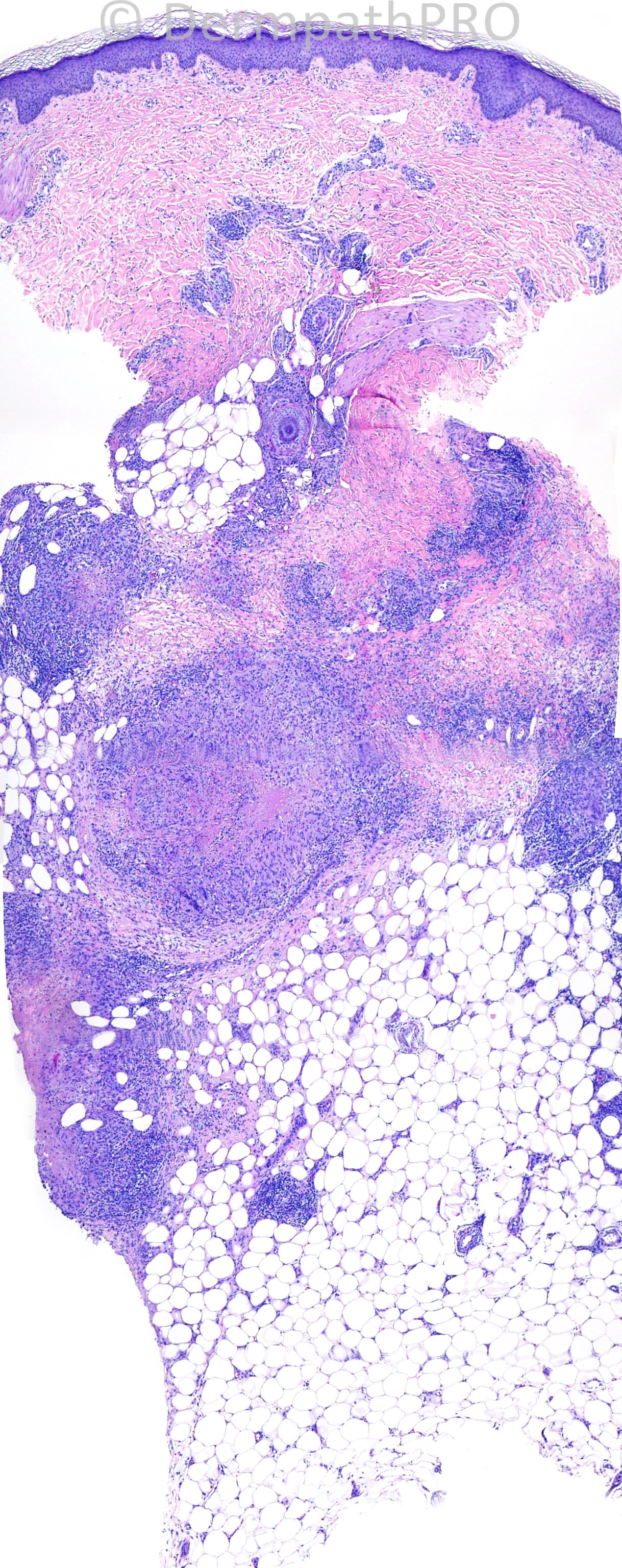
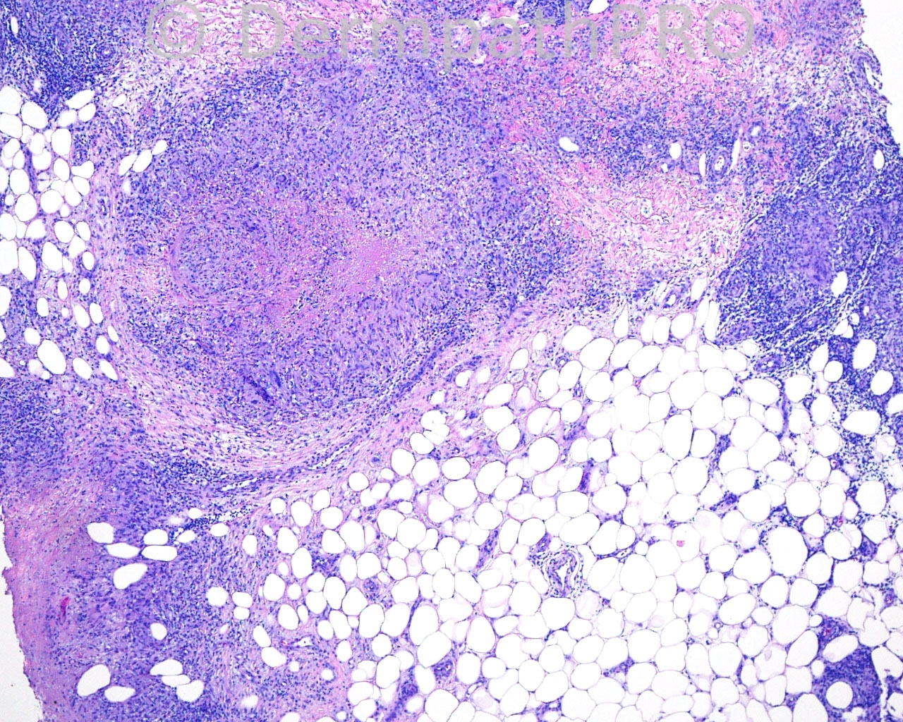
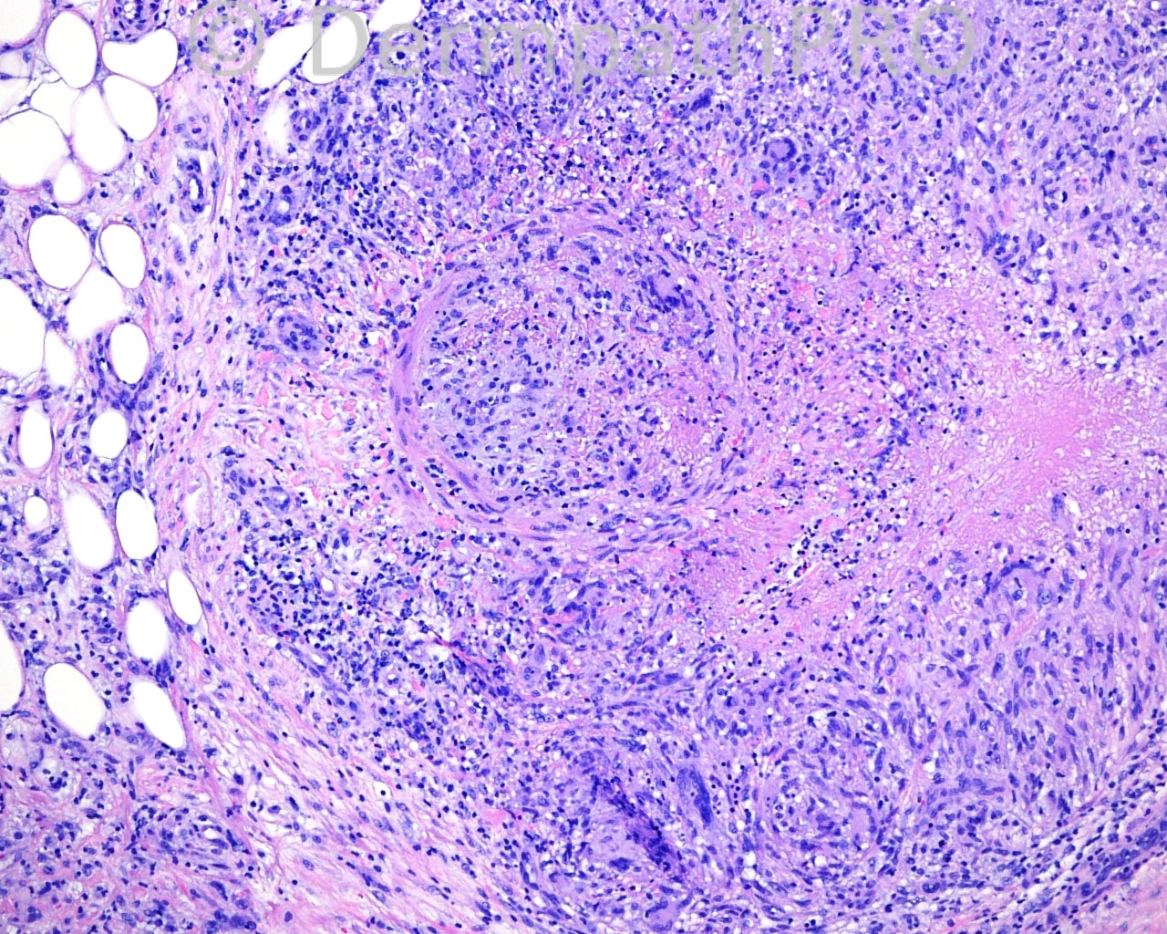
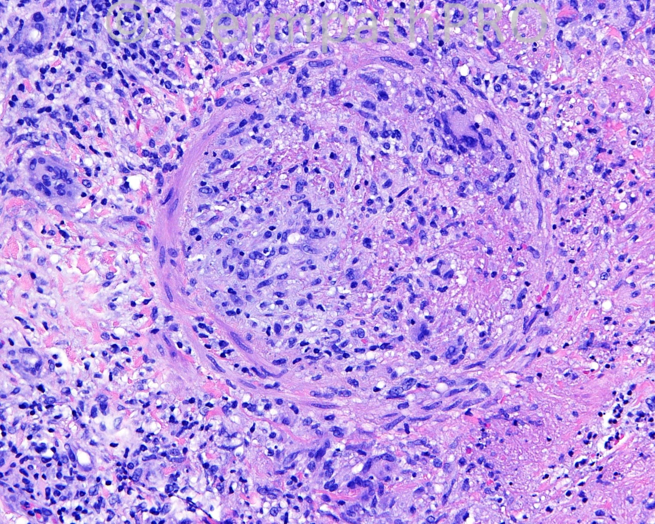
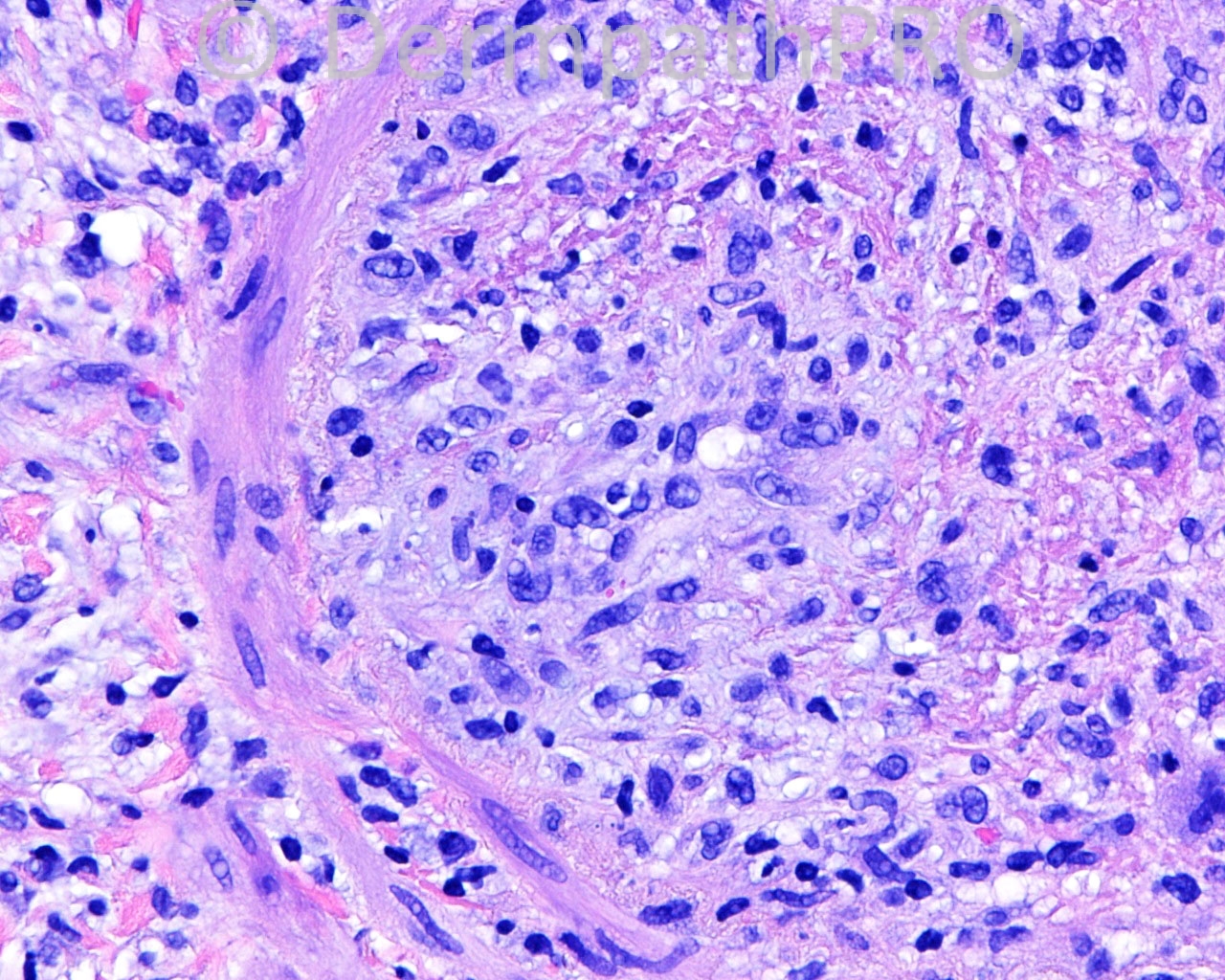
46 year old female with a biopsy taken from the medial aspect of the left knee.

Case posted by Dr. Mark Hurt.


  Report Record

User Feedback


The infiltrate is predominantly septal with vasculitis with some neutrophils at the periphery and neovascularization.
The lobules are not affected by the inflammatory infiltrate and there´s no necrosis.
I suspect of cutaneous polyarteritis nodosa. The classical differential diagnosis is with superficial trombophlebitis and an elastic stain would be very helpfull to differentiate between artery and vein.

Share this comment


Link to comment
share_externally

Robledo F. Rocha

Posted

Granulomatous lobular panniculitis associated with vasculitis. I go with nodular vasculitis / erythema induratum of Bazin.

Share this comment


Link to comment
share_externally

Sorry, I did not see the granulomatous inflammation has Robledo did, and he is right.
I have to chenge my opinion.
I still not believe it is a Nodular vasculitis/EIB, because I would expect to see more lobular inflammation and necrosis. The fact that the panniculitis is located on lower left knee doesn´t help too much.
It is a difficult case and now I think of Wegener´s granulomatosis or Vasculitis associated with Crohn´s disease (I once had one case with Crohn).

Share this comment


Link to comment
share_externally

Guest Jim Davie MD

Posted

I favor Wegener's granulomatosis over erythema induratum/nodular vasculitis. Fungal and mycobacterial stains would be helpful to eliminate an infectious panniculitis. Although unlikely, Takayasu arteritis [giant-cell arteritis] may also present initially on the lower extremities. I think clinical correlation (history and serologies) will be critical to the diagnosis.

There is an obliterated pannicular large vessel with giant-cell granulomatous component, adjacent palisading necrobiotic fibrinous granulomas with giant-cell component, subtle leukocytoclastic component, red cells, and (presumed) absence of significant eosinophils.

Share this comment


Link to comment
share_externally

Dr. Richard Carr

Posted

My first impression was nodular vasculities or erythema induratum of Bazin. I think it is a vein and the elastic supports this impression (although the vessels is heavily disrupted I perceive finer elastic between concentric layers of muscle. Would have liked a more lobular predominance but such is life! Any exposure to TB (I am guessing yes).

Share this comment


Link to comment
share_externally


Join the conversation

You can post now and register later. If you have an account, sign in now to post with your account.

Guest
Add a comment...

×   Pasted as rich text.   Paste as plain text instead

  Only 75 emoji are allowed.

×   Your link has been automatically embedded.   Display as a link instead

×   Your previous content has been restored.   Clear editor

×   You cannot paste images directly. Upload or insert images from URL.

Loading...

×
×
  • Create New...