Jump to content
In this section we have spot diagnoses posted on a daily basis since June 2010, now over 4000! You can review the archived cases and read the suggested diagnoses by users and the final comment by the contributors.
Case are uploaded each week day by 10 am UK time with the correct diagnosis will generally be posted at 8 pm UK time. Why not view the most recent spot diagnosis and proffer a diagnosis?

Case Number : Case 844 - 11th September Posted By: Guest

Please read the clinical history and view the images by clicking on them before you proffer your diagnosis.
Submitted Date :
   (0 reviews)

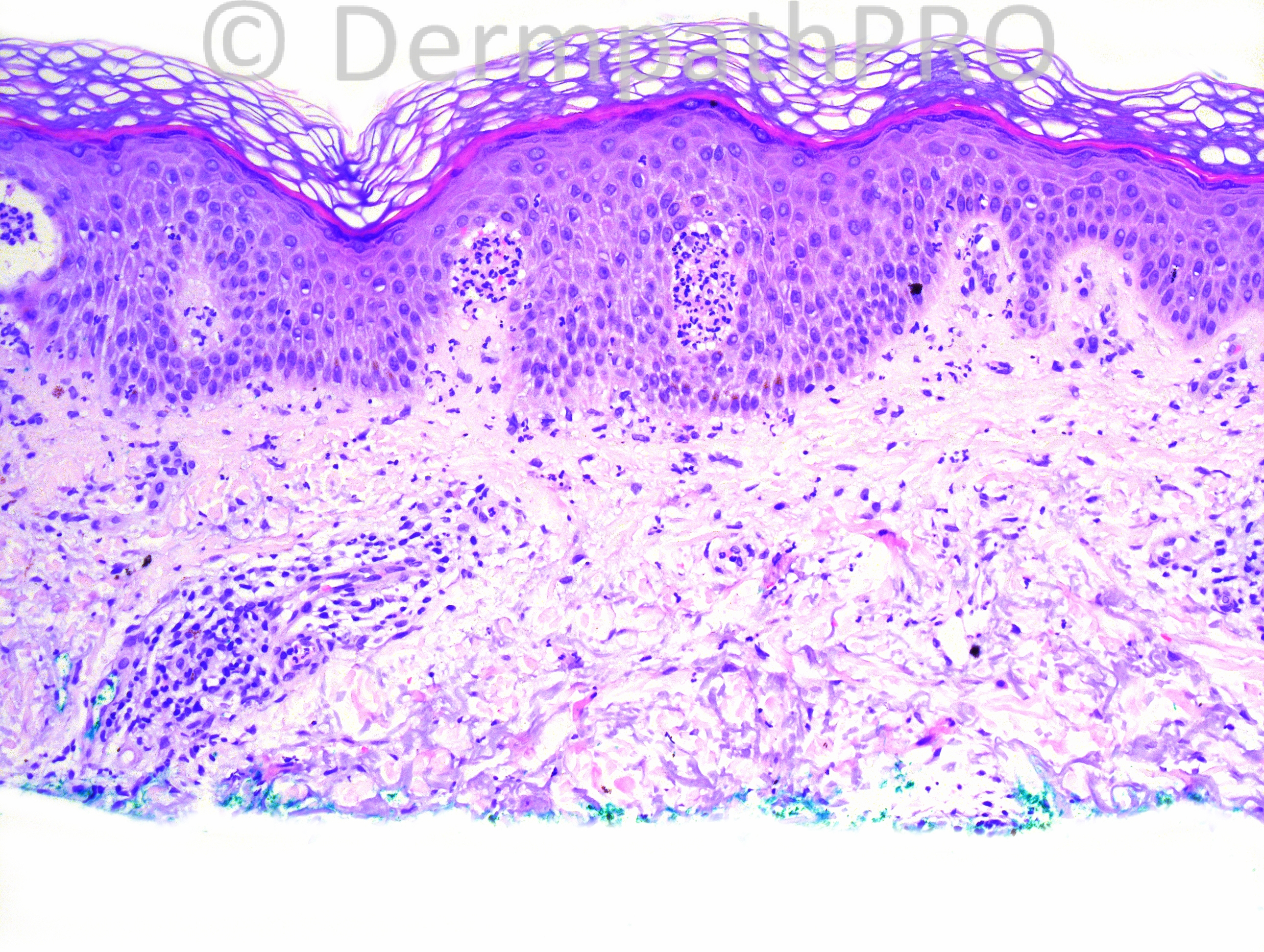
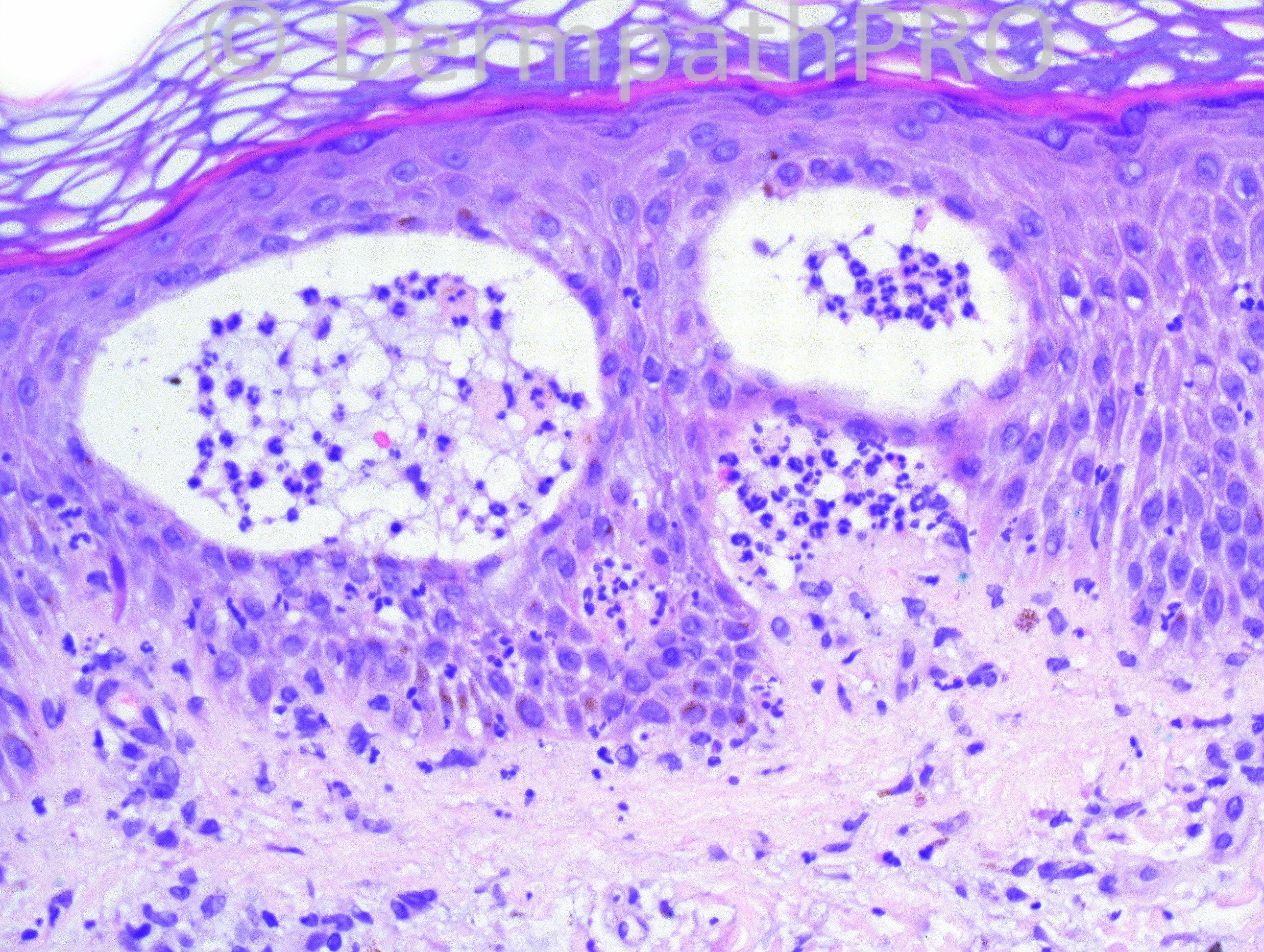
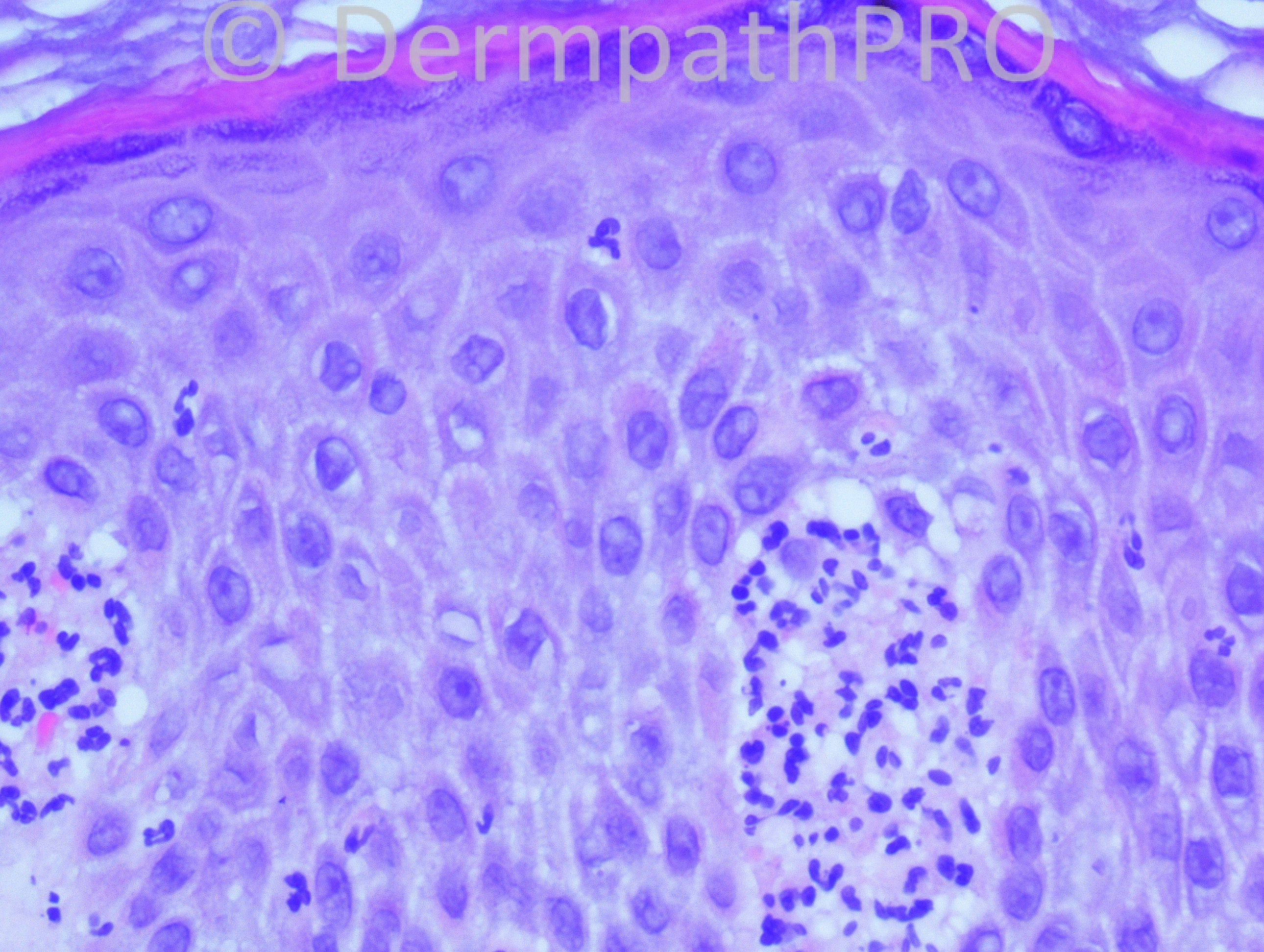
61-year-old female with biopsy from elbow.

Case posted by Dr. Hafeez Diwan.


  Report Record

User Feedback


Guest Yüksel OKUMUŞ, MD

Posted

Agree with Dermatitis Herpetiformis (Duhring Disease)

Share this comment


Link to comment
share_externally

[b]Subepidermal blisters with neutrophils[/b]
Dermatitis herpetiformis
Papulovesicles have a characteristic herpetiform grouping and a predilection for extensor surfaces such as the elbows, usually in symmetrical distribution. Excoriations are almost always present. Direct immunofluorescence is expected to show granular deposits of IgA in the dermal papillae of perilesional and uninvolved skin, and the deposition is not uniform (IgA deposits haphazard in papilla).

Share this comment


Link to comment
share_externally

Guest Romualdo

Posted

Vesicular subepidermal dermatitis with predominance of neutrophils. The differential includes dermatitis herpetiformis, linear IgA dermatosis, acquired epidermolysis bullosa and systemic lupus erythematosus. I can't see dermal mucin deposits to think in the last diagnosis. Although clinical information and, principally, direct immunofluorescence testing, are absolutely necessary, my prefered diagnosis is IgA linear dermatosis.

Share this comment


Link to comment
share_externally

The histopathological distinction between dermatitis herpetiformis and linear IgA disease is almost impossible in most cases. Direct immunofluorescence is essential for diagnosis.

Share this comment


Link to comment
share_externally

Hopping there´s moderate to intense pruritus, my diagnosis is DH.

Share this comment


Link to comment
share_externally

Robledo F. Rocha

Posted

I favor dermatitis herpetiformis over linear IgA dermatosis just because the patient's age and the affected site.

Share this comment


Link to comment
share_externally

Guest Jim Davie MD

Posted

[font=arial,helvetica,sans-serif][size=4]I agree with the above comprehensive differentials for a neutrophil-predominant subepidermal bullous dermatosis. Most [i]likely[/i] DH or Linear IgA disease (assuming that the photos represent a non-solitary lesion).

The presence of very sparse eosinophils in the infiltrate in some photos is non-specific; however, I believe it warrants addition of bullous pemphigoid, cicatricial pemphigoid, or epidermolysis bullosa acquisitia to the differential dx. These can uncommonly be neutrophil-predominant instead of eosinophil-predominant. (This looks like a recent lesion, with no significant upper epidermal changes, so I doubt this is the case).

Direct immunofluorescence and/or indirect immunofluorescence studies should pare down the long differential to one or two entities, if clinical presentation is not helpful. [/size][/font]

Share this comment


Link to comment
share_externally

Guest Romualdo

Posted

It may well be dermatitis herpetiformis but age does favor the diagnosis of linear IgA dermatosis over that of dermatitis herpetiformis. According to McKee's book dermatitis herpetiformis occurs in all ages, "but particularly people in their second to fourth decades" and about linear IgA dermatosis of adults: "there are peaks in teenagers and young adults and in patients in their sixties".

Share this comment


Link to comment
share_externally

Dermatitis herpetiformis , but the age of the pt will make think in bullous pemphigoid - neutrophilic infiltrate , epidermolysis bullosa aquisita or bullous lupus

immunofloresence will tell the difference

Share this comment


Link to comment
share_externally

Guest Saleem Taibjee

Posted

As mentioned above, immunofluorescence is needed, although DH seems the most likely.

Share this comment


Link to comment
share_externally

Dr. Hafeez Diwan

Posted

Dermatitis herpetiformis. This was confirmed with immunofluorescence.

Share this comment


Link to comment
share_externally


Join the conversation

You can post now and register later. If you have an account, sign in now to post with your account.

Guest
Add a comment...

×   Pasted as rich text.   Paste as plain text instead

  Only 75 emoji are allowed.

×   Your link has been automatically embedded.   Display as a link instead

×   Your previous content has been restored.   Clear editor

×   You cannot paste images directly. Upload or insert images from URL.

Loading...

×
×
  • Create New...