Jump to content
In this section we have spot diagnoses posted on a daily basis since June 2010, now over 4000! You can review the archived cases and read the suggested diagnoses by users and the final comment by the contributors.
Case are uploaded each week day by 10 am UK time with the correct diagnosis will generally be posted at 8 pm UK time. Why not view the most recent spot diagnosis and proffer a diagnosis?

Case Number : Case 847 - 16th September Posted By: Guest

Please read the clinical history and view the images by clicking on them before you proffer your diagnosis.
Submitted Date :
   (0 reviews)

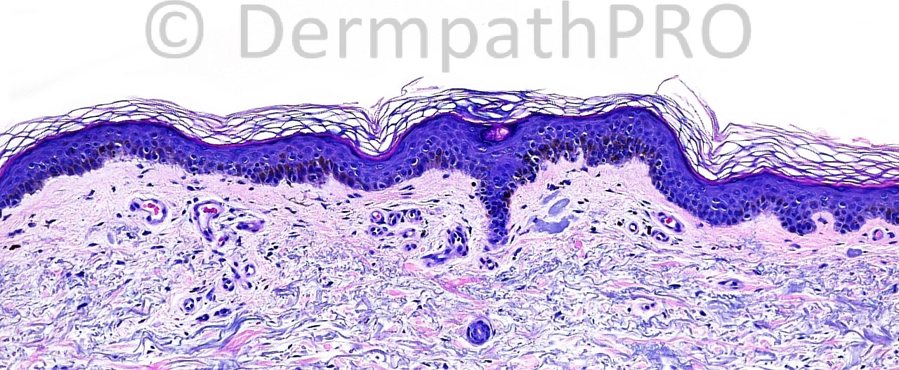
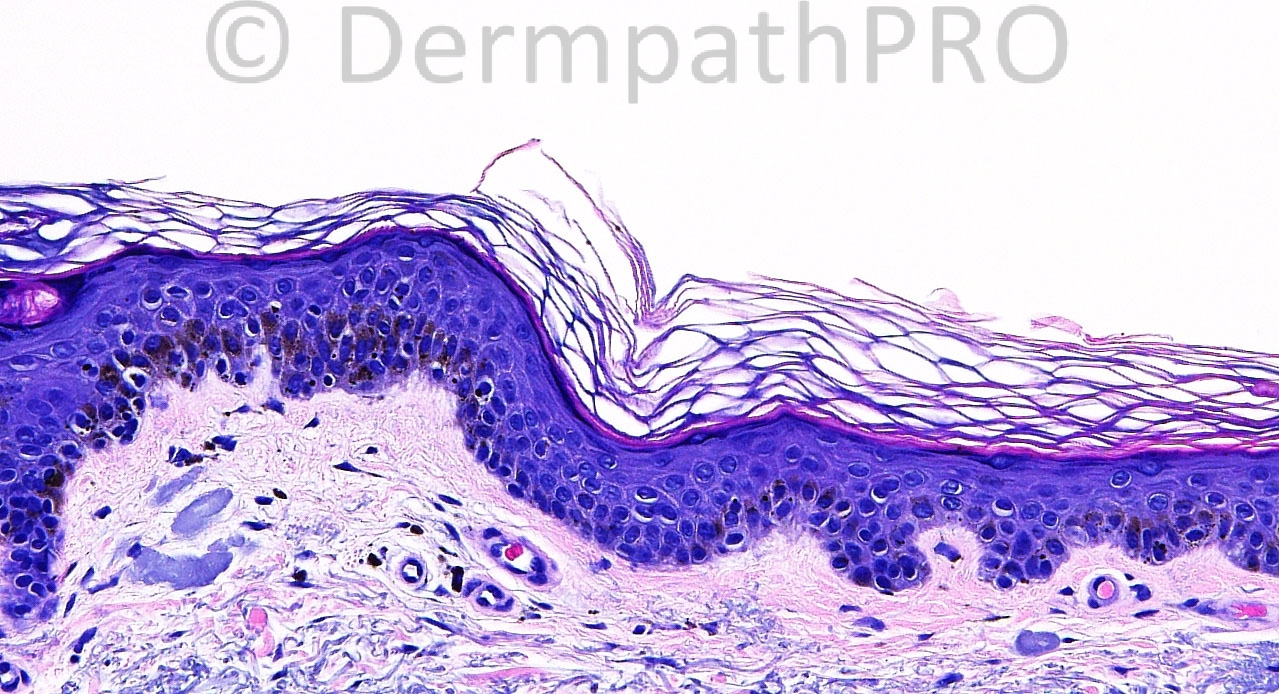
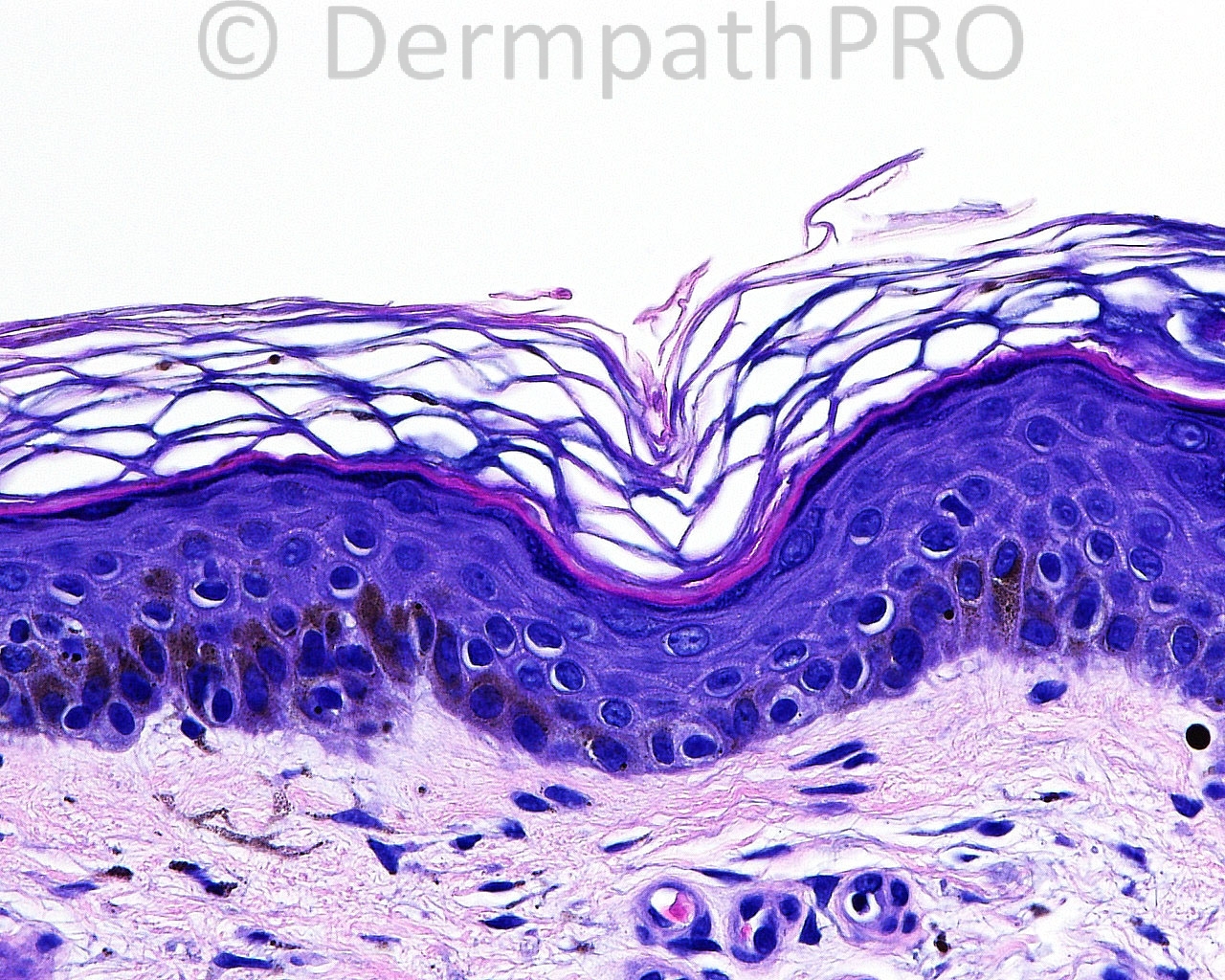
The patient is a 53 year old man with shave biopsies taken from the right ear.

Case posted by Dr. Mark Hurt.


  Report Record

User Feedback


Guest Tiberiu Tebeica

Posted

Very broad junctional melanocytic lesion on sun damaged skin, highlighted by Mart-1. There is some brown stuff in the papillary dermis, but from low magnification I guess they are melanophages. Since Mart-1 often picks up activated keratinocytes too, I prefer to use it in conjunction with MiTF to better characterize the pagetoid spread and confluent proliferation of melanocytes. This is certainly early melanoma in situ, lentigo maligna type.

Share this comment


Link to comment
share_externally

Guest Jim Davie MD

Posted

I favor lentigo simplex.

Although there is marked solar elastosis, the junctional melanocytes appear cytologically bland (no enlargement, pleomorphism, hyperchromatism, angulated nuclear/cytoplasmic contours, or 'starburst' multinucleated giant cells). No significant architectural atypia is present (neither high level intraepidermal pagetoid scatter, nor downward pagetoid spread along eccrine or follicular adnexa on Melan-A immunostain; there is no lentiginous confluence, and no irregular junctional nesting at least in the images provided). This biopsy shows heavy melanin in basal keratinocytes and in stratum corneum, which I imagine might clinically correspond with an 'inkspot lentigo' or dark macule.

Share this comment


Link to comment
share_externally

Guest Giovanni Falconieri

Posted

De novo lentiginous melanocytic proliferation in sun damaged skin, adult patient. Of course melanoma in situ goes on top of differential although I share the doubts of dr Davie,and I am not able to dispell matter on the posted images. Unfortunately, Melan A may not be always helpful in these circumstances inasmuch as it is often picked up by keratinocytes, either single or in nests hence the word of caution by Graz people (AJD: 2004 Oct;26(5):364-6; AJD 2009 May;31(3):305-8) to use it in combination with other markers including HMB45 and S100 protein.

Share this comment


Link to comment
share_externally

Guest Jim Davie MD

Posted

I find the use of Giemsa counterstain (in place of hematoxylin counterstain) invaluable when using DAB (brown chromogen) immunohistochemistry on pigmented lesions; I find it a [b]much [/b]preferred alternative to melanin bleaching.
Melanin granules will stain a distinct dark green-brown, whereas DAB is a pure brown; this makes identification of melanophages and pigmented keratinocytes much easier, and does not suffer the cytology-obscuring, overstaining artefact often seen when using ACE (red) chromogen as an alternative to DAB (brown), which makes it easy to see both melanin granules and positive DAB staining in the same cells. [Not to mention, DAB is more durable than ACE over the years.]

Share this comment


Link to comment
share_externally


Join the conversation

You can post now and register later. If you have an account, sign in now to post with your account.

Guest
Add a comment...

×   Pasted as rich text.   Paste as plain text instead

  Only 75 emoji are allowed.

×   Your link has been automatically embedded.   Display as a link instead

×   Your previous content has been restored.   Clear editor

×   You cannot paste images directly. Upload or insert images from URL.

Loading...

×
×
  • Create New...