Jump to content
In this section we have spot diagnoses posted on a daily basis since June 2010, now over 4000! You can review the archived cases and read the suggested diagnoses by users and the final comment by the contributors.
Case are uploaded each week day by 10 am UK time with the correct diagnosis will generally be posted at 8 pm UK time. Why not view the most recent spot diagnosis and proffer a diagnosis?

Case Number : Case 848 - 17th September Posted By: Guest

Please read the clinical history and view the images by clicking on them before you proffer your diagnosis.
Submitted Date :
   (0 reviews)

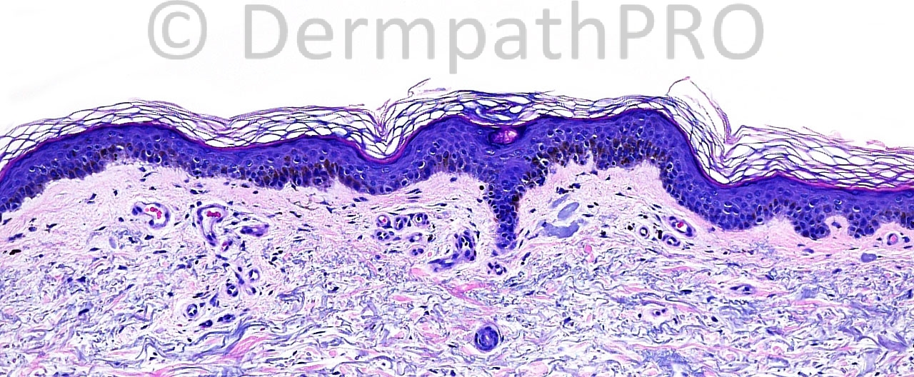
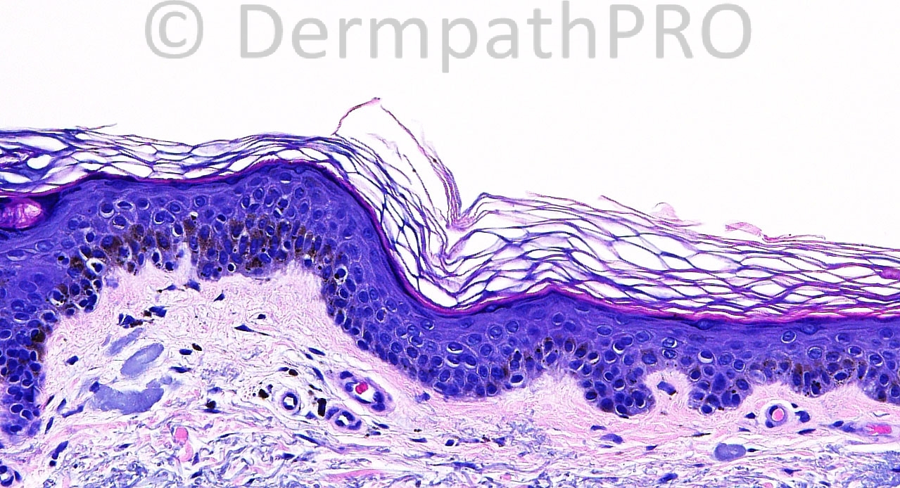
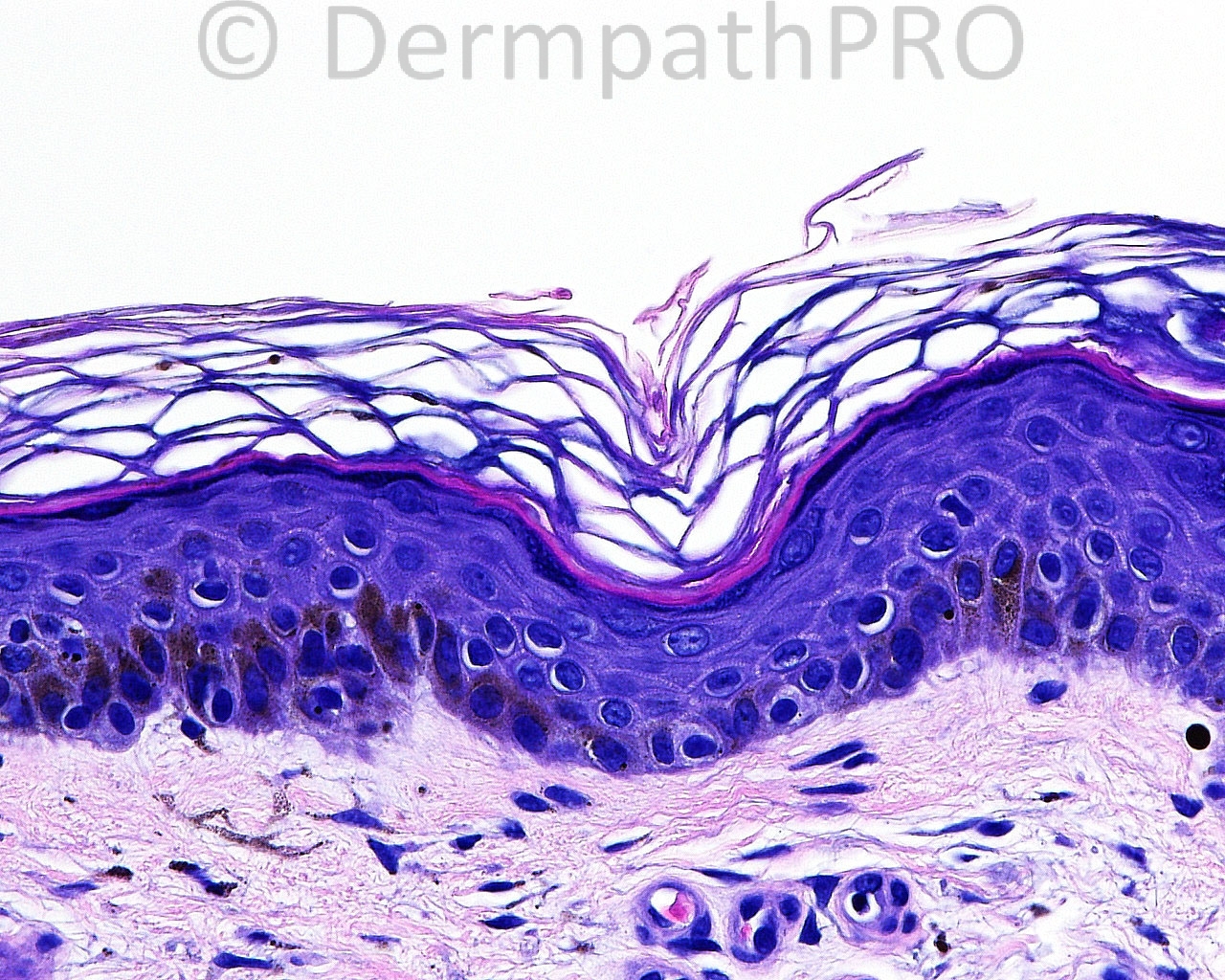
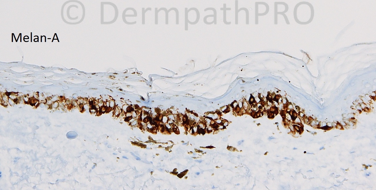
The patient is a 79 year old white woman with shave biopsies taken from the right calf.

Case posted by Dr. Mark Hurt.

  Report Record

User Feedback


Hanan Vaknine MD

Posted

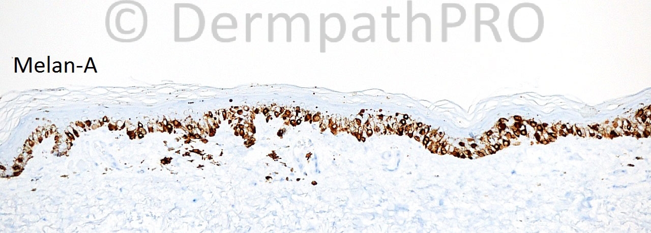
There is a prominent discrepancy between the H&E-stained histology (which does not show extensive melanocytic proliferation) and the Melan A (which appears to stain many cells); therefore one should wonder whether he is dealing with overstained keratinocytes (or more correctly stated picked up by keratinocytes). MITF, being a nuclear stain, may resolve this dilemma. I favor a non-malignant pigmented process.

Share this comment


Link to comment
share_externally

Guest Saleem Taibjee

Posted

Agree. Would be nice to see additional melanocytic markers or dual staining to clarify if this is predominantly keratinocyte staining with Melan-A (favoured), although no doubt some of the crisper staining does appear to relate to dendritic melanocytes, versus genuine melanocytic increase.

Share this comment


Link to comment
share_externally

Clinical description will definitely help noting the [b]size, colour, border, any surface changes and any skin markings alterations[/b] preferably with the use of dermatoscope. There is [b]basal hyperpigmentation[/b]. [b]Melanocyte number seems to be increased[/b] (but [b]Melan-A can recognise the antigen transferred from melanocytes to keratinocytes[/b]). The papillary dermis contains a sparse [b]lymphoid infiltrate, solar elastosis and scattered melanophages[/b]. Lentigo is a possibility. Lentigo is a risk factor for skin cancer and sunburn. Lentiginous melanoma is another possibility.

Share this comment


Link to comment
share_externally

Dr. Mona Abdel-Halim

Posted

Same as Sasi, feeling confused, waiting to be enlightened!!!!

Share this comment


Link to comment
share_externally

Guest Jim Davie MD

Posted

[i]This case looks familiar! There is a website glitch where there are two cases labeled #847 from yesterday....today's case is partially copied there as the first of the two cases #847 (with fewer immunostain images). I will copy over my opinions from that earlier case, to here...[/i]

Share this comment


Link to comment
share_externally

Guest Jim Davie MD

Posted

[quote name='Jim Davie MD' timestamp='1379372689']
I favor lentigo simplex.

Although there is marked solar elastosis, the junctional melanocytes appear cytologically bland (no enlargement, pleomorphism, hyperchromatism, angulated nuclear/cytoplasmic contours, or 'starburst' multinucleated giant cells). No significant architectural atypia is present (neither high level intraepidermal pagetoid scatter, nor downward pagetoid spread along eccrine or follicular adnexa on Melan-A immunostain; there is no lentiginous confluence, and no irregular junctional nesting at least in the images provided). This biopsy shows heavy melanin in basal keratinocytes and in stratum corneum, which I imagine might clinically correspond with an 'inkspot lentigo' or dark macule.
[/quote]

Share this comment


Link to comment
share_externally

Guest Jim Davie MD

Posted

[quote name='Jim Davie MD' timestamp='1379430098']
I find the use of Giemsa counterstain (in place of hematoxylin counterstain) invaluable when using DAB (brown chromogen) immunohistochemistry on pigmented lesions; I find it a [b]much [/b]preferred alternative to melanin bleaching.
Melanin granules will stain a distinct dark green-brown, whereas DAB is a pure brown; this makes identification of melanophages and pigmented keratinocytes much easier, and does not suffer the cytology-obscuring, overstaining artefact often seen when using ACE (red) chromogen as an alternative to DAB (brown). This makes it easy to see both melanin granules and positive DAB staining in the same cells. [Not to mention, DAB is more durable than ACE over the years.]
[/quote]

Share this comment


Link to comment
share_externally

Dr. Richard Carr

Posted

As usual Jim pretty much read my own thought processes! I was thinking of a lesion akin to dark flat solar lentigo or ink-spot lentigo-like pigmented macular lesion. I don't see any increase in melanocytes on the H&E's (so did not go for a simple lentigine / lentigo simplex) and presumed the IHC is mainly DAB but spuriously expressed in the keratinocytes. Will give Jim's suggestion a try in future but I usually find a very thin good quality H&E is preferable to melanin bleach (or IHC) in any case for darkly pigmented lesions.

Share this comment


Link to comment
share_externally

Robledo F. Rocha

Posted

I think there's no melanocytic proliferation, but just epidermic hyperpigmentation characterized by alternating areas of heavy hyperpigmented keratinocytes and areas of light hyperpigmented keratinocytes, what may impart a reticulate appearance to the lesion on clinical examination. Following this line of reasoning, I could think of late Kitamura's reticulate acropigmentation or related disorders.
Aditional clinical information certainly will be helpful.

Share this comment


Link to comment
share_externally

Weedon points out that [b]a subsequent excision[/b] may show more pronounced features than the original biopsy specimen. This is particularly so for lesions on actinically damaged skin, in which up to 40% of excisions may show more pronounced changes.

Share this comment


Link to comment
share_externally

On a different note, I would like to thank Dr McKee for the extremely useful site he had. I have been an occasional visitor to the new DermpathoPro site and I wish it the best.

Share this comment


Link to comment
share_externally


Join the conversation

You can post now and register later. If you have an account, sign in now to post with your account.

Guest
Add a comment...

×   Pasted as rich text.   Paste as plain text instead

  Only 75 emoji are allowed.

×   Your link has been automatically embedded.   Display as a link instead

×   Your previous content has been restored.   Clear editor

×   You cannot paste images directly. Upload or insert images from URL.

Loading...

×
×
  • Create New...