Jump to content
In this section we have spot diagnoses posted on a daily basis since June 2010, now over 4000! You can review the archived cases and read the suggested diagnoses by users and the final comment by the contributors.
Case are uploaded each week day by 10 am UK time with the correct diagnosis will generally be posted at 8 pm UK time. Why not view the most recent spot diagnosis and proffer a diagnosis?

Case Number : Case 856 - 27th September Posted By: Guest

Please read the clinical history and view the images by clicking on them before you proffer your diagnosis.
Submitted Date :
   (0 reviews)

85 year old female with an erythematous scale measuring 10mm on the leg.

Case posted by Dr. Richard Carr.


  Report Record

User Feedback


Guest Maria George

Posted

Is this a melanoma which also destruct the hair follicle or a melanoma with herpetic folliculitis.Is the patient on chemotherapy or radiotherapy? .Need more clinical info.

Share this comment


Link to comment
share_externally

Robledo F. Rocha

Posted

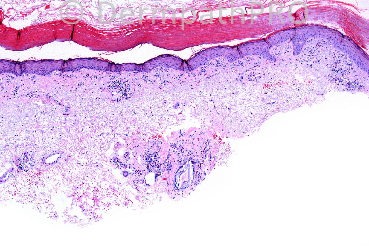
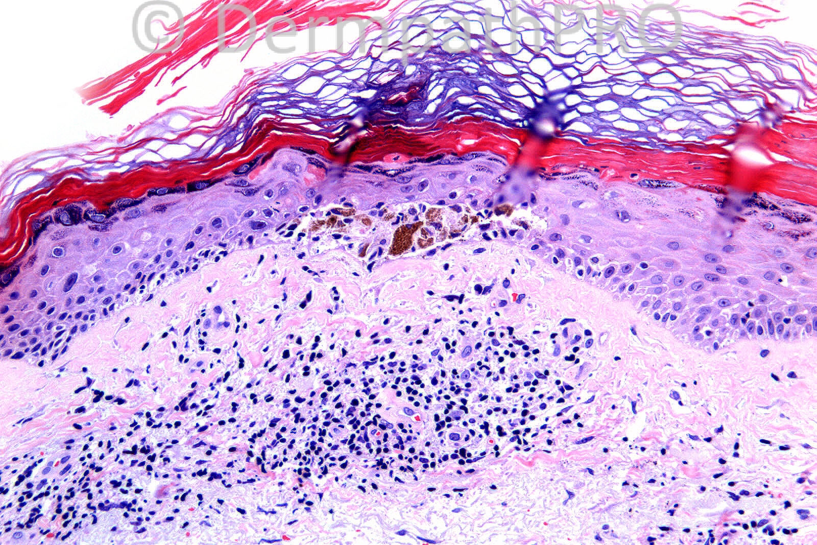
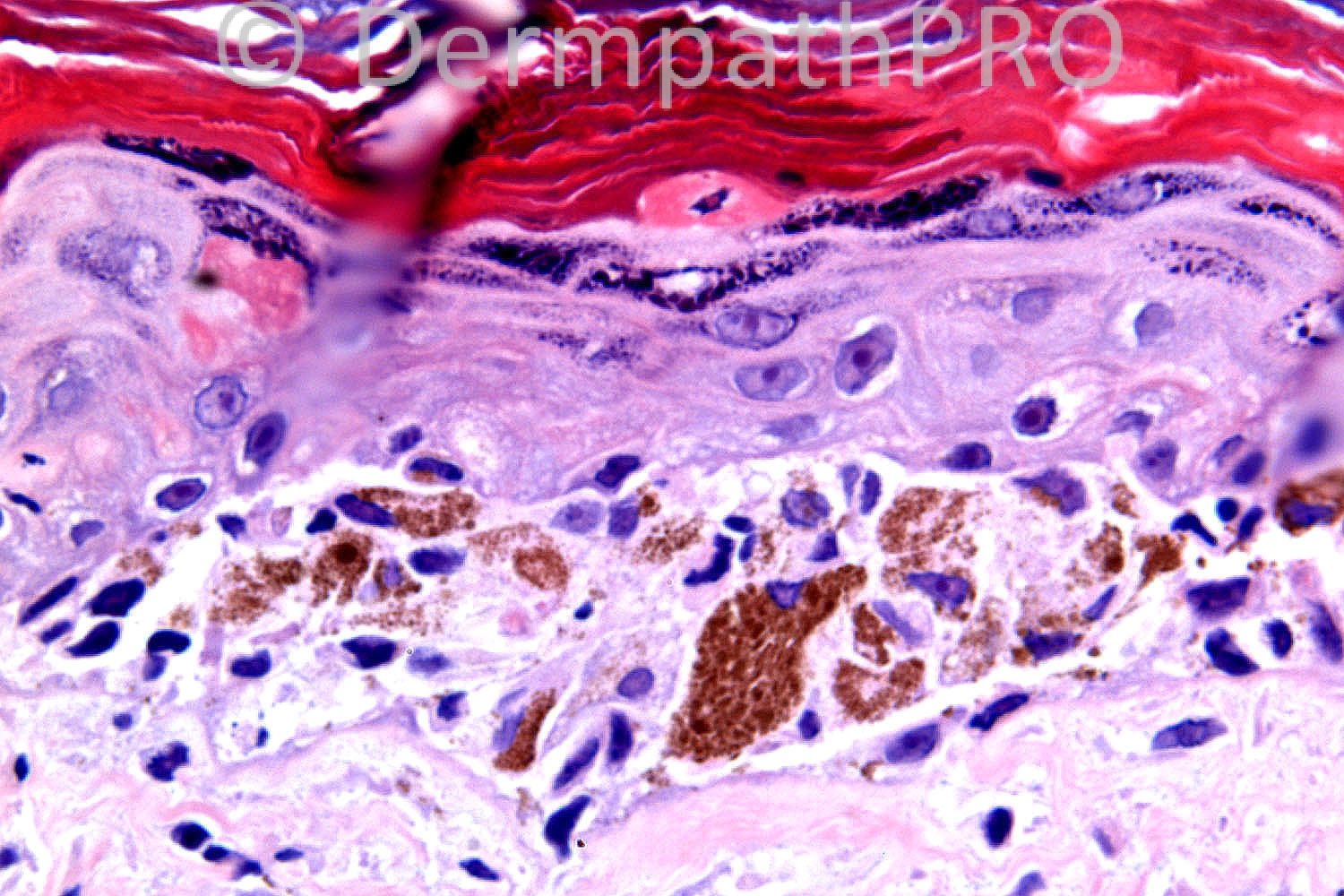
[color="#000000"][size="4"]A few dyskeratotic keratinocytes restricted to upper spinous and granular layers that overlie a subepidermal microvesicle containing melanophages and [/size][/color][color="#000000"][size="4"]acantholytic[/size][/color][color="#000000"][size="4"] keratinocytes (related to [/size][/color][color="#000000"][size="4"]acrotrichium?[/size][/color][color="#000000"][size="4"]). There are also degenerative changes in a deep adnexal (follicle?) portion. These findings suggest viral infection, probably [/size][/color][color="#000000"][size="4"]herpes simplex folliculitis, although I didn’t find any persuasive [/size][/color][color="#000000"][size="4"]specific virus-induced cytopathic change.[/size][/color]
[color="#000000"][size="4"]In addition to the case history of a solitary non-blistering lesion, the presence of a [/size][/color][color="#000000"][size="4"]sandwich, whose strata are parakeratosis[/size][/color][color="#000000"][size="4"] / basket-weave [/size][/color][color="#000000"][size="4"]orthokeratosis[/size][/color][color="#000000"][size="4"] / [/size][/color][color="#000000"][size="4"]parakeratosis /[/size][/color][color="#000000"][size="4"] granular layer, point to [/size][/color][color="#000000"][size="4"]intermittent activity or healing lesion.[/size][/color]

Share this comment


Link to comment
share_externally

Guest Jim Davie MD

Posted

Favor discoid lupus. (differential: dermatomyositis, atrophic lichen planus, lichenoid drug reaction)

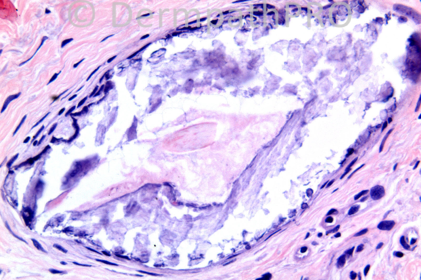
The epidermis shows features of interface dermatitis resembling lupus, with reactive keratinocyte enlargement and pleomorphism, effacement of rete pegs, slightly thickened basement membrane, coarse hypergranulosis, and compact orthokeratosis. There are scattered lymphocytes at the junction. The transition between basketweave orthokeratosis and the more recent, underlying compact orthokeratosis in the stratum corneum suggests abrupt onset of the inflammatory process. The pigmented cells at the junction appear to be melanophages. I doubt there is a melanocytic proliferation, given the above features (I've heard anecdotally of cases of lupus masquerading convincingly as a melanoma in situ, especially in small biopsies). There is a sparse perivascular chronic inflammatory infiltrate with polarity towards the epidermis, and periadnexal infiltrate. The 4th picture shows a central hair shaft surrounded by a bit of orthokeratotic plugging, and an outer concentric eggshell-type dystrophic calcification, possibly secondary to complete obliteration of the follicular epithelium by prior periadnexal inflammation.

Share this comment


Link to comment
share_externally

Dr. Richard Carr

Posted

A small turn-out today so far. What a shame as this case is a little tricky. Jim and Mona doing very well as always! I agree lichenoid with pseudomelanocytic nests of macrophages - so very well done on that. I want you to take it further though as you have all missed something. Well done to Jim who spotted the vellus hair shaft in the calcified follicle. Looking forward to more comments - this could run till Monday! I am currently in Florence at the ISDP symposium - wow!! what a day amazing lectures and up-dates - what they are finding in melanocytic lesions is incredible. Wondering if an old morphologist can make it to retirement - ?worth a blog!

Share this comment


Link to comment
share_externally

Dr. Mona Abdel-Halim

Posted

Thanks Dr Carr for your nice comment. I will push it further: thickening of the stratum corneum with both orthokeratosis and parakeratosis. Necrotic keratinocytes with some high up. Perifollicular infiltrate. Evident thickening and deposition of collagen in the upper dermis as well as around the hair follicles. The follicle changes seem to have been severe with secondary calcification. Could be dealing with a lesion of GVHD ?? Subacute evolving to chronic?? Esinophils are also seen, but they r not against GVHD...

Share this comment


Link to comment
share_externally

Dr. Mona Abdel-Halim

Posted

I think the clinical description will not be favoring GVHD, so I will be pushing it again and considering the clinical description of a 10 mm erythematous scale, which suggests a solitary lesion, and will consider lichen planus like keratosis (lichenoid keratosis).

Share this comment


Link to comment
share_externally

Guest Jim Davie MD

Posted

If we are missing something else, I offer the following observations (with some equivocation based on the detail limits of the photos):
1. Possible hyperchromatic, angulated lymphocytes in the dermis, with junctional lymphocytes showing halos. This could merely represent processing or photo artefact. But if real, could represent pseudo-MF (drug), or MF. MF is deceptive, and can mimick a multitude of inflammatory patterns.

2. Extravasated red cells with possible granular debris (pigment?) in dermis. This raises consideration of a lichenoid pigmented purpura if the clinical fits; however, red cells and pigment are non-specific on lower extremity, and can be seen in multiple inflammatory processes, including lupus. Or just be artefactual to biopsy procedure.

Additional clinical clues (e.g. single or multiple lesions) will be very useful to pare down the differential diagnosis list.

Share this comment


Link to comment
share_externally

Dr. Richard Carr

Posted

Just a solitary 10mm scaly lesion. Also what is the significance of the follicular calcification. The latter may or may not be totally incidental.

Share this comment


Link to comment
share_externally

Robledo F. Rocha

Posted

What about lupus erythematosus-like lichenoid keratosis? It fits on the clinical presentation and Weedon’s book mention subepidermal clefting and follicular involvement at a slightly deeper level. Unfortunately, I didn’t find any information about follicular calcification in this entity.

Share this comment


Link to comment
share_externally

Dr. Richard Carr

Posted

Okay a little more help. I deliberately did not make this easy although it was tricky looking at the glass slides blind. The main pathology (i.e. the scaly lesion) is not a rare thing. The follicular calcification (may be totally incidental) but is in my experience exceedingly rare (I would be interested in others views - try to think if you have seen this before) and I wanted you to be aware follicular calcification can be a clue to a rare diagnosis. So three questions. 1. What is the main diagnosis of the scaly lesion. 2. Can you have incidental follicular calcification. 3. What rare diagnosis has follicular calcification as a clue? Will this run till Monday? Unfortunately I have to leave the conference early today because I cannot get across to the airport easily due to the world cycling championships in Florence today!! I have to leave early to cross the cycling course to be on the same side of the city as the airport!! I will therefore miss the self assessment case presentations which I am sure will be stunning.

Share this comment


Link to comment
share_externally

Alistair Robson

Posted

Dear All. Buongiorno from the ISDP meeting in Florence! This looks like a case of porokeratosis with pseudo-melanocytic nest. Regards Alistair

Share this comment


Link to comment
share_externally

Arti Bakshi

Posted

Wow, what a discussion!
After all the clues which Dr Carr has given and a bit of reading, I too arrived at a diagnosis of porokeratosis. (glad that Dr Robson thinks so too).
Follicular involvement may occur in porokeratosis and maybe the follicular calcification is a result of this.

Share this comment


Link to comment
share_externally

Dr. Richard Carr

Posted

I will let this run till this evening - hope Mark does not mind me dragging it out a little.

Share this comment


Link to comment
share_externally

Guest Saleem Taibjee

Posted

Since no-one else has mentioned this yet, and I think this is what Richard may be hinting at:
I have seen a paediatric case of Conradi-Hunermann-Happle syndrome (diagnosed clinically by an experienced paediatric dermatologist) in which the histology showed ichyosis and follicular calcification which was felt to be supportive of the clinical diagnosis. In cases of icthyosis, calcification of the stratum corneum and follicle seems to be specific for CHH to distinguish from other icthyoses.
But I presume in this case it is an incidental finding, although might still be prudent to check serum calcium, parathyroid hormone?

Share this comment


Link to comment
share_externally

Sasi Attili

Posted

I am glad I am not yet late!

I see a focal lichenoid infiltrate with few atypical lymphocyte and melanophages. I am not sure I can explain the hair follicle calcification, but don't have an answer o what it is a clue for.

Given that this is a solitary lesion Lichenoid purpura (which can overlap with MF) and resolving lichenoid keratosis (diagnosis of exclusion) would be my first thoughts. I would favour the former, though solitary MF is a rare and difficult diagnosis to make. I don't think that this is a genetic syndrome, given the patients age.

Share this comment


Link to comment
share_externally

Dr. Richard Carr

Posted

The answer is porokeratosis with pseudomelanocytic nests and (probably) incidental follicular dystrophic calcification. Saleem is quite right I wanted to bring your attention to follicular calcification in ichthyosis as a clue to Conradi-Hunermann-Happle syndrome (I am assuming Saleem's spelling is correct and it was Saleem who taught me on this condiation in any case!). Alastair made the correct diagnosis from my mobile phone (in Florence) on rather small images so it could be done. I think his "educated" guess was partly based on the clinical but it was still a very very impressive call!! Thank you all for a great discussion and differentials on the case.

Share this comment


Link to comment
share_externally

Sasi Attili

Posted

Thanks for the interesting case Richard. I find it a bit difficult though, to accept the diagnosis of porokeratosis here (esp without the clinical picture), unless the clinical was very suggestive? The cornoid lamella (if you can call it that) in this picture is certainly not something that one would photograph for a text book.

Can you please explain the following:

1. Did you think the lymphocytes (I presume confirmed on immunostains) in the 'pseudomelanocytic nests' are atypical? Has this been described before?
2. Dyskeratosis in PK is usually confined to the area under the cornoid lamella. However, as others have pointed out I think the epidermis surrounding this also shows dyskeratotic keratinocytes associated with parakeratosis? Is this true or an illusion? (we are not seeing much of the surrounding epidermis). If this is true- can the cornoid lamella be an incidental finding (as it can be) in a possibly dysplastic in-situ keratinocytic tumor?
3. Hypergranulosis is not a feature seen under cornoid lamella, as seen here (hypogranulosis is).
4. What is your view on the nature of pseudo melanocytic nests which have actually been [url="http://www.ncbi.nlm.nih.gov/pubmed/21812802"]shown to stain with SOX-10 and MITF[/url] in a recent paper? These might not be pseudo after all!

Thanks a lot once again. Regards.

Sasi

Share this comment


Link to comment
share_externally

Dr. Richard Carr

Posted

Thanks Sasi,
The clinical was porokeratosis (I with-held that info deliberately) and there were several other nice cornoid lammella in the biopsies. I deliberately only photographed the edge of one (upper right and lower left) but don't expect them to read the textbooks. The high dyskeratotic cells and focally interupted granular layer is clearly evident as is the slanting alternation in the cornfiication. The compact hyperkeratosis overlying effaced rete ridges in top left image presumably is secondary lichenification. I was a bit surprised by all the suggestions to be honest! The "nest-like" arrangement in this case is a few lymphocytes (atypia is often in the eye of the beholder but with this clinical CTCL is just not even going to be considered) and predominantly histiocytes and melanophages. I did not do Sox-10 or MITF but the cells were S100 and MelanA negative if that helps.

Share this comment


Link to comment
share_externally


Join the conversation

You can post now and register later. If you have an account, sign in now to post with your account.

Guest
Add a comment...

×   Pasted as rich text.   Paste as plain text instead

  Only 75 emoji are allowed.

×   Your link has been automatically embedded.   Display as a link instead

×   Your previous content has been restored.   Clear editor

×   You cannot paste images directly. Upload or insert images from URL.

Loading...

×
×
  • Create New...