Jump to content
In this section we have spot diagnoses posted on a daily basis since June 2010, now over 4000! You can review the archived cases and read the suggested diagnoses by users and the final comment by the contributors.
Case are uploaded each week day by 10 am UK time with the correct diagnosis will generally be posted at 8 pm UK time. Why not view the most recent spot diagnosis and proffer a diagnosis?

Case Number : Case 1003 - 28th April Posted By: Guest

Please read the clinical history and view the images by clicking on them before you proffer your diagnosis.
Submitted Date :
   (0 reviews)

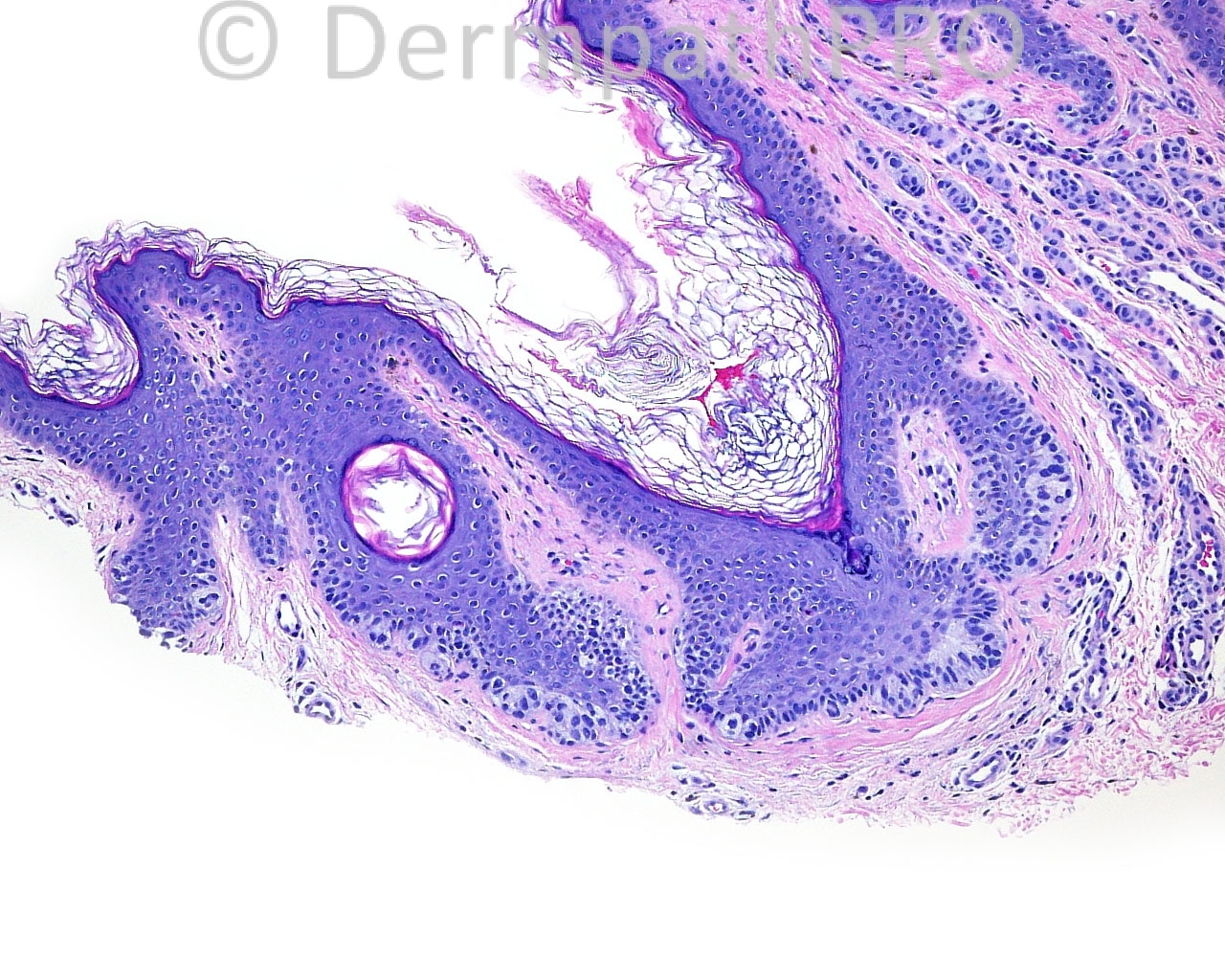
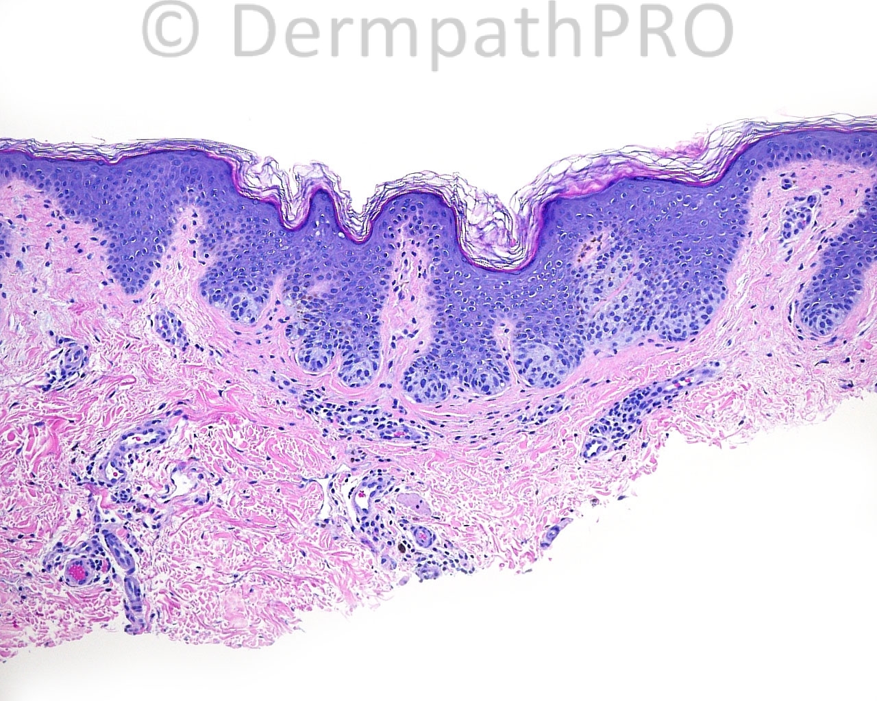
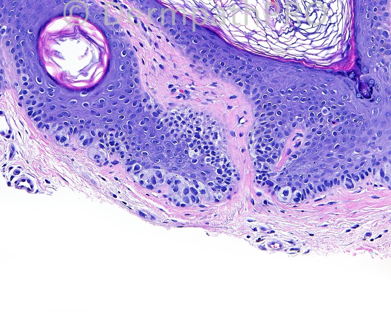
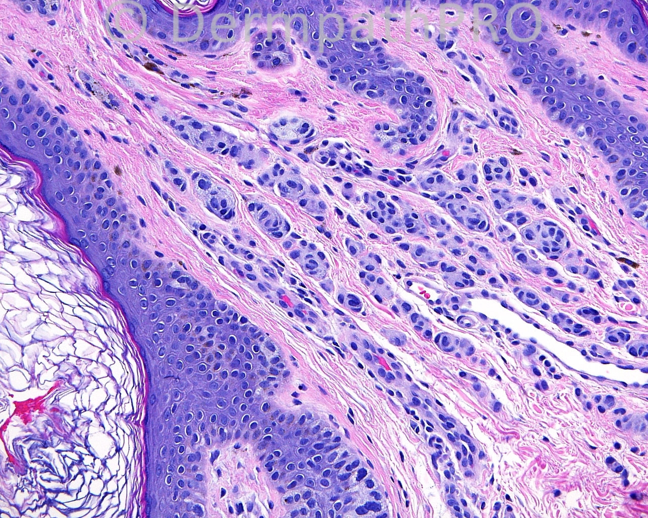
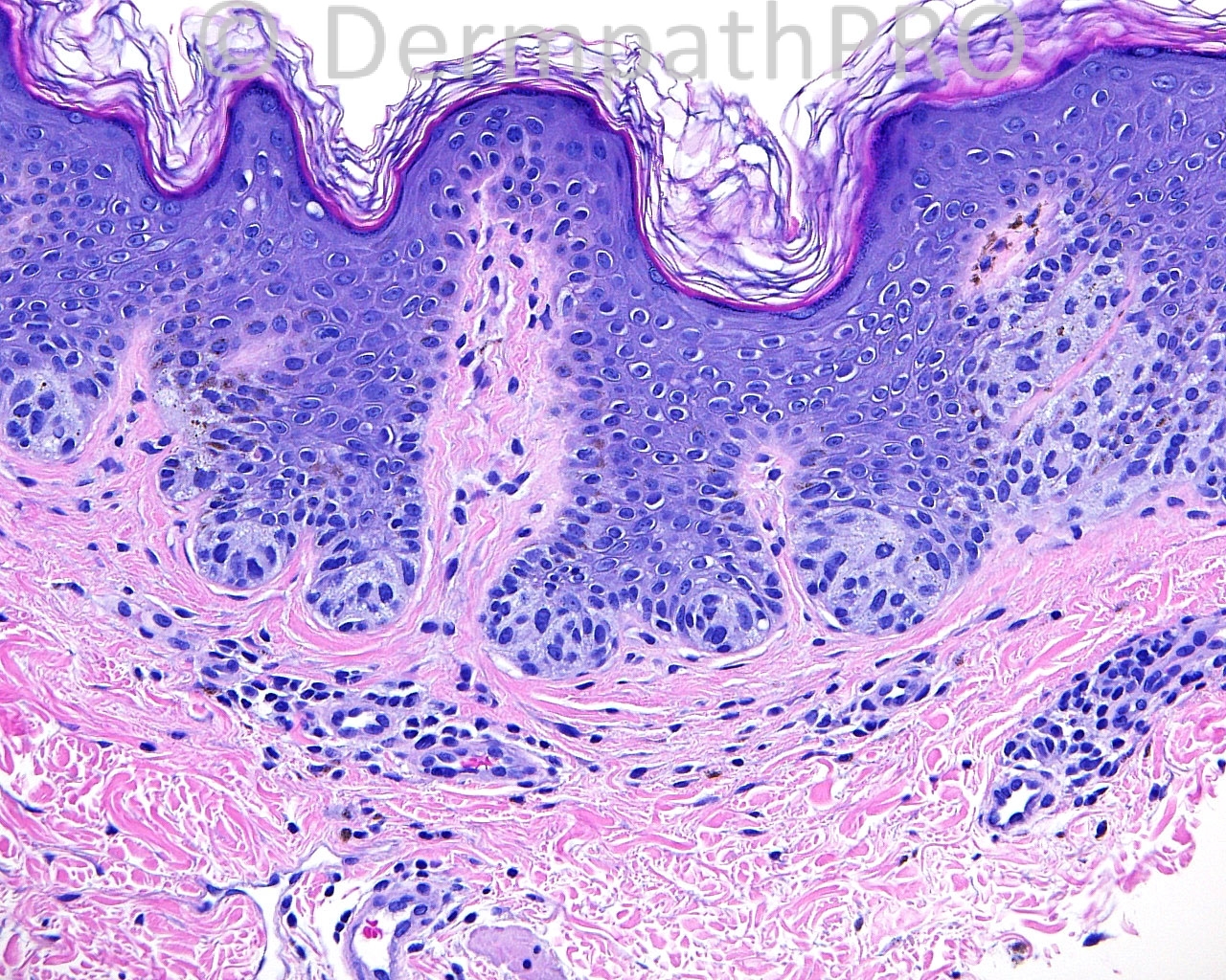
The patient is a 34 year old woman with shave biopsies of a lesion on the left groin.

Case posted by Dr. Mark Hurt.


  Report Record

User Feedback


Dr. Mona Abdel-Halim

Posted

Dysplastic nevus (moderate/severe dysplasia)

Share this comment


Link to comment
share_externally

nick turnbull

Posted

I think it might be Atypical genital or flexural nevi. The associated epidermis is a bit papillomatous. The junctional component quite lintigenous and nested with some architectual atypia. Mild cytological atypia is present also.
Dermal componet looks bland wih maturation, i think. Considering site and age, I guess this is benign

Share this comment


Link to comment
share_externally

Robledo F. Rocha

Posted

Nevus of inguinal region comprising enlarged, irregularly shaped and unevenly spaced junctional nests on the flanks of the lesion and intradermal small nests and cords, but inconspicuous junctional component, on the center of the lesion. Cytological features of the melanocytes are bland and mitotic figures were not identified.
I’d call this special site nevus and I’d alert for the likely recurrence of the lesion in a few months due to incomplete excision.

Share this comment


Link to comment
share_externally

Dr. Richard Carr

Posted

Looks benign and agree with above but might want to add naevus spilus (agminate naevus) as there may be more than one focus of naevus here.

Share this comment


Link to comment
share_externally

Guest Tiberiu Tebeica

Posted

The Ki67 is low in the dermal component. I don't see pagetoid spread. Even though there is some architercural disorder, I think it is related mostly to the anatomical localization. My diagnosis would be special site nevus. It is benign, but I would probably advise the clinician that the lesion might persist / recur with a worrisome look.

Share this comment


Link to comment
share_externally


Join the conversation

You can post now and register later. If you have an account, sign in now to post with your account.

Guest
Add a comment...

×   Pasted as rich text.   Paste as plain text instead

  Only 75 emoji are allowed.

×   Your link has been automatically embedded.   Display as a link instead

×   Your previous content has been restored.   Clear editor

×   You cannot paste images directly. Upload or insert images from URL.

Loading...

×
×
  • Create New...