Jump to content
In this section we have spot diagnoses posted on a daily basis since June 2010, now over 4000! You can review the archived cases and read the suggested diagnoses by users and the final comment by the contributors.
Case are uploaded each week day by 10 am UK time with the correct diagnosis will generally be posted at 8 pm UK time. Why not view the most recent spot diagnosis and proffer a diagnosis?

Case Number : Case 986 - 3rd April Posted By: Guest

Please read the clinical history and view the images by clicking on them before you proffer your diagnosis.
Submitted Date :
   (0 reviews)

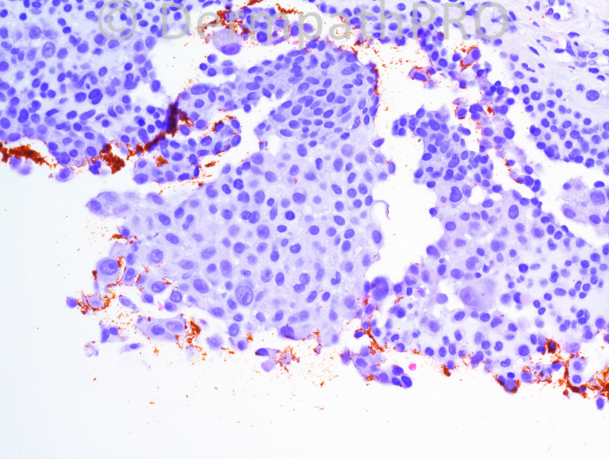
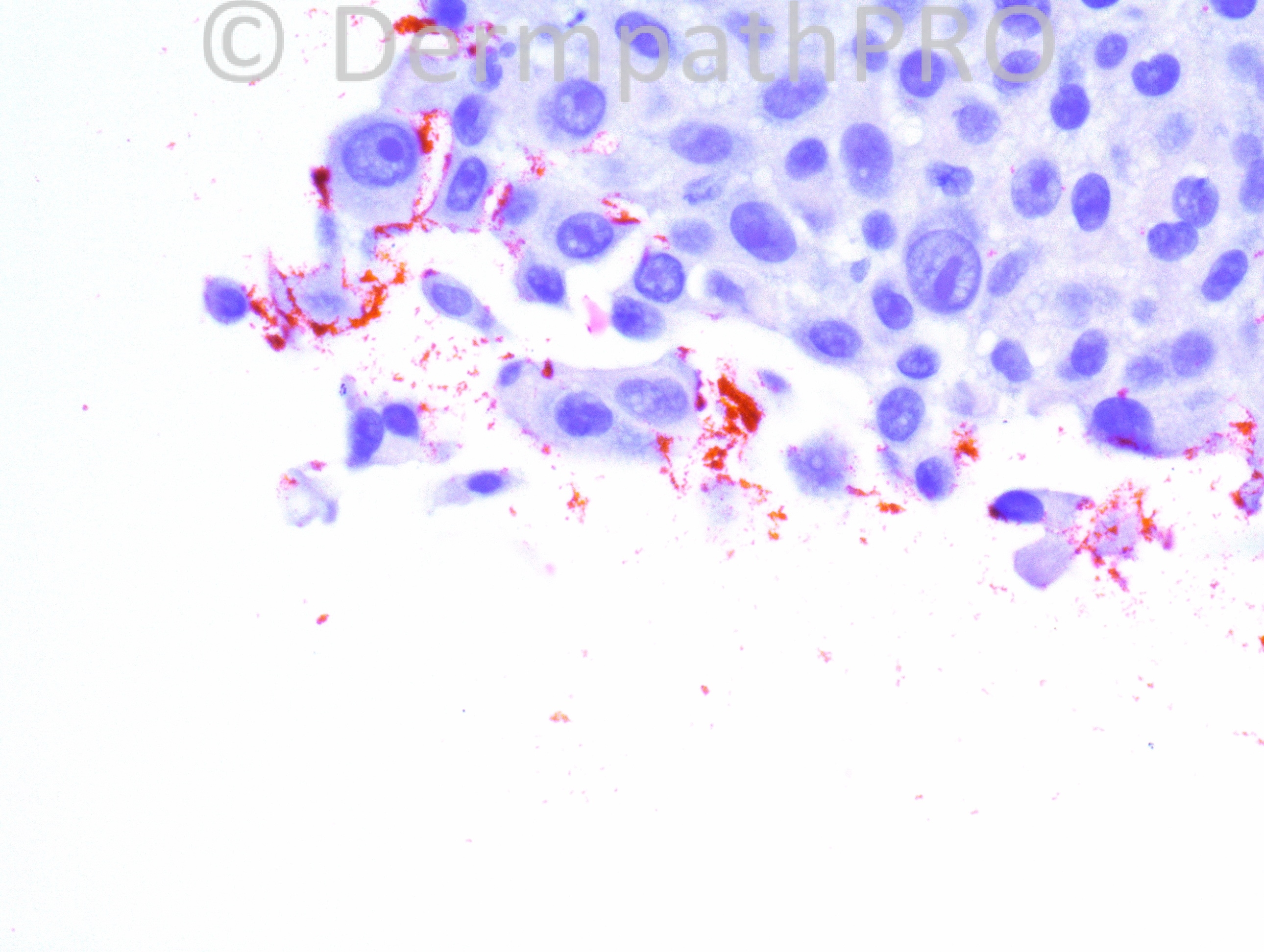
80 year-old Hispanic female with a lesion on her left shin. Clinical impression: rule out BCC.

Case posted by Dr. Hafeez Diwan.


  Report Record

User Feedback


Dr. Mona Abdel-Halim

Posted

I think this is a cutaneous metastases. The specimen shows only small part of the lesion. I think we should ask for complete excision and do IHC to verify.

Share this comment


Link to comment
share_externally

Robledo F. Rocha

Posted

A very small sample of the lesion makes this case hard. My initial impression is adenocarcinoma due to the pleomorphic cells showing vesicular nuclei with prominent nucleoli and slight foamy cytoplasm, and to some subtle tubular structures.

Share this comment


Link to comment
share_externally

Hard to say. Superficial sample with a neoplastic infiltration. Agree with metastatic adenocarcinoma as first impression.

Share this comment


Link to comment
share_externally

Dr. Richard Carr

Posted

Does look quite "epithelioid". A rare suggestion to add in the differential is a symplastic glomus (fried egg cells). Would also consider epithelioid haemangioendothelioma (long shot).

Share this comment


Link to comment
share_externally

Guest Tiberiu Tebeica

Posted

agree with all the above differentials. i would add metastatic melanoma. curious what immunos show

Share this comment


Link to comment
share_externally

Dr. Hafeez Diwan

Posted

This turned out to be melanoma, presumably metastatic; there is scar and so it could be a recurrent or residual, but we didn't have a history of anything at this site. The tumor was not present on levels, and so we destained one of the H&E slides. The tumor cells were Melan-A positive. There was no prior history of melanoma.

Share this comment


Link to comment
share_externally


Join the conversation

You can post now and register later. If you have an account, sign in now to post with your account.

Guest
Add a comment...

×   Pasted as rich text.   Paste as plain text instead

  Only 75 emoji are allowed.

×   Your link has been automatically embedded.   Display as a link instead

×   Your previous content has been restored.   Clear editor

×   You cannot paste images directly. Upload or insert images from URL.

Loading...

×
×
  • Create New...