Jump to content
In this section we have spot diagnoses posted on a daily basis since June 2010, now over 4000! You can review the archived cases and read the suggested diagnoses by users and the final comment by the contributors.
Case are uploaded each week day by 10 am UK time with the correct diagnosis will generally be posted at 8 pm UK time. Why not view the most recent spot diagnosis and proffer a diagnosis?

Case Number : Case 988 - 7th April Posted By: Guest

Please read the clinical history and view the images by clicking on them before you proffer your diagnosis.
Submitted Date :
   (0 reviews)

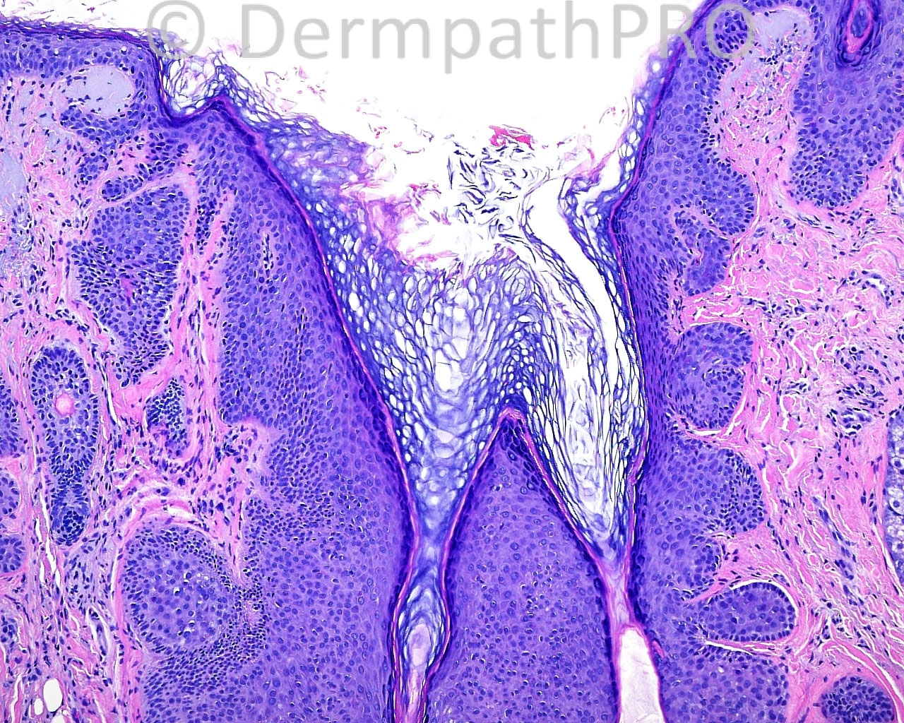
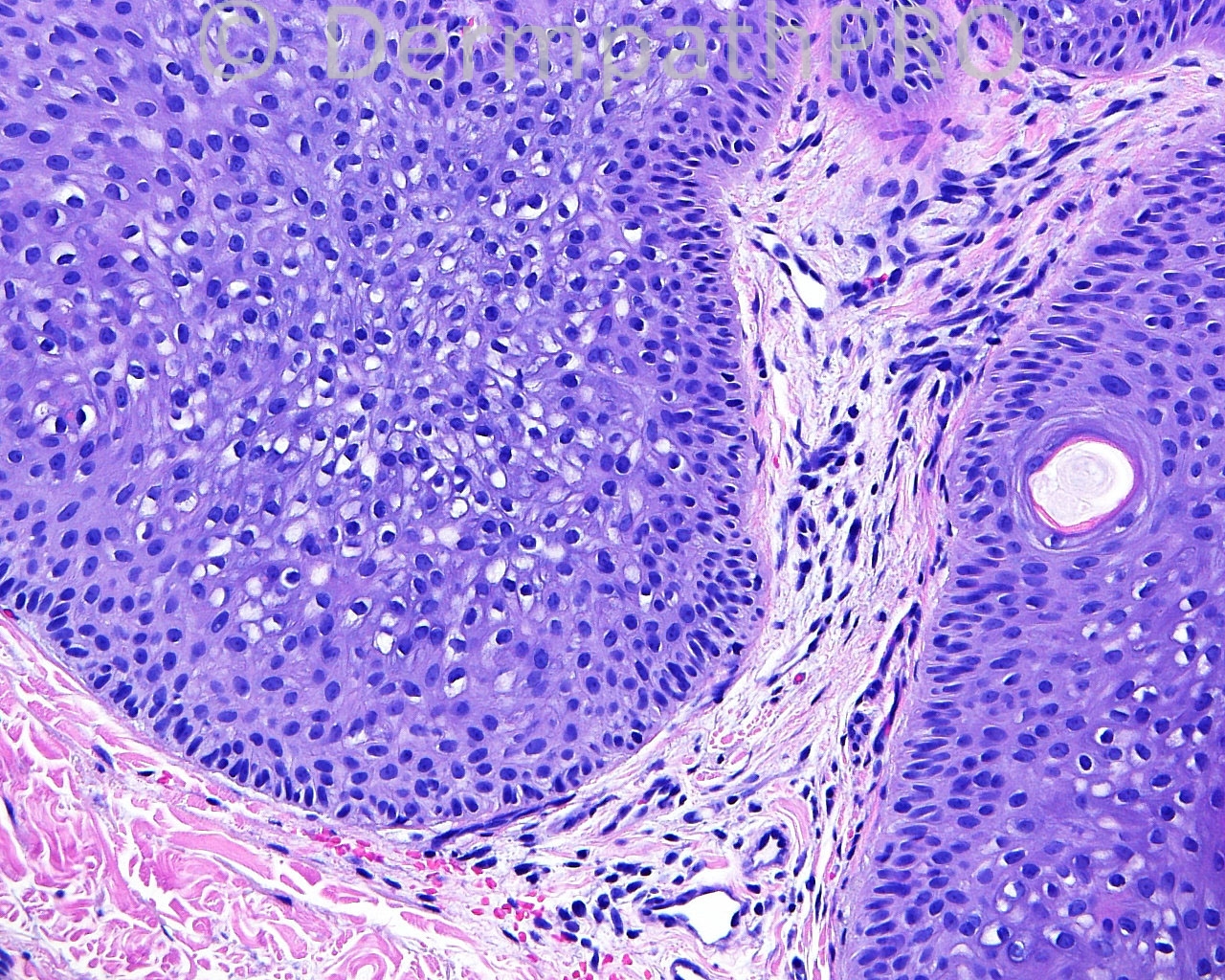
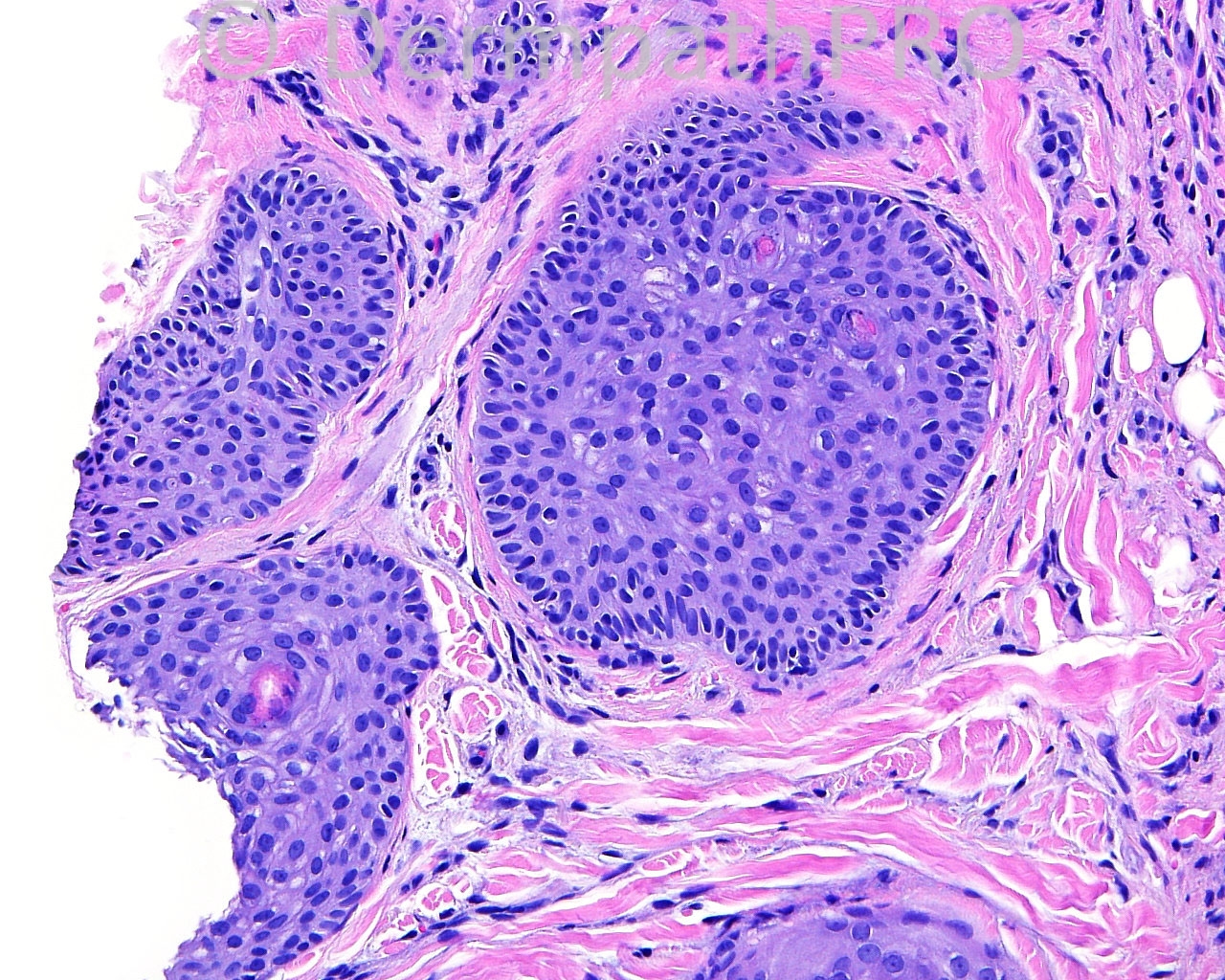
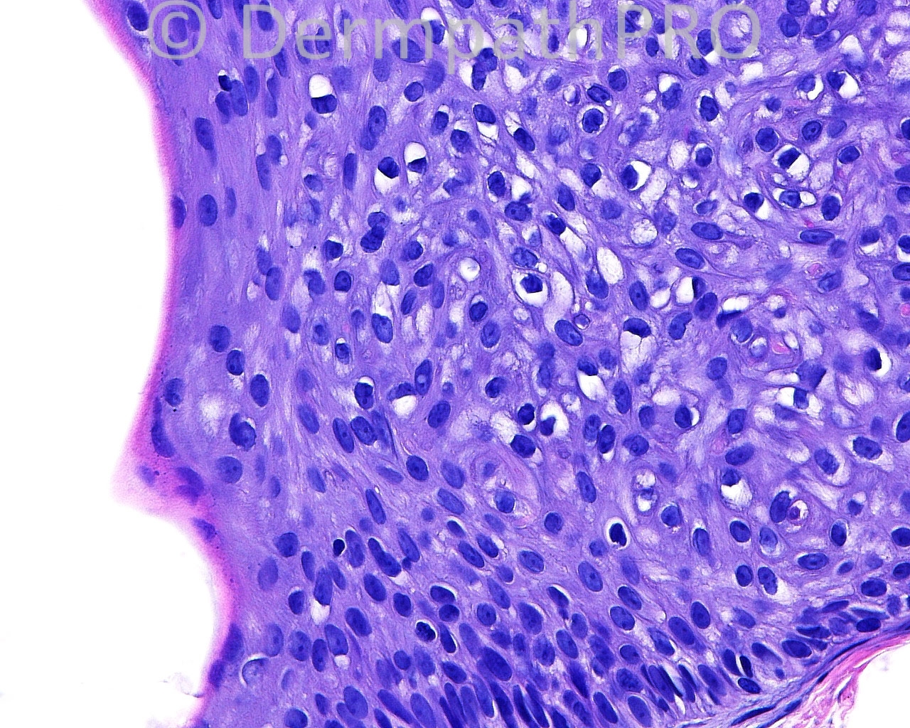
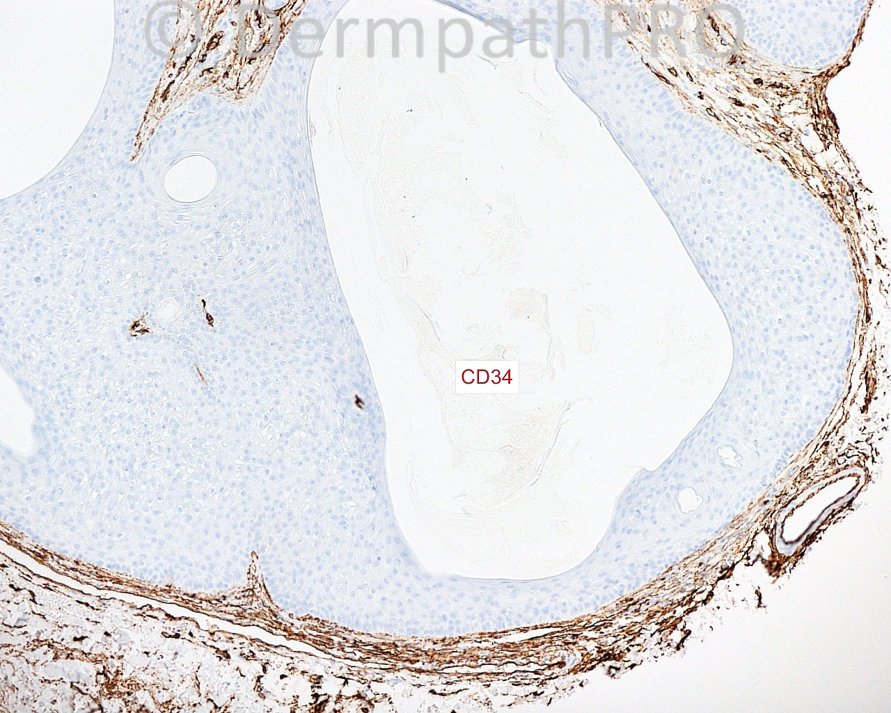
The patient is a 52 year old white man with a punch biopsy of a changing, small, ulcerated lesion on the right upper lip.

Case posted by Dr. Mark Hurt.


  Report Record

User Feedback


Robledo F. Rocha

Posted

Lobular infundibuloisthmicoma, just to bring an alternative name [url="http://journals.lww.com/amjdermatopathology/Citation/1996/08000/Pilar_Sheath_Acanthoma__Lobular.24.aspx"]proposed by Dr. Hurt[/url].

Share this comment


Link to comment
share_externally

Dr. Mona Abdel-Halim

Posted

Pilar sheath acanthoma, interesting alternative name !!!

Share this comment


Link to comment
share_externally

Mark A. Hurt MD

Posted

Yes, this is a [url="http://www.markahurt.com/files/documents/20060157hurt-Pilar-Sheath-Acanthoma.pdf"]pilar sheath acanthoma[/url], which I affectionately term "lobular infundibuloisthmicoma."

The back-story of my proposed name is that I thought the lesion looked like elements of the infundibulum and isthmus more than it did the stem and bulb; thus, my abstract in 1996, which Robledo kindly referenced.

One cannot, from the name as such, determine the nature of this lesion, because the term "tricholemmoma" (Greek - Hair Sheath Tumor) and the phrase "Pilar Sheath Acanthoma" (Latin - Hair Sheath Tumor) would seem to refer to neoplasms with the same properties -- yet those names actually refer to neoplasms with [i]different [/i]properties. The lesion we know as "tricholemmoma" differentiates toward the outer sheath at the bulb and is positive with CD34, while the "pilar sheath acanthoma," as in the example above, differentiates mainly toward the infundibulum and isthumus and lacks CD34 positivity.

If, today, one were to rename the lesion historically termed "tricholemmoma," it might be, more specifically, "follicular bulb outer sheath acanthoma" or some more stylized name, which, at present eludes me.

Thanks, everyone, for your participation!

Mark

Share this comment


Link to comment
share_externally

Dr. Richard Carr

Posted

The outer root sheath passes from the lower outer bulb, up around the entire follicular stem and up around the isthmus and blends imperceptibly with the infundibulum. You could simply argue tricholemmoma (common) and pilar sheath acanthoma (quite uncommon) are morphological variants (with actually more in common than not - I note this case is from the upper lip a typcial site for tricholemmoma) of "outer root sheath-oma". Whether the infundibular component (seen in both tumours in my view) is reactive or neoplastic I do not know but the cellular "boundaries" between isthmus and infundibulum are not perceptible in normal follicles, cysts (we have all seen examples of hybrid epidermal - infundibular - and pilar - outer root sheath of the isthmus/upper stem - cysts) and neoplasms like tricholemmoma and especially the common (infundibular-tricholemmal) follicular SCC where most tumours have both components (contrary to some literature implying differently).

Share this comment


Link to comment
share_externally

Mark A. Hurt MD

Posted

Richard,

Your points are well taken; I am merely attempting to classify on the dominant elements of the lesions, recognizing that there is considerable (and expected) variation. Although I don't think I have ever seen a "combined" pilar sheath acanthoma-tricholemmoma in a single specific lesion, it would not surprise me at all if one were "out there" to be discovered. Have you seen one?

Mark

Share this comment


Link to comment
share_externally

Dr. Richard Carr

Posted

Turns out I have not but I have only diagnosed 5 typical pilar sheath acanthomas in nearly 15 years at Warwick (compared with probably >100 tricholemmoma). I did have 3 other cases that were a little interesting but were morphologically overlapping with trichoepithelioma or trichofolliculoma. Having said that if any had been CD34 positive I might have called them tricholemmoma anyway although I don't appear to have recorded them.

Share this comment


Link to comment
share_externally


Join the conversation

You can post now and register later. If you have an account, sign in now to post with your account.

Guest
Add a comment...

×   Pasted as rich text.   Paste as plain text instead

  Only 75 emoji are allowed.

×   Your link has been automatically embedded.   Display as a link instead

×   Your previous content has been restored.   Clear editor

×   You cannot paste images directly. Upload or insert images from URL.

Loading...

×
×
  • Create New...