Jump to content
In this section we have spot diagnoses posted on a daily basis since June 2010, now over 4000! You can review the archived cases and read the suggested diagnoses by users and the final comment by the contributors.
Case are uploaded each week day by 10 am UK time with the correct diagnosis will generally be posted at 8 pm UK time. Why not view the most recent spot diagnosis and proffer a diagnosis?

Case Number : Case 989 - 8th April Posted By: Guest

Please read the clinical history and view the images by clicking on them before you proffer your diagnosis.
Submitted Date :
   (0 reviews)

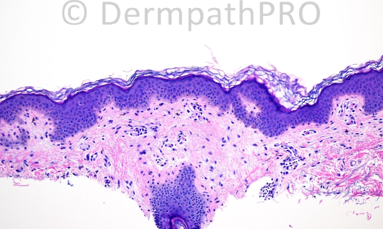
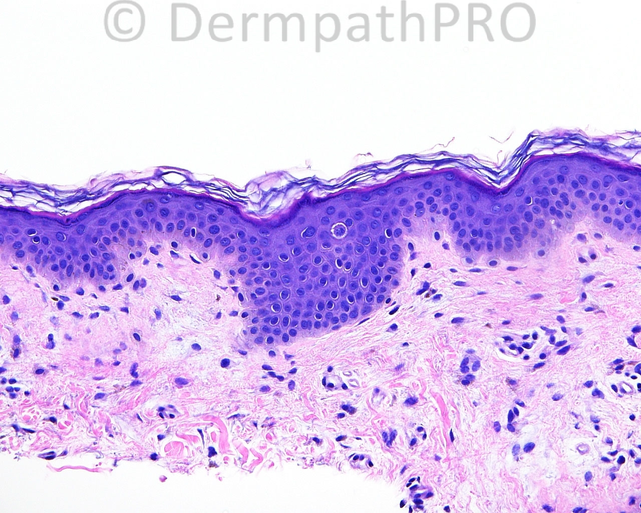
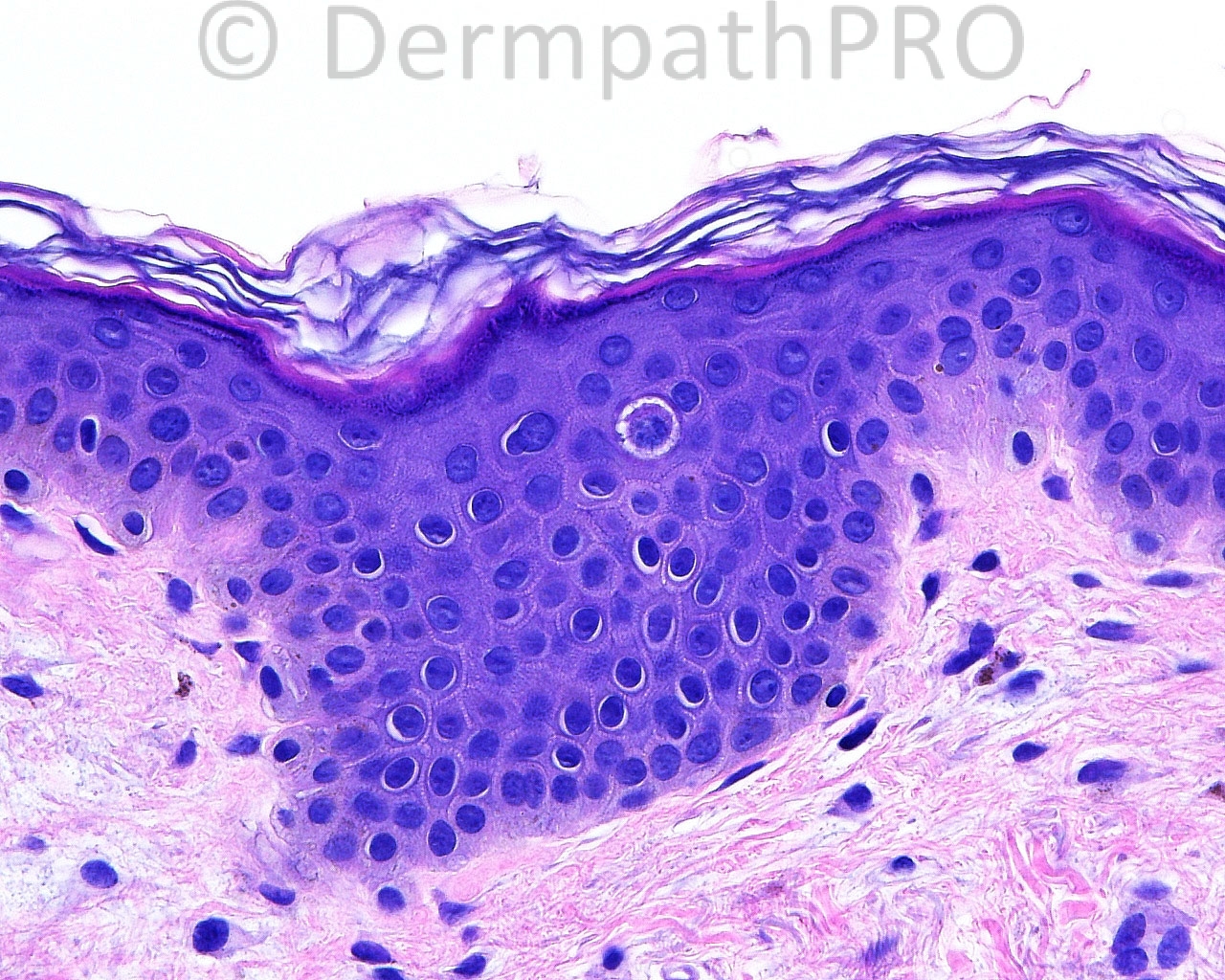
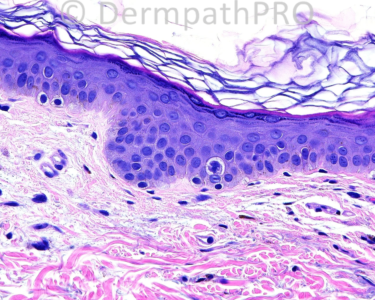
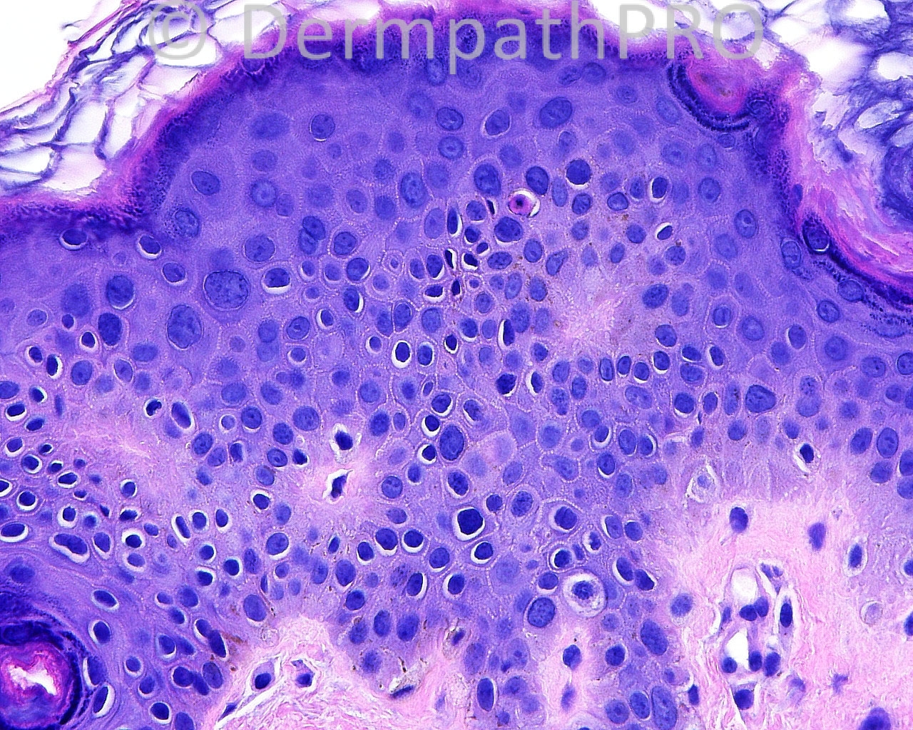
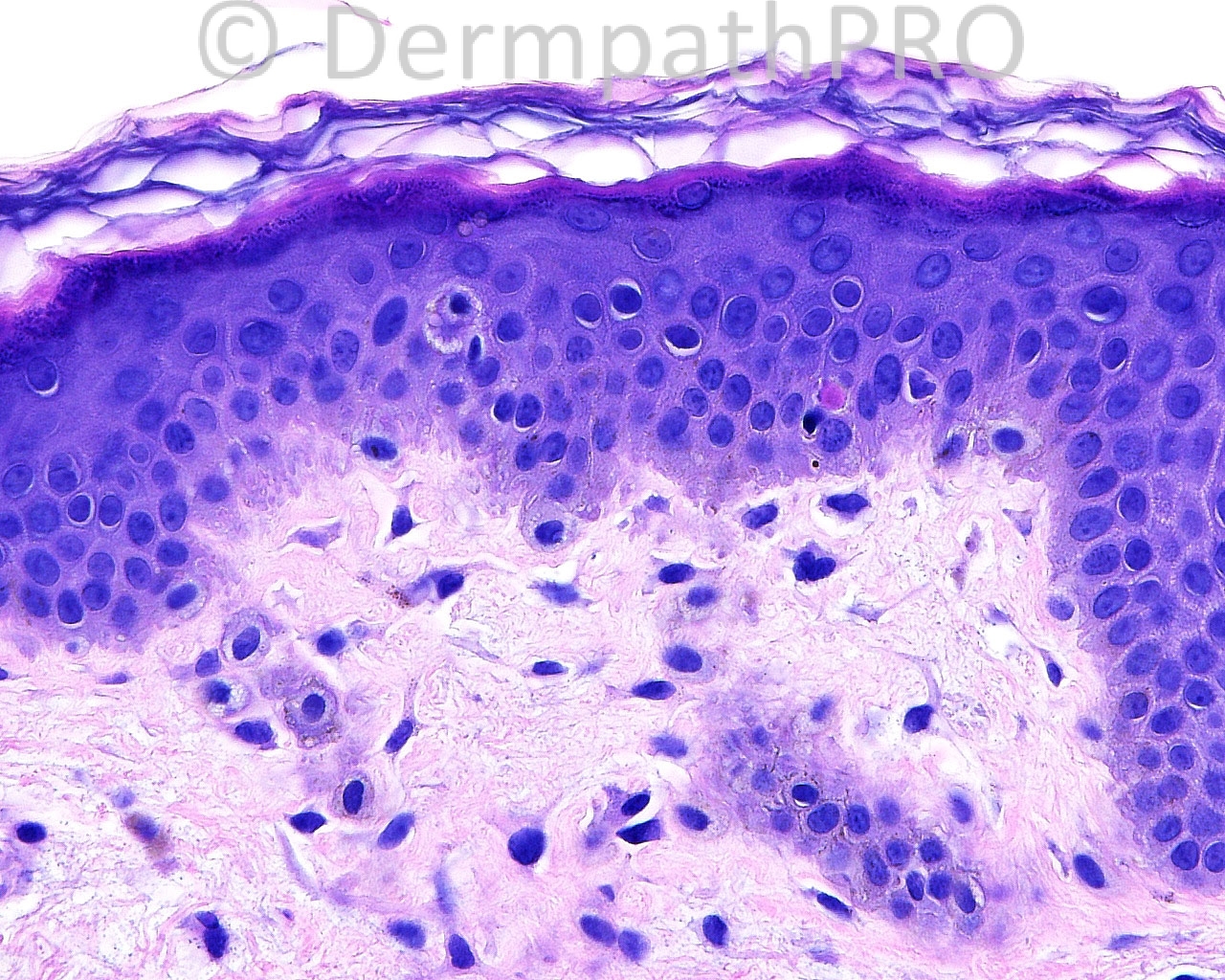
The patient is a 57 year old woman with a biopsy of a lesion on the posterior neck.

Case posted by Dr. Mark Hurt.


  Report Record

User Feedback


amiratawdy

Posted

I can see only some focal thickening of the collagen bundles and some sort of elastic tissue disturbance in the upper dermis so my d.d based as well on the site of the lesion together with the age and sex of the patient: white fibrous papulosis of the neck versus papillary dermal elastolysis for elastic tissue stain to differentiate

Share this comment


Link to comment
share_externally

I see low number of melanocytes, foci of pigmentary incontinence and rare necrotic keratinocytes. Maybe figure 5 represents a large melanocyte at the border of the lesion. I think this may be Vitiligo.

Share this comment


Link to comment
share_externally

Robledo F. Rocha

Posted

There are some signs of photosensitivity, namely, scattered necrotic keratinocytes, solar elastosis, and papillary dermis with stellate fibroblasts and telangiectatic vessels. Moreover, epidermis is atrophic, inflammatory infiltrate is minimal, subtle incontinence of melanin can be found (better seen in image 4), basement membrane seems to be thickened (better seen in image 7), and dermis on the left of image 1 appears clear (suggesting increased mucin). So, my first impression is dermatomyositis.

Share this comment


Link to comment
share_externally

Dr. Mona Abdel-Halim

Posted

Agree with Robledo that this could be DM

Share this comment


Link to comment
share_externally

Eman El-Nabarawy

Posted

[quote name='Robledo F. Rocha' timestamp='1396963346']
There are some signs of photosensitivity, namely, scattered necrotic keratinocytes, solar elastosis, and papillary dermis with stellate fibroblasts and telangiectatic vessels. Moreover, epidermis is atrophic, inflammatory infiltrate is minimal, subtle incontinence of melanin can be found (better seen in image 4), basement membrane seems to be thickened (better seen in image 7), and dermis on the left of image 1 appears clear (suggesting increased mucin). So, my first impression is dermatomyositis.
[/quote]
Agree.

Share this comment


Link to comment
share_externally


Join the conversation

You can post now and register later. If you have an account, sign in now to post with your account.

Guest
Add a comment...

×   Pasted as rich text.   Paste as plain text instead

  Only 75 emoji are allowed.

×   Your link has been automatically embedded.   Display as a link instead

×   Your previous content has been restored.   Clear editor

×   You cannot paste images directly. Upload or insert images from URL.

Loading...

×
×
  • Create New...