Jump to content
In this section we have spot diagnoses posted on a daily basis since June 2010, now over 4000! You can review the archived cases and read the suggested diagnoses by users and the final comment by the contributors.
Case are uploaded each week day by 10 am UK time with the correct diagnosis will generally be posted at 8 pm UK time. Why not view the most recent spot diagnosis and proffer a diagnosis?

Case Number : Case 990 - 9th April Posted By: Guest

Please read the clinical history and view the images by clicking on them before you proffer your diagnosis.
Submitted Date :
   (0 reviews)

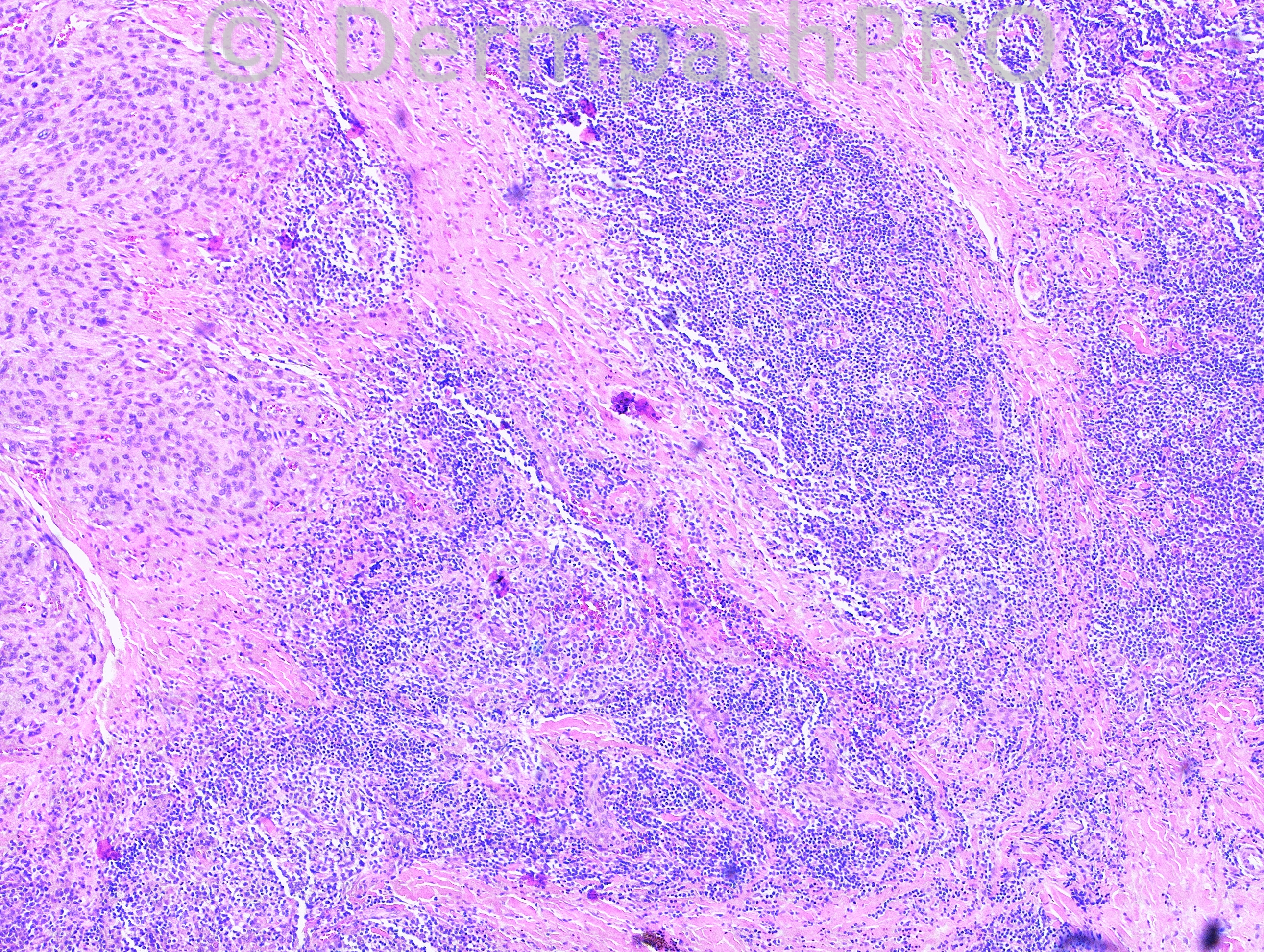
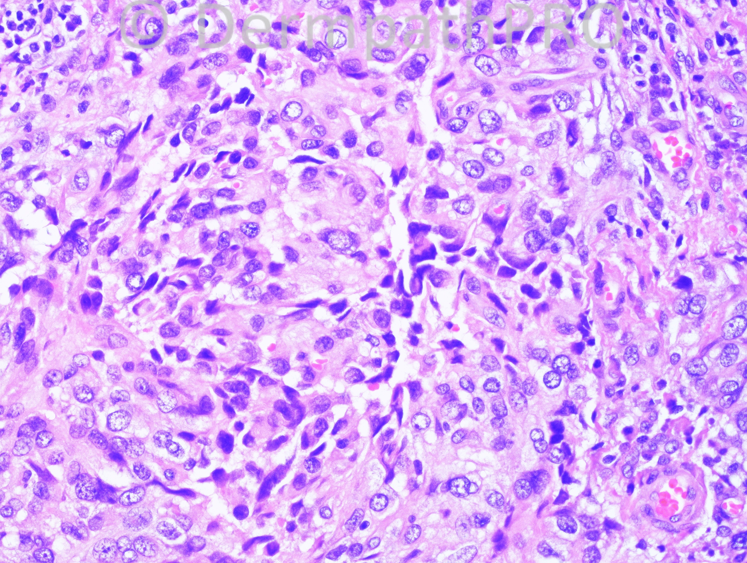
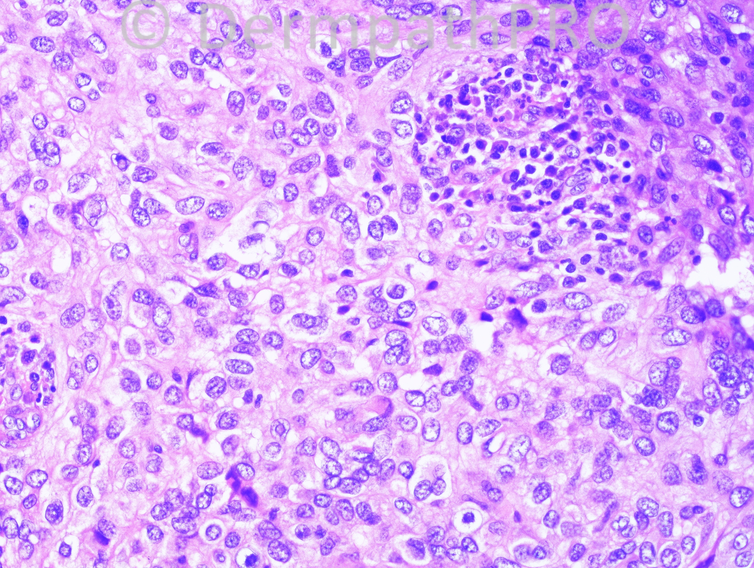
8 year-old boy with right upper arm soft tissue mass.

Case posted by Dr. Hafeez Diwan.

  Report Record

User Feedback


Cellular epithelioid haemangioma vs. epithelioid sarcoma.

Share this comment


Link to comment
share_externally

Eman El-Nabarawy

Posted

I can see Reed Sternberg cells in the last image so I will go for Hodgkin's lymphoma.

Share this comment


Link to comment
share_externally

amiratawdy

Posted

Inflammatory myofibroblastic tumor!!!!

Share this comment


Link to comment
share_externally

Dr. Richard Carr

Posted

Don't know but epithelioid haemangioendothelioma and plexiform fibrohistiocytic tumour were my thoughts.

Share this comment


Link to comment
share_externally

Dr. Richard Carr

Posted

Could also add Kaposi's and follicular dendritic cell tumour.

Share this comment


Link to comment
share_externally

Robledo F. Rocha

Posted

My impression is plexiform fibrohistiocytic tumor. Images show well-circumscribed nodules composed of plump histiocytic cells, some of them with dark wrinkle nuclei, embedded in a fibrous background. Age and site are also typical of this entity.

Share this comment


Link to comment
share_externally

Guest Romualdo

Posted

This neoplasm is composed of nodules of oval and round cells with moderate to abundant pale eosinophilic cytoplasm and uniform vesicular nuclei without mitotic figures or significant atypia. A fibrous capsule circunscribes the lesion and separates the neoplastic nodules. Lymphocytes and plasma cells are seen inside and around the tumor, mostly around it. Red blood cells are present too. These findings are typical of a angiomatoid fibrous histocytoma, solid variant. Location and age are also typical.

Share this comment


Link to comment
share_externally

Eman El-Nabarawy

Posted

I like the idea of follicular dendritic cell sarcoma by Dr Carr!!

Share this comment


Link to comment
share_externally

Dr. Hafeez Diwan

Posted

Angiomatoid fibrous histiocytoma. EWSR1/CREB1 translocation was present.

Share this comment


Link to comment
share_externally

Robledo F. Rocha

Posted

[quote name='Romualdo' timestamp='1397058337']
This neoplasm is composed of nodules of oval and round cells with moderate to abundant pale eosinophilic cytoplasm and uniform vesicular nuclei without mitotic figures or significant atypia. A fibrous capsule circunscribes the lesion and separates the neoplastic nodules. Lymphocytes and plasma cells are seen inside and around the tumor, mostly around it. Red blood cells are present too. These findings are typical of a angiomatoid fibrous histocytoma, solid variant. Location and age are also typical.
[/quote]
Dear Romualdo,
Glad to see you back here. I missed your awesome comments.
Robledo

Share this comment


Link to comment
share_externally

Dr. Richard Carr

Posted

Yes thanks Romualdo for correcting us today and Thanks to Hafeez for sharing this case with us. I have only one example in my slide collection and have never seen a case in routine sign-out but it is certainly one that can appear in exams.

Share this comment


Link to comment
share_externally


Join the conversation

You can post now and register later. If you have an account, sign in now to post with your account.

Guest
Add a comment...

×   Pasted as rich text.   Paste as plain text instead

  Only 75 emoji are allowed.

×   Your link has been automatically embedded.   Display as a link instead

×   Your previous content has been restored.   Clear editor

×   You cannot paste images directly. Upload or insert images from URL.

Loading...

×
×
  • Create New...